Recent advances in DNA methylation in tumorigenesis and diagnosis
Authors
Shikang Liu, Youli Yao, Zhongjun Li, Zhiyi Wang*
- aSpin-X Institute, School of Chemistry and Chemical Engineering, South China University of Technology, Guangzhou, China
- bCollege of Electronic and Information Engineering, Shandong University of Science and Technology, Qingdao, China
- cInstitute of Systems Engineering, Macau University of Science and Technology, Macau, China
- dState Key Laboratory of Optoelectronic Materials and Technologies, School of Materials, Shenzhen Campus of Sun Yat-Sen University, Shenzhen, China.
* Correspondence: Address: Zhiyi Wang, State Key Laboratory of Optoelectronic Materials and Technologies, School of Materials, Shenzhen Campus of Sun Yat-Sen University, Shenzhen 518107, Guangdong Province, China. Email: wangzhy379@mail.sysu.edu.cn (Z. Wang).
MedMat · 2026 · Vol. 3 · No. 1 · pp. 27-45
Abstract
DNA methylation is the process of adding a methyl group to the 5’-carbon of the cytosine residue in the CpG dinucleotide sequence, and it is one of the key components of epigenetic modifications. DNA methylation occurs alongside various biological processes. Abnormal DNA methylation is often associated with the onset of various severe diseases. As a century-old problem that threatens human life, in-depth studies of tumors have revealed abundant evidence of dysregulated DNA methylation. Numerous studies have indicated that DNA hypermethylation tends to impact the transcription of many tumor suppressor genes, leading to the immortalization of tumor cells. In this review, we systematically summarize the progression of DNA methylation in tumor types with current high incidence rates. We also summarize the current clinical methods of DNA methylation detection and treatment and provide an in-depth analysis of the advantages and limitations of these methods. In addition, we discuss the future limitations and challenges faced by DNA methylation research, aiming to advance its clinical application in tumor diagnosis and treatment.
Translations
Long abstracts in additional languages. The English article is the version of record.
中文zh-Hans
DNA甲基化是表观遗传修饰的关键组成部分,指在CpG二核苷酸序列的胞嘧啶残基5'碳原子上添加一个甲基的过程。这一生物学过程伴随多种生命活动发生,其异常往往与各类严重疾病的 onset 密切相关。作为威胁人类生命的世纪性难题,肿瘤研究已积累了大量关于DNA甲基化失调的证据。本综述旨在系统梳理高发病率肿瘤的DNA甲基化演变进程,深入探讨其在肿瘤发生发展中的核心作用,并明确当前临床检测与治疗方法的现状,为后续研究提供理论框架与方向指引。
本文采用系统性文献回顾的方法,对近年来关于DNA甲基化在肿瘤中作用的最新研究成果进行了全面整合与分析。文章重点考察了多种高发病率肿瘤的病理特征及其伴随的表观遗传改变机制,特别是针对CpG位点异常修饰的具体分子路径。研究框架涵盖了从基础生物学过程到临床转化应用的完整链条,详细梳理了当前用于DNA甲基化检测的主流技术手段及相应的治疗策略。通过对比分析不同方法的原理与操作流程,本文构建了评估现有诊断与治疗体系优劣的综合性理论模型,为理解肿瘤表观遗传学提供了多维度的视角。
综述发现,异常DNA高甲基化是导致多种抑癌基因转录沉默的关键机制之一,进而促使肿瘤细胞获得无限增殖能力并实现“永生”。大量研究证据表明,这种特定的表观遗传修饰模式在各类高发肿瘤中普遍存在,且与疾病的发生发展紧密相关。当前临床检测技术已能较为精准地识别这些甲基化标志物,为肿瘤的早期诊断提供了有力工具;同时,针对DNA甲基化的干预策略也在不断拓展。然而,现有方法在具体应用中的敏感性与特异性仍存在差异,不同肿瘤类型对甲基化改变的响应机制也表现出显著的异质性,这要求我们在解读数据时必须结合具体的生物学背景进行审慎分析。
尽管DNA甲基化研究在肿瘤诊断与治疗领域展现出巨大潜力,但仍面临诸多挑战与局限性。当前检测技术的标准化程度、成本效益比以及临床转化的实际可行性仍是亟待解决的问题。此外,不同个体间及不同组织微环境下的甲基化动态变化机制尚不完全明确,限制了其作为通用生物标志物的广泛应用前景。未来研究需致力于克服这些技术瓶颈,优化现有检测方法,并深入探索DNA甲基化调控网络在肿瘤治疗中的具体作用靶点。通过持续的创新与跨学科合作,有望进一步提升其在临床实践中的应用价值,最终实现更精准的肿瘤早期筛查、个性化诊断及靶向治疗的突破。
Françaisfr
La méthylation de l'ADN constitue un composant clé des modifications épigénétiques, définie par l'ajout d'un groupe méthyle au carbone en position 5 du résidu cytosine dans la séquence dinucléotidique CpG. Ce processus biologique se déroule parallèlement à diverses fonctions cellulaires essentielles, et son altération est fréquemment associée à l'apparition de maladies graves. En tant que problème centenaire menaçant la vie humaine, les études approfondies sur les tumeurs ont révélé des preuves abondantes d'une méthylation de l'ADN dérégulée. L'objectif de cette revue systématique est de synthétiser les avancées récentes concernant le rôle de la méthylation dans les types tumoraux à forte incidence, tout en évaluant rigoureusement les méthodes cliniques actuelles de détection et de traitement pour identifier leurs avantages et limites respectifs.
Cette analyse repose sur une synthèse exhaustive des données scientifiques publiées récemment, structurée autour d'un cadre conceptuel intégrant la biologie fondamentale et l'application clinique. L'étude examine en détail les mécanismes moléculaires sous-jacents aux modifications anormales de la méthylation dans divers cancers à haute incidence, en se concentrant spécifiquement sur l'impact des hyperméthylations sur la transcription des gènes suppresseurs de tumeurs. Les auteurs ont méthodiquement recensé et comparé les techniques diagnostiques actuelles utilisées pour détecter ces marques épigénétiques, ainsi que les stratégies thérapeutiques émergentes ciblant ce processus. Le design de cette revue permet une évaluation critique de l'état actuel des connaissances, en mettant en lumière la complexité des interactions entre le profil méthylique et la progression tumorale.
Les résultats principaux indiquent clairement qu'une hyperméthylation de l'ADN tend à affecter négativement la transcription de nombreux gènes suppresseurs de tumeurs, conduisant inévitablement à l'immortalisation des cellules cancéreuses. De nombreuses études confirment que ce phénomène est une caractéristique centrale du développement tumoral dans les cancers courants. Les méthodes cliniques actuelles permettent désormais d'identifier ces anomalies avec un degré croissant de précision, offrant ainsi de nouvelles perspectives pour le diagnostic précoce et la surveillance thérapeutique. Cependant, l'interprétation scientifique souligne également que chaque type tumoral présente des profils méthyliques distincts, ce qui nécessite une approche personnalisée pour maximiser l'efficacité diagnostique et éviter les faux positifs ou négatifs dans un contexte clinique complexe.
La signification de ces travaux réside dans leur capacité à clarifier le rôle central de la méthylation de l'ADN comme outil potentiellement révolutionnaire en oncologie, tout en soulignant honnêtement les défis persistants. Les limitations actuelles incluent des contraintes techniques liées aux méthodes de détection et une compréhension encore incomplète des mécanismes dynamiques à long terme dans différents microenvironnements tumoraux. L'avenir de ce domaine dépendra de la capacité à surmonter ces obstacles, notamment en développant des technologies plus sensibles et accessibles pour le dépistage clinique. Les recherches futures devront se concentrer sur l'intégration de ces marqueurs épigénétiques dans les protocoles thérapeutiques standardisés, afin d'avancer vers une application clinique robuste qui améliore significativement la prise en charge du diagnostic et du traitement des tumeurs malignes à travers le monde.
Españoles
La metilación del ADN es el proceso de añadir un grupo metilo al carbono en la posición 5' del residuo de citosina dentro de la secuencia dinucleotídica CpG, constituyendo uno de los componentes clave de las modificaciones epigenéticas. Este fenómeno ocurre simultáneamente con diversos procesos biológicos fundamentales, y su alteración se asocia frecuentemente con el inicio de diversas enfermedades graves. Como un problema que amenaza la vida humana desde hace un siglo, estudios profundos sobre tumores han revelado abundante evidencia de una metilación del ADN desregulada. El objetivo de esta revisión es resumir sistemáticamente la progresión de la metilación en tipos tumorales con altas tasas de incidencia actuales, analizar las metodologías clínicas existentes para su detección y tratamiento, y proporcionar un análisis profundo sobre sus ventajas y limitaciones específicas.
Este trabajo adopta una metodología de revisión exhaustiva que integra los hallazgos más recientes sobre el papel de la metilación del ADN en la oncogénesis. El diseño se centra en examinar cómo las alteraciones anómalas afectan específicamente a genes supresores tumorales, utilizando como base material científico evidencia acumulada durante décadas de investigación tumoral. Se ha realizado una síntesis crítica de los métodos clínicos actuales para detectar cambios epigenéticos y evaluar estrategias terapéuticas dirigidas a este mecanismo molecular. El marco analítico permite comparar sistemáticamente las diferentes técnicas disponibles, destacando sus principios operativos y su aplicabilidad en contextos clínicos reales, proporcionando así una visión completa del estado actual de la investigación en este campo.
Los hallazgos principales indican que la hipermetilación del ADN tiende a afectar negativamente la transcripción de numerosos genes supresores tumorales, lo que conduce directamente a la inmortalización de las células tumorales. Numerosos estudios confirman que esta alteración es una característica central en tumores con alta incidencia actual, estableciendo un vínculo claro entre el desregulación epigenética y la progresión maligna. Las metodologías clínicas actuales permiten identificar estos patrones anómalos con creciente precisión, ofreciendo nuevas herramientas para el diagnóstico temprano; sin embargo, cada técnica presenta ventajas distintivas junto con limitaciones inherentes que deben considerarse cuidadosamente al interpretar los resultados en diferentes contextos biológicos y clínicos.
La importancia de este trabajo radica en su capacidad para clarificar el papel central de la metilación del ADN como herramienta diagnóstica y terapéutica potencial, mientras reconoce honestamente los desafíos pendientes. Las limitaciones actuales incluyen barreras técnicas relacionadas con las metodologías de detección y una comprensión aún incompleta de los mecanismos dinámicos en diversos microambientes tumorales. El futuro de esta área dependerá de la capacidad para superar estos obstáculos mediante el desarrollo de tecnologías más sensibles y accesibles, así como por investigaciones que profundicen en los mecanismos regulatorios subyacentes. Se espera que abordar estas limitaciones permita avanzar significativamente hacia una aplicación clínica robusta que mejore sustancialmente el diagnóstico temprano y las estrategias terapéuticas personalizadas para pacientes oncológicos a nivel global.
日本語ja
DNAメチル化は、CpGジヌクレオチド配列内のシトシン残基の5'炭素原子にメチル基が付加する過程であり、エピジェネティック修飾の主要な構成要素の一つです。この生物学的プロセスは様々な生命活動と並行して起こり、異常なDNAメチル化は多くの重篤な疾患の発症に関連しています。人類の生命を脅かす世紀にわたる問題である腫瘍に関する深入りの研究により、DNAメチル化の調節不全に関する豊富な証拠が明らかになりました。本レビューでは、現在高い罹患率を持つ腫瘍種におけるDNAメチル化の進行を体系的に要約し、現在の臨床的検出法および治療法の現状を整理するとともに、これらの手法の利点と限界について深入りした分析を提供することを目的としています。
本研究は、最新の文献に基づき、高罹患率腫瘍種におけるDNAメチル化の役割に関する知見を包括的に統合・再構築する体系的レビューアプローチを採用しています。対象とした材料および設計においては、CpG配列内のシトシンへのメチル基付加という分子レベルの機構に焦点を当て、これがどのように転写調節に影響を与えるかを詳細に検討しました。特に、腫瘍抑制遺伝子の発現制御におけるDNA高メチル化の影響と、それが細胞の不死化へと至る経路について重点的に分析しています。また、現在の臨床現場で用いられている検出技術および治療戦略を網羅的にレビューし、各手法のプロトコルや原理に基づいた比較評価を行い、包括的な枠組みの中でその有効性と課題を浮き彫りにしました。
主要な知見として、DNAの高メチル化は多くの腫瘍抑制遺伝子の転写に影響を与え、結果として腫瘍細胞の不死化をもたらすことが多数の研究によって示されています。この現象は、現在高い罹患率を持つ種々の腫瘍において普遍的に見られる特徴であり、疾患の発症と進行に深く関与しています。現在の臨床検出法はこれらのメチル化マーカーを特定する能力を持っており、診断における有用性が確認されていますが、各手法には固有の利点と限界が存在します。科学的解釈においては、腫瘍の種類や状態によってメチル化プロファイルが異なる可能性があり、単一の普遍的なアプローチではなく、個別の文脈に応じた慎重な分析が必要であることが強調されます。
本レビューの意義は、DNAメチル化研究が腫瘍診断および治療における臨床応用を推進する上で重要な役割を果たすことを示しつつも、直面している課題と限界についても率直に論じている点にあります。現在の研究方法には技術的な制約や標準化の欠如といった問題があり、これらが広範な臨床利用を妨げている要因となっています。今後の研究では、これらの制限を克服し、より敏感で特異性の高い検出法の開発や、メチル化動態に関する理解の深化が求められます。将来的には、腫瘍診断と治療におけるDNAメチル化の研究の限界と課題に対処することで、その臨床応用をさらに進め、より効果的な早期発見および個別化医療の実現に向けた新たな道筋を開くことが期待されています。
العربيةar
ميثيلة الحمض النووي هي عملية إضافة مجموعة ميثيل إلى ذرة الكربون في الموضع 5' من بقية السيتوزين ضمن تسلسل الدي نوكليوتيد CpG، وتُعد أحد المكونات الرئيسية للتعديلات اللاجينية. تحدث هذه العملية بالتزامن مع عمليات بيولوجية متنوعة، وغالبًا ما يرتبط حدوث ميثلة الحمض النووي بشكل غير طبيعي بظهور أمراض خطيرة مختلفة. وباعتبارها مشكلة تهدد الحياة البشرية منذ قرن من الزمان، كشفت الدراسات المتعمقة حول الأورام عن أدلة وفيرة على اضطراب ميثيلة الحمض النووي. يهدف هذا الاستعراض إلى تلخيص التقدم في مسار ميثلة الحمض النووي بشكل منهجي عبر أنواع الأورام ذات معدلات الإصابة المرتفعة حاليًا، كما يلخص الأساليب السريرية الحالية للكشف والعلاج، ويقدم تحليلًا متعمقًا لمزاياها وعيوبها.
يعتمد هذا العمل على نهج استعراضي شامل يجمع أحدث النتائج العلمية حول دور ميثلة الحمض النووي في نشوء الأورام وتطورها. تم تصميم الدراسة لاستكشاف الآليات الجزيئية التي تؤثر من خلالها التعديلات غير الطبيعية، مع التركيز تحديدًا على كيفية تأثير فرط الميثيلة على نسخ العديد من جينات كبت الورم. يغطي الإطار التحليلي سلسلة كاملة تتراوح بين العمليات البيولوجية الأساسية والتطبيقات السريرية، حيث تم مراجعة وتقييم الأساليب التشخيصية والعلاجية الحالية المستخدمة في هذا المجال. يتيح تصميم هذه الدراسة مقارنة نقدية للمناهج المختلفة المتاحة، مما يوفر رؤية شاملة حول حالة المعرفة الحالية وكيفية تطبيقها عمليًا.
تُظهر النتائج الرئيسية أن فرط ميثيلة الحمض النووي يميل إلى التأثير سلبًا على نسخ العديد من جينات كبت الورم، مما يؤدي حتمًا إلى خلود الخلايا السرطانية. تشير دراسات عديدة إلى أن هذه الظاهرة شائعة في أنواع الأورام ذات معدلات الإصابة العالية، وتلعب دورًا محوريًا في تطور المرض. تسمح الأساليب السريرية الحالية بالكشف الدقيق عن هذه العلامات البيولوجية، مما يوفر أدوات واعدة للتشخيص المبكر؛ ومع ذلك، فإن كل طريقة لها مزاياها الخاصة وتحدياتها التي يجب أخذها بعين الاعتبار عند تفسير النتائج في سياقات سريرية مختلفة لضمان دقة التشخيص والفعالية العلاجية.
تكمن أهمية هذا العمل في قدرته على توضيح الدور المركزي لدراسات ميثلة الحمض النووي كأداة محتملة للثورة في مجال الأورام، مع الاعتراف الصريح بالتحديات الحالية. تشمل القيود الحالية عقبات تقنية تتعلق بطرق الكشف وفهم غير مكتمل للآليات الديناميكية عبر بيئات الورم المختلفة. يعتمد مستقبل هذا المجال على القدرة على التغلب على هذه العقبات من خلال تطوير تقنيات أكثر حساسية وقابلة للوصول، بالإضافة إلى أبحاث تركز على فهم آليات التنظيم الجيني بشكل أعمق. يُتوقع أن معالجة هذه القيود ستسمح بتقدم كبير نحو تطبيق سريري قوي يحسن بشكل ملحوظ الكشف المبكر والعلاجات المخصصة للمرضى المصابين بالأورام حول العالم.
Keywords
Full Text
1. Introduction
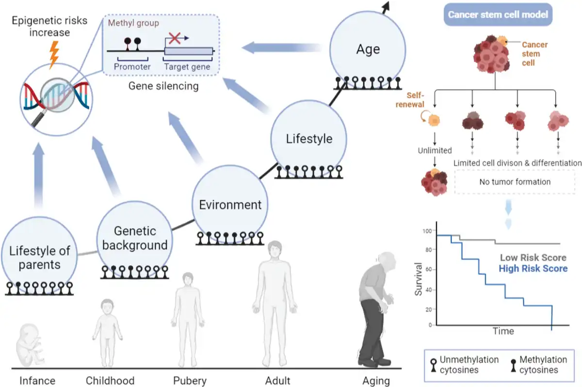
Epigenetics is a discipline that reveals how life activities achieve heritable changes through modifications of the genomic sequence[1]. This process does not result in changes in the genome sequence, but brings about dynamic regulation of life activities, such as regulating biological growth[2], disease[3,4], and death. DNA methylation, as a form of epigenetic modification, has captured the attention of numerous researchers. During genome transcription, functional group modifications of DNA sequences exert different roles in cellular activities, such as the activation and deactivation of cis-acting elements and the stabilization of mRNA. Specifically, DNA methylation performs a crucial role in embryonic development, imprinting, and X-chromosome inactivation[5–6–7]. The DNA methylation process requires specific enzymatic reactions, namely DNA methyltransferases (DNMTs). These enzymes transfer the methyl group from S-adenosylmethionine (SAM) to the cytosine bases in CpG dinucleotides within promoter and regulatory regions[8]. CpG dinucleotides are concentrated in short CpG islands (CGIs), commonly referred to as “CGIs.” In humans, CGIs account for approximately 60% of gene promoters, which will undergo methylation during the development of life, leading to long-term gene silencing. A typical example of naturally occurring CGI methylation is X-chromosome inactivation and imprinted genes[9,10]. Unfortunately, DNA methylation has been widely reported to be associated with the silencing of tumor suppressor genes and differentiation genes in various cancers[11].
In 1925, DNA methylation was initially identified in bacteria. Over the subsequent decades, the biological significance of DNA methylation was not given significant attention by researchers[10]. It was not until the advancement of high-throughput sequencing technologies that a deeper understanding of the regulatory mechanisms of DNA methylation was established[12]. As research into DNA methylation has been progressing, it is involved in various biological processes, ranging from gene regulation to cell growth[13], and the mechanisms underlying major diseases[14,15]. DNA methylation regulates gene expression by interacting with cis-acting elements. In normal cells, DNA methylation maintains genomic stability by suppressing the transcription of transposons and other repetitive elements. The dynamic regulatory mechanisms of DNA methylation are also crucial for maintaining normal stem cells and progenitor cells[16,17]. Analysis of embryonic stem cells has revealed that even in embryonic stem cells completely lacking DNA methylation, over 98% of methylated genes remained inactive, suggesting the presence of a potential mechanism that helps maintain their silence in somatic cells[18]. This mechanism appears to utilize SAM as a methyl donor, carried out by 3 DNMTs: DNMT1, DNMT3a, and DNMT3B[19,20]. DNA methylation patterns are initially established by DNMTs DNMT3A and DNMT3B and are subsequently maintained by DNMT DNMT1 during DNA replication. In tumors, dysregulation of DNMT activity increases the likelihood of genomic instability, thereby conferring the ability for unlimited proliferation to cells[21–22–23]. Of note, as it is a problem at the genetic level, DNA methylation brings about many challenges in the process of cancer treatment.
Figure 1.
Illustration of epigenetic modifications in cancer.
In this review, we provide an in-depth summary of the role of DNA methylation at different stages of tumor formation. Moreover, based on its properties, we describe the current status of DNA methylation-related diagnostic and therapeutic applications as well as the inevitable challenges. To the best of our knowledge, we further prospect for more advanced and effective therapeutic strategies based on the combination of conventional therapeutic means and some current novel scientific technology, such as nanotechnology.
2. DNA methylation in cancer
Tumors tend to be classified as low, intermediate, and high grades based on their different invasive potentials, which also reflect the malignant nature of the tumor tissue. In this review, we concentrate on the division of tumors into 2 categories, benign and malignant. Noncancerous growths in the body are referred to as benign tumors. Their biological characteristics are more akin to hyperplastic tissue, which does not spread to other organs of the body. In comparison, malignant tumor cells with strong self-renewal capabilities present a more dangerous scenario. Due to their uncontrolled cell division, they continuously produce poorly differentiated offspring cells. Malignant tumors are able to disrupt the cellular connections of adjacent tissues through the enzymes they produce, enabling cellular metastasis. They may also spread or metastasize to other parts of the body via the bloodstream and lymphatic system, forming new tumor loci[24]. In the following subsections, we will summarize the research progress of DNA methylation in benign and malignant tumors, respectively.
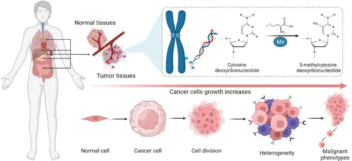
Figure 2.
The intrinsic mechanism of DNA methylation in tumorigenesis.
2.1 DNA methylation and tumorigenesis
In tumor cells, we have observed alterations in the modification of genetic material, some of which appear to directly influence tumor growth and other characteristics[25]. Over the past few decades, numerous studies have shown that alterations in the distribution pattern of 5-methylcytosine (5mC) can be used to distinguish cancer cells from normal cells (Figure 1). At least 3 major pathways have been identified. The first is through widespread hypomethylation of the cancer genome. The second is the local hypermethylation of tumor suppressor gene promoter regions. Third, sequences containing 5mC may undergo mutations directly through deamination, ultraviolet exposure, or other carcinogen exposures[26–27–28]. Notably, these 3 alterations often occur simultaneously to trigger cancer, suggesting that alterations in epigenetic homeostasis are central to cancer evolution.
Hypermethylation of promoter regions has been observed in nearly all types of human cancers. As shown in Figure 2, these hypermethylated phenomena suppress the transcription of important tumor suppressor genes, including cell cycle regulators, DNA repair proteins, and antiapoptotic factors, some of which play functional roles in normal cell development[29,30]. Substantial evidence suggests that DNA methylation observed in cancer may originate from a small subset of cells. In the early stages of human colorectal carcinogenesis, not only are target sites in normal tissues partially methylated, but target sites in polyps are also extensively modified[31,32]. Colonic epithelium is formed by adult stem cells located in crypts, and this tissue is capable of rapid self-renewal. Therefore, the methylation patterns observed in normal colon reflect those in stem cells. Although DNA methylation on individual molecules from a single crypt appears relatively uniform, methylation levels vary significantly between different crypts[33]. This suggests that within each tissue, there may be cells with very low methylation levels, while other cells are highly methylated in these CGI regions, promoting subsequent dominance in growth selection.
On the other hand, DNA methylation modifications also provide a potential factor for genomic mutations in tumor cells. Similar modifications may induce an irreversible constitutive heterochromatin state, but were unable to activate key differentiation genes. Therefore, these cells acquired a relatively proliferative state. While it may not be sufficient to generate tumor tissues, it is likely to provide the necessary circumstances for cells undergoing transformation through prior genetic susceptibility or spontaneous mutations. The concept that these cells accumulate DNA methylation during the aging process and become priority targets for transformation is supported by the following observations. Hematopoietic stem cells from humans indeed undergo a natural aging process, characterized by an increase in cell number and a decline in lymphoid differentiation capacity, ultimately resulting in a myeloid-dominant phenotype[34]. This aging process is associated with methylation and demethylation events commonly observed in tumors[35]. This suggests that these methylation changes may exert a role in promoting stem cell renewal and inhibiting differentiation. The most significant and earliest identified change in DNA methylation patterns in cancer cells is a reduction in methylated regions[36], which is currently classified as genome-wide hypomethylation through genome-wide analysis[37–38–39]. Although the full consequences of these losses remain to be clarified, DNA demethylation may lead to genomic instability and increased chromosomal aneuploidy, which are hallmark features of cancer.
Indeed, DNA hypomethylation actively contributes to increased chromosome fragility[40,41]. The loss of DNA methylation appears to be coupled with transcriptional activation, facilitating the transcription of repetitive sequences, transposable elements, and oncogenes[42,43]. Comparatively, the most well-known mechanism by which DNA methylation drives cancer is focal hypermethylation of tumor suppressor gene promoters. Typically, DNA hypermethylation occurs in CpG-rich regions or CGIs near the transcriptional start sites of abnormally silenced genes. In tumors, promoter hypermethylation can disrupt hundreds of genes, a mechanism that holds for almost all types of cancer[25,26,28,44]. In fact, through more in-depth DNA methylation analysis of various tumor types, the incidence of epigenetic changes seems to exceed the number of genetic mutations in human tumors[45,46]. Furthermore, promoter regions’ hypermethylation appears in almost all genes involved in signaling pathways concerning tumorigenesis. The participation of such a massive number of genes presents an unprecedented challenge in the field of cancer epigenetics: which silencing events are truly critical to the tumorigenesis process? Detecting the critical role of each gene in tumorigenesis and progression through functional knockout experiments is a challenging task.
2.2 DNA methylation facilitating malignant tumor phenotypes
With the progress and development of society, the contradiction between tumor diseases and human health has become increasingly prominent. Cancer has become the principal cause of mortality in 172 countries worldwide[47]. In China, cancer has gradually developed into one of the most common causes of death among the general public and a serious public health problem. Lung cancer, colorectal cancer (CRC), gastric cancer, liver cancer, and breast cancer were the 5 most common malignancies, accounting for 57.4% of recent cancer cases, meanwhile, the 5 cancers with the highest mortality rates were, respectively, lung cancer, liver cancer, gastric cancer, CRC, and esophageal cancer, accounting for 69.3% of all deaths[48]. As a malignant disease has not been cured, epigenetic modifications, especially DNA methylation, exert a critical and nonnegligible role in many factors that promote its development. In this subsection, we summarize some recent research advances on DNA methylation associated with common malignant tumors.
2.2.1 Lung cancer
Lung cancer is the most common cancer with a high mortality rate, exhibiting a significant gender preference, possibly because it is closely related to an individual’s lifestyle. Previous studies have reported that CGIs of some specific genes are frequently methylated in lung cancer, including CDKN2A, RASSF1A, RARβ, GSTP1, APC, DAPK, and TIMP3[48–49–50–51–52–53]. The frequency of CGI methylation in specific genes in malignant tumors typically ranges from 10% to 80% (Figure 3A, B). Studies have shown that methylation of the SHOX2 and RASSF1A genes is strongly associated with lung cancer[54–55–56]. Kneip et al.[57] found that SHOX2 gene methylation demonstrated 90% specificity and 60% sensitivity in distinguishing normal tissues from lung cancer. Meanwhile, Schmidt et al.[55] analyzed that SHOX2 gene methylation appears more suitable for diagnosing lung adenocarcinoma and small cell lung cancer (SCLC), with diagnostic sensitivities of 82% and 97%, respectively. The RASSF1A gene is an experimental tumor suppressor gene whose methylation is closely associated with non-SCLC (NSCLC)[58,59]. Furthermore, researchers extracted lung tissue from normal subjects, NSCLC patients with cancer metastasis, and NSCLC patients without cancer metastasis for comparison (Figure 3C). Yu et al.[60] found that the EPHB6 gene undergoes methylation in lung cancer patients, and the higher the methylation level of the EPHB6 gene, the higher the risk of cancer metastasis. Besides, Ma et al. found that the TMEM88 gene exhibits hypermethylation in 12 NSCLC samples. Compared with the corresponding 12 noncancer samples, patients with higher methylation levels had shorter survival times[61]. Analysis suggests that the TMEM88 gene may act as a tumor suppressor gene, with its methylation closely associated with poor prognosis in NSCLC. These studies imply that methylation of tumor suppressor genes is an essential factor in the progression of lung cancer, indicating that demethylation of tumor suppressor genes can prevent the malignant development of lung cancer.
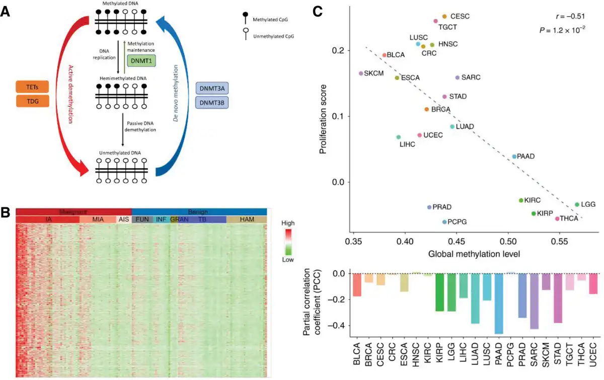
Figure 3.
Patterns of DNA methylation in lung cancer. (A) Schematic illustration of DNA methylation dynamics of DNMTs and TETs[62]. Copyright 2022, MDPI. (B) Heatmap showing randomly selected 1000 hypermethylation regions for representative lung cancer and benign tissue samples. Subtypes from left to right are IA (n = 33), MIA (n = 19), AIS (n = 8), FUN (n = 11), INF (n = 9), GRAN (n = 4), TB (n = 25), and HAM (n = 21) [63]. Copyright 2019, Ivyspring International Publisher. (C) Correlation between genomic methylation levels and cell proliferation markers[64]. Copyright 2019, Springer Nature. TDG, AIS, adenocarcinoma in situ; FUN, fungal infection; GRAN, inflammatory granuloma; HAM, hamartoma; IA, invasive adenocarcinoma; INF, inflammation; MIA, minimally invasive adenocarcinoma; TB, tuberculosis; TDG, thymine–DNA glycosylase; TETs, 10 to 11 translocation enzymes
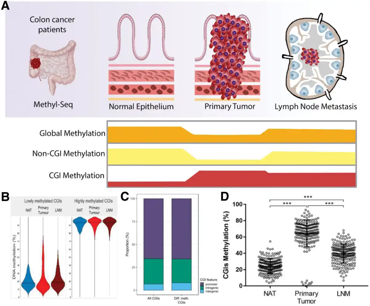
Figure 4.
Modes of DNA methylation within CRC. (A) Methylation levels of global, non-CGIs, and CGIs at different periods in CRC. (B) CGI methylation levels in NAT, primary tumor, and LNM samples of CRC patients. (C) The proportion of different CGI features represented in all analyzed CGIs (n = 7009) and differentially methylated CGIs (n = 246). (D) Methylation percentage of 204 CGIs in NAT, primary tumor, and LNM samples (1.5 kb upstream to coding genes) (***P < 0.001, Kruskal–Wallis followed by Dunn test)[65]. Copyright 2020, MDPI. LNM, lymph node metastasis; NAT, normal adjacent tissue.
2.2.2 Colorectal cancer
Colorectal cancer is the third most common malignancy worldwide, which has a high mortality rate. Alterations in gene expression, including the activation of oncogenes and the inactivation of tumor suppressor genes, are the primary causes of CRC formation and progression[66,67]. In addition to cumulative changes in DNA sequences, the dysfunction of epigenetic regulatory systems also results in abnormal formation of carcinogenesis in colonic epithelial cells[68,69]. Recent studies have reported the differences in DNA methylation between primary and metastatic cancers (Figure 4A-D)[70,71]. The CDKN2A gene is more frequently methylated in poorly differentiated[72], lymph node-metastatic CRC[73], and advanced CRC[74]. Methylation of the APC and ESR1 promoters may significantly affect the metastatic potential of colon cancer. Methylated APC is more common in liver metastasis than in primary CRC[75], while ESR1 methylation has been detected in lymph nodes resected from Union for International Cancer Control stage I and II CRC patients[76]. Hibi et al. observed that HACE1 functions as an E3 ubiquitin ligase with the potential to confer a malignant phenotype. An association has been noted between increased methylation of HACE1 and tumor size. Interestingly, tumors with methylated HACE1 tend to preferentially undergo lymph node metastasis[77]. Specifically, metastatic tumor cells utilize DNA hypermethylation to suppress the expression of specific tumor suppressor genes to evade cell apoptosis and acquire a metastatic phenotype. Moreover, INF regulatory factor 8 (IRF8) is a transcription factor of the IRF family, known as a regulator of the Fas-mediated apoptosis pathway and a metastasis suppressor in solid tumors[78]. It is silenced through epigenetic mechanisms in various cancers, including CRC[79]. The IRF8 promoter is methylated in metastatic CRC cell lines but not in primary CRC cells. Its gene expression is negatively correlated with antiapoptotic and metastatic phenotypes in vitro[78]. These results suggest that metastatic tumors employ DNA hypermethylation to suppress IRF8 expression, thereby evading apoptotic cell death and acquiring a metastatic phenotype. RARRES1 is also a tumor suppressor gene whose expression is frequently downregulated in various malignant tumors due to DNA hypermethylation. Downregulation of RARRES1 is thought to be associated with the progression of CRC[80]. However, while many genes are inactivated in CRC cell lines, there is significant variability between different cell lines, suggesting that not all inactivated genes are directly related to tumorigenesis.
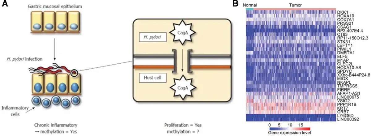
Figure 5.
DNA methylation in gastric cancer. (A) Infectious condition and pathogenicity of Helicobacter [81]. Copyright 2014, WJG Press. (B) The expression profile of the most significant 30 methylation-related differentially expressed genes between normal (n = 32) and gastric cancer samples (n = 375)[82]. Copyright 2020, BioMed Central.
2.2.3 Gastric cancer
Gastric cancer is one of the most common malignant tumors worldwide and a leading cause of cancer-related deaths in Asia and certain European countries[83]. It has been well-known that the main etiological risk factor for gastric cancer is Helicobacter pylori infection[84]. Previous studies have suggested that chronic inflammation induced by H. pylori infection may lead to abnormal DNA methylation, rather than bacteria themselves (Figure 5A)[85]. Another study utilizing the Mongolian gerbil model further confirmed that both H. pylori infection and abnormal methylation of the gastric mucosa can promote the development of gastric cancer[86]. Several studies have also provided evidence that epigenetic alterations facilitate gastric tumorigenesis. Various tumor suppressors and tumor-associated genes, including APC, CDH1 (E-cadherin), CHFR, DAPK, GSTP1, p16, and RUNX3, are known to be silenced in gastric cancer due to hypermethylation[87–88–89] In addition, methylation is frequently observed in the precancerous stages of gastric cancer, suggesting that aberrant methylation occurs early in the multistep process of gastric carcinogenesis[90–91–92]. Accumulation of aberrant methylation is thought to promote carcinogenesis by activating common cancer pathways (Figure 5B). Some negative regulators of Wnt signaling, including SFRP1, DKK2, and WIF1, are frequently methylated in gastric cancer[[93,94]].Methylation of RASSF family genes is considered an alternative mechanism to replace KRAS mutations in signaling pathways, leading to the activated Ras signal pathway[95]. Commonly, the development of gastric cancer results from a combination of external factors, infection, and epigenetic inheritance, whose in-depth study is critical to the survival of gastric cancer patients.
2.2.4 Hepatocellular carcinoma
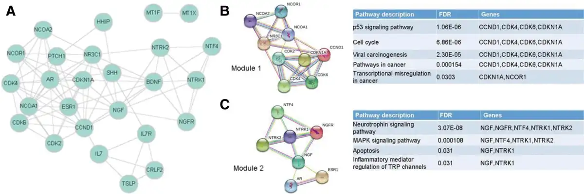
Hepatocellular carcinoma (HCC) is a primary malignancy of the liver, which is the third leading cause of cancer-related deaths for all humankind[97]. An increasing trend in the incidence and mortality rates of HCC has been observed in most countries[98]. It is well known that high methylation of the E-cadherin gene is commonly reported in HCC cases[99,100]. Jiang et al. demonstrated that high methylation of the CpG sites in the E-cadherin gene promoter region is associated with multidrug resistance in HCC. Indeed, E-cadherin expression is implicated in altered doxorubicin uptake, reduced P-glycoprotein expression, and promotion of apoptosis. Therefore, inhibition of E-cadherin facilitates cancer cells’ evasion of apoptosis and acquisition of multidrug resistance (Figure 6A)[101]. Another pathway participating in HCC development is the Wnt/β-catenin signaling pathway. Hypermethylation of regions encoding Wnt regulatory factors (such as SRY box 1 [SOX1] and SRY box 17 [SOX17]) leads to aberrant activation of the Wnt/β-catenin signaling pathway, thereby enhancing the likelihood of cell proliferation and survival[100,102,103]. Shu et al. found that the PRDM5 gene was silenced in 63% of HCC cases. As a stress response gene, PRDM5 counteracts the Wnt/β-catenin signaling pathway, which acts as an epigenetic modifier to suppress the expression of multiple oncogenes[104]. Additionally, SFRP1 is another antagonist of the Wnt/β-catenin pathway, and its methylation levels are higher in HCC specimens than in nontumor tissues. Epigenetic suppression of SFRP1 in HCC results in enhanced cellular proliferative potential and overexpression of oncogenes such as c-Myc and cyclin D1[105]. Growing evidence supports that methylation profiling of HCC reveals significant differences between tumorous liver tissue and adjacent noncancerous liver tissue (Figure 6B, C)[100]. Multiple studies have reported aberrant hypermethylation of some tumor suppressor genes (for instance, GSTP and SOCS1) in HCC, which leads to the loss of cell cycle checkpoints and activation of cell proliferation[106,107]. During hepatocarcinogenesis, many tumor suppressor genes are silenced by DNA methylation, for which hypermethylation of CGIs at promoters is an important mechanism for their inactivation.
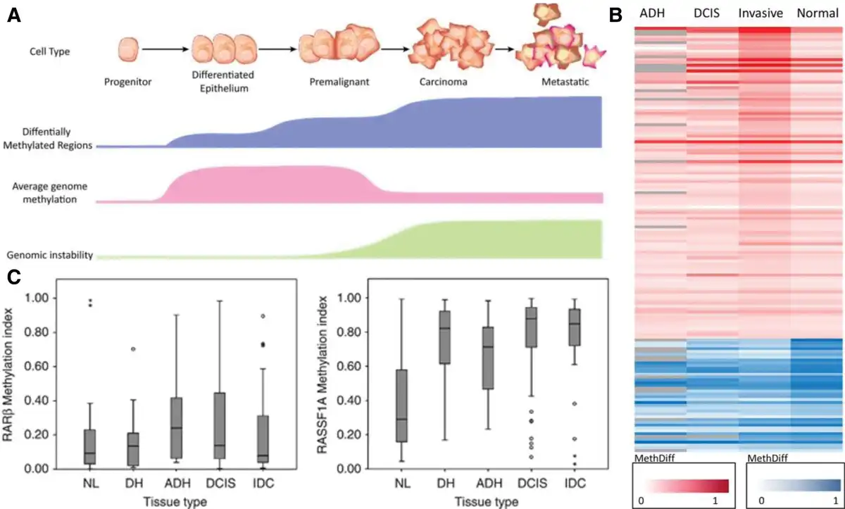
Figure 7.
DNA methylation in breast cancer. (A) Progression of breast cell epigenome from progenitor cells to malignancy[108]. Copyright 2012, BioMed Central. (B) Differentially methylated genes in ADH, DCIS, and invasive breast cancer compared with healthy tissue[109]. Copyright 2020, Springer Nature. (C) RARβ2 and RASSF1A vs tissue type (normal breast epithelia [NL], ductal hyperplasia [DH], atypical ductal hyperplasia [ADH], ductal carcinoma in situ [DCIS], and invasive ductal cancer [IDC])[110]. Copyright 2013, Springer Nature.
2.2.5 Breast cancer
The progression of breast cancer is the result of a multistage carcinogenic process. Clinical data indicate that breast cancer begins as a less invasive hormone-dependent type, which gradually progresses to a highly aggressive hormone-dependent phenotype (Figure 7A-C)[111]. Among the methylated genes in breast cancer, there are plenty of tumor suppressor genes, such as p16, whose methylation is reported to silence the gene and block cell growth regulatory signals[112,113]. Methylation of p16 prepared from the plasma of breast cancer patients has been reported to be involved in the lymph node metastasis process[114]. Damage response genes are another type of methylated genes reported in breast cancer, such as BRCA1[115] and mismatch repair genes hMLH1 and hMSH2[116]. Disruption of repair genes may elevate sporadic mutation frequency, a hallmark of tumorigenesis. Moreover, members of the steroid receptor gene family, including the estrogen receptor[117] and RARβ2 receptors, have been reported to undergo methylation in a subset of breast cancer cases[118]. The interaction of RARβ2 receptor with retinoic acid may resist tumor proliferation, whose silencing provides a selective benefit to late-stage breast cancer cells. E-cadherin and TIMP3 have also been detected to be methylated in breast cancer, thereby facilitating the metastasis of breast cancer cells[119,120]. APC gene, as a tumor suppressor gene with multiple cellular functions, plays a crucial role in the Wnt signaling pathway, cell–cell adhesion, and apoptosis[121]. DAPK1 is a proapoptotic serine/threonine protein kinase gene, acting as a positive mediator of γ-interferon (IFN-γ) mediated programmed cell death[122]. GSTP1 is a gene that plays a key role in detoxifying exogenous substances, carcinogens, pesticides, and various environmental pollutants. In the MAPK signaling pathway, GSTP1 participates in the regulation of cell survival and death signals[123]. Phosphatase and tensin homolog (PTEN), acting as a dual lipid and protein phosphatase, regulates the PI3K/AKT signaling pathway through its target PIP3 in breast cancer[124]. DCR1 and DCR2 are genes encoding membrane receptors that bind TNF-related apoptosis-inducing ligand and inhibit the TNF-related apoptosis-inducing ligand apoptosis pathway[125]. Through extensive analysis of clinical samples, researchers discovered that the aforementioned tumor-related genes were abnormally methylated in breast cancer cells. For the reason that tumorigenesis involves the coordinated expression and repression of multiple genes, there must be common mechanisms responsible for coordinating these changes. Understanding the mechanisms involved in this project is a considerable issue in breast cancer biology and treatment.
2.2.6 Others
Following the rapid advancement of epigenetics research in recent years, several unique DNA methylation patterns have been identified as playing a significant role in the progression of malignant tumors. The first type involves alterations in DNA methylation patterns caused by gene mutations. In 2010, Noushmehr et al. defined a CGI methylator phenotype (G-CIMP) in glioblastoma. The methylation pattern is highly correlated with mutations in isocitrate dehydrogenase 1 (IDH1) in tumor cells[126]. Mutated IDH1 catalyzes the conversion of α-ketoglutarate (α-KG) to D-2-hydroxyglutarate, structurally similar compounds. The latter competitively inhibits α-KG-dependent epigenetic modifiers (eg, histone and DNA demethylases), leading to genome-wide hypermethylation[127]. Studies indicate that G-CIMP⁺ patients tend to be younger, exhibit longer overall survival, and possess favorable prognostic expectations[128]. IDH1 mutations are also prevalent in hematologic malignancies, including myelodysplastic syndrome (MDS) and T-cell lymphoma[129,130], as well as solid tumors such as chondrosarcoma[131] and cholangiocarcinoma[132]. Meanwhile, dysregulation of noncoding RNAs (ncRNAs) has also been identified as a vital factor influencing multiple cellular characteristics in tumor cells. The ncRNAs are able to exert a synergistic effect by interregulating with DNMT3B to influence the progression of tumor cells[133]. Recent comprehensive studies indicate that the miR-29 family plays a pivotal role in the pathogenesis of glioma[134]. Xu et al. demonstrated that DNMT3B expression in the U87MG glioblastoma cell line was effectively inhibited by miR-29-mediated mRNA degradation. Exogenous miR-29 introduction significantly impedes U87MG cell proliferation and migration while stimulating apoptosis[135]. Wang et al.[136] reached similar conclusions when investigating the relationship between miR-29 and DNMT3B in pancreatic cancer. Besides that, the event that DNA methylation induced by infected viruses promotes tumor cell progression is gradually confirmed. Viral infection also induces aberrant DNA methylation in the human genome leading to carcinogenesis[137], such as HCC related to hepatitis B virus and hepatitis C virus[138] as well as cervical carcinoma[139] and head and neck squamous cell carcinoma[140], both of which are associated with human papillomavirus. Most of these aberrant DNA methylation events are induced by viral infection. Consequently, understanding the mechanisms of epigenetic regulation in cancer therapy is critical for optimizing existing therapeutic strategies, improving the prognosis of cancer patients, and exploiting the reversible properties of epigenetic modifications against cancer (Table 1).
Table 1
Genes with aberrant methylation in various cancers.
| Gene | Tumor type | Molecular regulation | Function | References |
|---|---|---|---|---|
| CDKN2A | Lung cancer, breast cancer, colorectal cancer, gastric cancer, thyroid cancer, hepatocellular carcinoma, bladder cancer, cervical cancer, leukemia | Hypermethylation | Cell cycle regulation, tumor metastasis | [48,88,141–142–143–144–145–146–147] |
| E-cadherin | Breast cancer, gastric cancer, hepatocellular carcinoma, cervical cancer, bladder cancer | Hypermethylation | Tumor metastasis, drug resistance | [88,100,119,142,148] |
| RARβ | Lung cancer, oropharyngeal squamous cell carcinoma, cervical cancer, colon cancer | Hypermethylation | Tumor suppressor genes | [12,149–150–151] |
| MGMT | Glioblastoma multiforme, colorectal cancer, pancreatic cancer | Hypermethylation | Drug resistance | [152–153–154–155] |
| SEPT9 | Gastric cancer, colorectal cancer, prostate cancer, cervical cancer, liver cancer | Hypermethylation | Diagnostic marker | [156–157–158–159–160] |
| SHOX2 | Prostate cancer, lung cancer, melanocytoma | Hypermethylation | Diagnostic marker | [57,158,161,162] |
| VIM | Urothelial carcinoma, bladder cancer | Hypermethylation | Diagnostic marker | [163,164] |
| DAPK | Lung cancer, breast cancer, gastric cancer, thyroid cancer, cervical cancer | Hypermethylation | Tumor suppressor genes | [50,88,165–166–167] |
| RASSF1A | Lung cancer, breast cancer, thyroid cancer, gastric cancer, bladder cancer | Hypermethylation | Tumor suppressor genes | [95,142,168–169–170] |
| GSTP1 | Lung cancer, breast cancer, gastric cancer, hepatocellular carcinoma, prostate cancer | Hypermethylation | Cell survival | [50,88,171–172–173] |
| APC | Lung cancer, colorectal cancer, breast cancer, cervical cancer | Hypermethylation | Tumor suppressor genes | [52,75,174,175] |
| PTEN | Breast cancer, thyroid cancer, hepatocellular carcinoma | Hypermethylation | Tumor suppressor genes | [166,171,176] |
| PAX1 | Cervical cancer, oral squamous cell carcinoma, esophageal squamous cell carcinoma, parathyroid adenomas | Hypermethylation or hypomethylation | Tumorigenesis | [177–178–179–180–181] |
| TIMP3 | Thyroid cancer, oral cancer, lung cancer, cavity cancer, head and neck cancer | Hypermethylation | Tumor metastasis | [182–183–184–185–186] |
| RUNX3 | Gallbladder cancer, renal cancer, breast cancer, prostate cancer, lung cancer | Hypermethylation | Tumor suppressor genes | [187–188–189–190–191–192] |
| SFRP1 | Colorectal cancer, ovarian cancer, pancreatic cancer, breast cancer, prostate cancer | Hypermethylation | Tumor metastasis | [193–194–195–196–197] |
| CDKN2B | Colorectal cancer, leukemia | Hypermethylation | Tumor suppressor genes | [147,198,199] |
| LOX | Melanoma, nasopharyngeal carcinoma | Hypermethylation | Tumor suppressor genes | [200,201] |
| SOCS1 | Prostate cancer, colorectal cancer, pancreatic cancer, liver cancer, myeloma, leukemia | Hypermethylation | Tumor metastasis | [106,202–203–204–205–206] |
| ESR1 | Breast cancer, ovarian cancer, bladder cancer, colorectal cancer | Hypermethylation | Diagnostic marker | [207–208–209–210] |
| TMEFF2 | Gliomas, colorectal cancer, lung cancer, bladder cancer, oral squamous cell carcinoma, breast cancer | Hypermethylation | Diagnostic marker | [193,211–212–213–214–215] |
| MAL | Cervical cancer, bladder cancer, cervical cancer, ovarian cancer, lymphoma | Hypermethylation | Tumor suppressor genes, drug resistance | [216–217–218–219–220] |
| BNC1 | Pancreatic cancer, liver cancer | Hypermethylation | Diagnostic marker | [221,222] |
| TBX2 | Bladder cancer, lung cancer | Hypermethylation | Tumor suppressor genes, drug resistance | [223,224] |
| SLIT2 | Gliomas, gastric cancer, lung cancer, breast cancer, colorectal cancer | Hypermethylation | Tumor suppressor genes | [225–226–227–228–229–230] |
| FOXL2 | Ovarian tumors | Hypermethylation | Diagnostic marker | [231] |
| PTGER4 | Lung cancer, colorectal cancer | Hypermethylation | Diagnostic marker | [232,233] |
| ALDH | Lung cancer, ovarian cancer | Hypermethylation | Diagnostic marker | [234,235] |
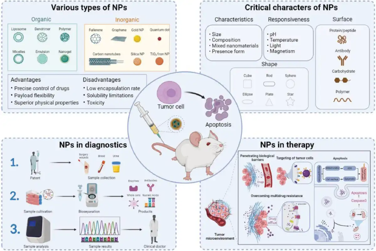
Figure 8.
Overview of nanomedicine in clinical practice for cancer diagnosis and treatment.
3. Present therapeutic strategies for tumors associated with DNA methylation
3.1 DNA methylation-related diagnosis
The occurrence and development of cancer are accompanied by various epigenetic alterations, including gene methylation and mutation[236–237–238]. The discovery that tumors can release epigenetic products into the bloodstream garnered significant attention, making it possible to utilize them as noninvasive liquid biopsy biomarkers for early cancer diagnosis[239]. As aberrant DNA methylation usually occurs early in cancer, DNA methylation has been recognized as the most promising biomarker for cancer diagnosis and prognostic monitoring[240,241]. Some of the important methods for detecting DNA methylation, such as bisulfite-converted DNA-based techniques, methylation-sensitive restriction endonuclease methods, CRISPR-based biosensor methods, and third-generation sequencing methods, are discussed in this subsection.
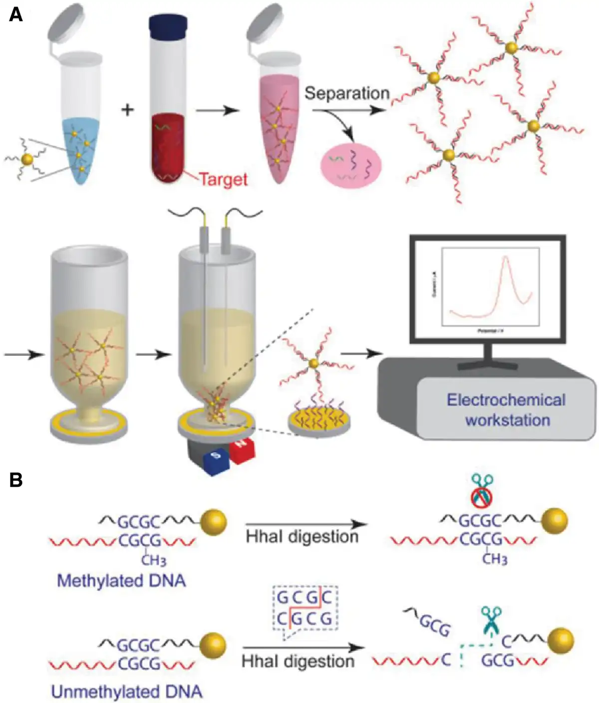
Figure 9.
Schematic diagram of DNA methylation detection based on nucleic acid hybridization on a gold-plated magnetic nanoparticle network. (A) Methylated DNA measurement workflow. (B) Principle of DNA-specific detection of methylation[242]. Copyright 2020, Elsevier.
3.1.1 Bisulfite sequencing
Bisulfite sequencing is regarded as the gold standard for detecting DNA methylation, widely used for this purpose[243]. The method is based on the fact that bisulfite specifically converts unmethylated cytosine to uracil, while methylated cytosine is unaffected. After bisulfite treatment, analysis of cytosine residues in DNA sequences by polymerase chain reaction (PCR) amplification and sequencing is able to distinguish between methylated and unmethylated cytosines[244,245]. The key advantage of this method is its high sensitivity and resolution, which enable the detection of low-frequency methylation events. Thus, it is widely used for genome-wide methylation analysis, especially for the study of gene regulation, disease epigenetics, and changes in gene methylation during development. Although this technique has achieved remarkable results in many fields, its drawbacks should not be ignored, including the complexity and high cost of the experimental process, and the need to strictly control the experimental conditions to avoid false-positive or false-negative results.
3.1.2 Methylation-sensitive restriction enzymes–based methods
The method based on methylation-sensitive restriction enzymes (MSREs) is one of the classical techniques for detecting DNA methylation. The method relies on the cleavage properties of certain restriction endonucleases, which are sensitive to methylated cytosine and are able to recognize and cleave unmethylated DNA sequences, but not methylated DNA[246,247]. This property makes the MSRE method an important tool for analyzing DNA methylation. Usually, the operation process of the MSRE assay includes DNA sample extraction, enzymatic reaction, PCR amplification, and subsequent analysis. By this method, the methylation level of a specific gene region or the whole genome can be assessed quantitatively or qualitatively. Compared with other methylation detection methods, the MSRE method has a simpler operating procedure and lower cost and is therefore widely used in small-scale experiments[248]. In addition, MSRE technology is often applied in combination with other methods, such as bisulfite sequencing, DNA microarrays, or high-throughput sequencing, to improve the coverage and resolution of detection[249,250]. However, a limitation of the MSRE method is its dependence on specific restriction endonucleases, which means that it can only detect methylation sites recognized by these enzymes and cannot fully reflect the methylation status of the entire genome.
3.1.3 CRISPR/Cas system-based methods
CRISPR-based biosensors for the detection of DNA methylation are a rapidly developing technology in recent years, taking full advantage of the properties of the CRISPR/Cas system in pinpointing DNA sequences[251]. Such methods mainly rely on variants such as CRISPR/Cas9, CRISPR/Cas12, or CRISPR/Cas13, which are systems capable of efficiently recognizing and cleaving specific DNA or RNA sequences. When combined with specific methylation markers, these systems can provide sensitive, specific, and efficient methylation detection[252]. Specifically, CRISPR/Cas systems recognize DNA sequences containing methylation marks by combining Cas proteins with a guide RNA that targets the methylation site. This process can be detected not only by signal outputs such as fluorescence and luminescence but also by the ability to further enhance the signal by combining it with other molecular probes, further improving the sensitivity and specificity of the detection. Currently, there are 2 typical application strategies for CRISPR-based biosensors: one is to take advantage of the cleavage activity of Cas12 or Cas13 proteins, which recognize a target sequence and cleave the single-stranded DNA or RNA next to it, thus generating an easily detectable optical signal[253]. Another approach is through the gene-editing function of the CRISPR/Cas9 system, which can trigger a downstream gene-editing reaction when a methylated DNA sequence is recognized, and then the methylation status can be sensed by fluorescence and chemical reaction. These 2 strategies are superior in terms of sensitivity, specificity, and ability to detect multiple methylation sites, making them widely applicable in epigenetic research, early disease diagnosis, and personalized medicine[254]. In addition, CRISPR-based biosensors offer greater operational simplicity and lower cost compared with traditional bisulfite sequencing methods and are particularly suitable for high-throughput and rapid on-site detection. However, this technology still faces many challenges, including the stability of the system, the amplification efficiency of the signal, and how to perform efficient signal discrimination in complex samples.
3.1.4 Third-generation sequencing
Third-generation sequencing–based methods have shown great potential for DNA methylation detection. Compared with traditional second-generation sequencing, third-generation sequencing has the advantages of longer read lengths, higher throughput, and real-time detection, especially in genome-wide DNA methylation analysis, which can provide a more refined and comprehensive perspective. Representative methods of third-generation sequencing technologies include single-molecule real-time (SMRT) sequencing and nanopore sequencing, both of which have unique application advantages in the detection of DNA methylation[255,256]. In SMRT sequencing, the technology developed by the PacBio Company identifies DNA sequences by sequencing in real-time at the single-molecule level and utilizing changes in the fluorescence signal of DNA polymerase. Because the SMRT sequencing technology is able to simultaneously detect differences between methylated and nonmethylated cytosines on the DNA strand, it is able to infer the methylation status directly from the signal changes without the need for tedious chemical modification steps. This allows SMRT sequencing to have extremely high resolution in methylation detection, effectively identifying rare methylation events and providing precise data to support epigenetic studies[257]. Another major third-generation sequencing technology is nanopore sequencing, with a representative platform being Oxford Nanopore Technologies[258]. Nanopore sequencing directly reads DNA sequences by measuring the changes in electrical currents induced by the passage of DNA molecules through nanopores. In the process, nanopore sequencing technology is able to directly sense how methylation modifications on the DNA strand affect the current, thus identifying the methylation status at a specific location. Compared with SMRT sequencing, nanopore sequencing technology offers greater portability and real-time performance, making it particularly suitable for clinical field testing and rapid diagnosis. By using appropriate algorithms, nanopore sequencing enables in-depth analysis of methylation patterns, further advancing its application in epigenetics and disease monitoring. Methylation detection methods based on third-generation sequencing have not only improved the accuracy of the assays but also extended their application to large-scale, complex samples. Especially in the field of oncology research and personalized medicine, third-generation sequencing technology can reveal early markers of disease and potential therapeutic targets by accurately depicting changes in methylation patterns. However, the challenges of high cost, data processing complexity, and relatively high error rate of third-generation sequencing technology remain bottlenecks in the current development of the technology. As the technology continues to mature, third-generation sequencing will play a greater role in a wider range of application scenarios in the future.
To date, most of the mainstream methods for DNA methylation analysis require PCR and sequencing[259,260]. Unfortunately, these mainstream DNA methylation analysis methods face a number of problems, including their cumbersome cloning and sequencing workflows, complex operational steps, reliance on sophisticated instruments, and high costs, which make them unable to meet the practical needs of real-time diagnosis and clinical practice. With the updating of science concepts, nanotechnology has opened up new avenues for traditional tumor diagnosis and treatment, especially for the application of epigenetic diagnosis in tumors (Figure 8)[261], such as the third-generation sequencing technology. Several physically and chemically based strategies for DNA methylation detection have been reported recently, such as DNA methylation assays based on single-base extension reactions and surface-enhanced Raman spectroscopy[262], surface plasmon resonance[263], methylation-specific microarrays[264], and methylation-specific fluorescence resonance energy transfer (Figure 9A, B)[265]. Generally, there are still some limitations in the application of nanotechnology for the identification of methylation levels in cancer patients, such as the design of detection materials, the requirements for the operation of detection equipment, and the fatal problem: the contradiction between the low detection limit and the quick analysis time. Commonly speaking, the lower the detection limit, the longer the analysis time required. Therefore, an essential milestone in applying DNA methylation detection to clinical practice is the development of rapid, highly sensitive, and selective sensing technologies for clinical samples, providing clinicians with a timely diagnostic basis to strive for more living space for patients.
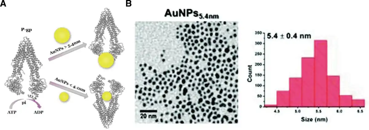
Figure 10.
Nanomaterials for multidrug resistance. (A) Principle of gold nanoparticles against multidrug resistance. (B) Transmission electron microscopy images (TSM) and particle size distributions of 5.4 nm AuNPs. [266]. Copyright 2020, Royal Society of Chemistry. AuNPs, Au naoparticles.
3.2 DNA methylation-related therapy
Presently, the main DNA methylation drugs for the treatment of tumors are demethylating agents, including 5-azadeoxyguanine (5-Aza-dC) and 5-azadeoxycytosine (5-Aza-C), which inhibit the activity of DNMTs by competitive binding to DNA sequences, reversing the abnormal DNA methylation in cancer cells. These drugs effectively decrease methylation levels in the promoter regions of tumor suppressor genes, thereby restoring the expression of these genes and inducing apoptosis in tumor cells. The demethylating agents 5-Aza-dC and 5-Aza-C have been approved for the treatment of hematologic tumors, such as acute myeloid leukemia and MDS[267]. Clinical studies have shown that these demethylating agents can significantly improve patient survival, although their side effects, such as myelosuppression and immunosuppression, remain a challenge during treatment. In addition to demethylating agents, drugs such as Vidaza (azacitidine) and decitabine have shown their potential in the clinic. By inhibiting DNMTs, these drugs reduce aberrant methylation patterns in tumor cells and activate the expression of tumor suppressor genes[268]. Tumor therapeutic drugs targeting DNA methylation have been applied mainly in hematological tumors, with relatively few applications in solid tumors[269,270]. One of the major challenges in applying these DNA methylation drugs is the specificity and durability of the treatment. Since DNA methylation alterations occur in many different genes and may result in effects on healthy cells, achieving more precise drug targeting and reducing side effects remains a focus of current research.
Nanomedicine is a research field that integrates advanced nanotechnology into medicine, with the aim of advancing disease prevention, diagnosis, and treatment[271]. Combining therapeutic agents with nanomaterials has been considered a promising future direction, as it can enhance the targeting precision of drugs at the cellular level, thereby effectively avoiding systemic side effects[272,273]. Depending on their chemical composition, these nanoparticles are able to encapsulate and protect hydrophilic or hydrophobic drugs from enzymatic, chemical, or physical degradation, maintaining stability during transport to target cells[274]. Researchers have utilized various drug-loaded nanospheres to enhance cancer treatment efficacy, including organic, inorganic, and hybrid nanoparticles (Figure 10A, B)[275–276–277]. The first-generation “demethylating agents” azacitidine and decitabine are the most successful epigenetic modulators, widely utilized in the clinical treatment of myeloid malignancies and highly favored as pretreatment drugs for advanced solid tumors. Unfortunately, the clinical response rate of these drugs is only 30% to 50%, and their clinical efficacy is limited by low bioavailability. The genomic instability caused by the activation of multiple oncogenes at high doses of similar drugs has led to their rare use in the clinic, especially in solid tumors. To improve the efficacy of these drugs, the introduction of nanodelivery carriers provides better targeting and biocompatibility.
Nanodelivery systems, especially nanoparticles (eg, lipid nanoparticles and polymer nanoparticles), can effectively carry DNA methylation drugs and enhance their stability. Nanocarriers can enhance drug accumulation in tumor tissues by altering drug release kinetics and reducing drug degradation in the blood circulation (Figure 11A). Nanodelivery carriers release drugs through external or internal stimuli (eg, pH changes and temperature changes) to achieve precise drug release and delivery (Figure 11B, C)[278]. The key advantage of nanodelivery carriers over conventional drug delivery systems is that the specific recognition of tumor cells and drug release can be achieved through surface modification, thus minimizing the toxic effects on normal tissues. For example, lipid nanoparticles not only efficiently carry DNA-methylated drugs but also promote drug endocytosis through the fusion of cell membranes, thus enabling precise delivery to tumor cells[279]. In addition, polymeric nanoparticles such as poly-lactide-co-glycolide (PLGA) nanoparticles have demonstrated superior drug delivery capabilities. PLGA nanoparticles can avoid premature drug clearance by modulating their surface properties and can control the rate of drug release[280,281]. Meanwhile, PLGA nanoparticles have better biodegradability, which enables the drug to be released gradually after delivery to the target site, reducing toxic side effects[282,283]. The advantage of the nanodelivery system is not only to improve the targeting of drugs but also to solve the solubility and bioavailability of DNA methylation drugs in the body. By combining nanotechnology, the efficacy of DNA methylation drugs has been significantly improved, and the side effects of the drugs have been effectively controlled.
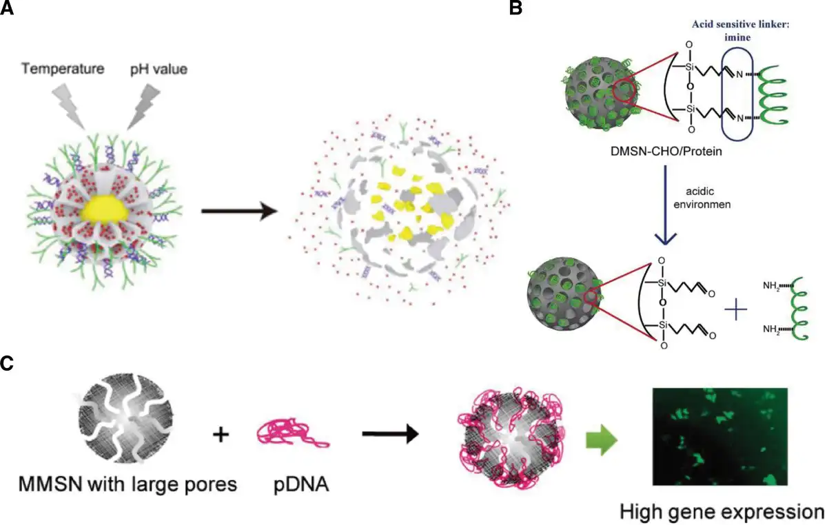
Figure 11.
Intelligent drug delivery system based on mesoporous silica nanoparticles. (A) Schematic illustration of the degradation and drug release of the nanozymes with the drug delivery system based on Cu@Fe2C@mSiO2-PEG/LA-R848-ICG-AS1411. [284]. Copyright 2022, American Association for the advancement of science. (B) pH-responsive protein drug delivery system based on dendritic mesoporous silica nanoparticles. [285]. Copyright 2017, Walter de Gruyter GmbH. (C) Mesoporous silica nanoparticles loading plasmid DNA. [286]. Copyright 2011, American Chemical Society.
3.3 Concerns related to DNA methylation therapy
As is well known, the methylation level of the vast majority of CpG sites in the genome (excluding CpG-enriched regions) is approximately 80%. In cancer, the average CpG methylation level ranges from 40% to 60%. Advances in gene sequencing technology have enabled researchers to map the patterns of genetic modifications more precisely, which has revealed that DNA hypomethylation covers about one-third of the genome[287,288]. The exact mechanism by which DNA methylation is lost from the cancer epigenome remains poorly understood. Moreover, the widespread occurrence of DNA hypermethylation in promoter regions poses unprecedented challenges in understanding the scope of these changes. Hypermethylation of specific gene promoters alone does not necessarily imply functional significance in cancer, similar to the case of gene mutations. Particularly, the hypermethylated genes are not known tumor suppressor genes, and there is no evidence that the gene is frequently methylated in cancer. Thus, the discussed genes must be investigated to determine the significance of their functional loss, including both biological processes regulated by encoded proteins and their impact on tumor progression.
Currently, the most critical issue faced by epigenetic therapy is specificity, meaning that its effects manifest at the genome-wide level rather than at the target-specific level. It may lead to the simultaneous targeting of tumor suppressor genes while also promoting the excessive activation of oncogenes, thereby triggering genomic instability[289,290]. Besides, poor performance was observed in solid tumor cases because DNMT inhibitors (DNMTis) are dependent on DNA participation, making them more effective in actively dividing cells, but not in solid tumors with relatively slow division rates. The instability of DNMTis also causes difficulties in their effective delivery to solid tumors. To date, the efficacy of epigenetic drugs has been primarily applied to hematological cancers[291], partly because solid tumors typically originate from cells with higher degrees of differentiation or even terminal differentiation, whose epigenetic reprogramming ability is low[292]. Furthermore, another important factor limiting efficacy is the intrinsic defects of the drug during treatment, which are attributed to cellular mechanisms affecting drug uptake and metabolism[293]. These drugs showed poor pharmacokinetic properties and difficulties in administration. Due to the chemical instability of the pyrimidine ring in water, DNMTis are not administered orally[294]. Meanwhile, the effect of DNMTis treatment seems to be transient, giving rise to the recovery of DNA methylation signatures after treatment is suspended[295]. Chronic or continuous treatment with DNMTis can engender side effects, including the effects of high-dose 5-azacytidine, which include fetal abnormalities and decreased male fertility[296]. Due to its cytotoxicity and instability, DNMTis cannot be used continuously in patients. To mitigate this effect, stable and safer alternatives to 5-aza-CR and 5-aza-CdR or efficient and precise drug delivery vehicles need to be developed[297,298]. Epigenetic mechanisms also lead to inter- and intratumoral heterogeneity, thereby affecting the efficacy of current epigenetic therapies. Besides, spatiotemporal clonal diversity can exist in the same tumor at the molecular level. Such differences may generate negative responses to therapies that rely on standard care rather than focusing on each individual’s unique characteristics.
4. Conclusion and outlooks
Cancer is a disease caused by epigenetic disorders of normal cells, and its increasing prevalence is threatening the survival and development of human beings, as well as increasing the burden on happy families, leading to a decline in the quality of life of individuals. Early screening of tumor patients is one of the ways to prevent the deterioration of tumor diseases, and DNA methylation diagnosis has become increasingly important in this regard with the improvement of detection technologies. The development of technologies from bisulfite sequencing to third-generation sequencing, as well as the fine-tuning of detection technologies with different disciplinary fields, has improved the precision and efficiency of the analysis to some extent. As described in the review, each assay has its own scope of application. Therefore, provided that the accuracy of the test is ensured, it is necessary to develop diverse testing platforms to meet the needs of clinicalization. Meanwhile, sophisticated operational requirements and high costs are unavoidable problems with the current commonly used detection technologies. In the future, simplifying the operation steps and reducing the cost of testing will be of positive significance in promoting DNA methylation testing. Regarding the treatment of DNA methylation in the field of oncology, it is currently only used in blood-related tumors, and its application in solid tumors has greater limitations, although the phenomenon of aberrant DNA methylation in tumors is already an indisputable fact. For the application of DNA methylation in tumor therapy, the combination with nanodelivery carriers is an effective way to address the targeting and side effects of DNA methylation drugs. Controlled release of the nanodelivery system can enable DNA methylation drugs to better exert therapeutic effects at the tumor site. In the practical application related to epigenetic drugs, the study of their theory is particularly important. At present, although there is a small amount of mechanism investigation in clinical practice based on DNA methylation, most of it exists in the form of a phenomenal association, especially in the process of diagnosis. There is a lack of in-depth investigation into the mechanisms of DNA methylation, which is the theoretical cornerstone for future improvements in DNA methylation diagnosis and treatment. Exploring the mechanisms of DNA methylation requires great efforts from researchers on the path from phenomena to the identification of molecular biological mechanisms, although it is an extremely taxing process. The in-depth exploration of the DNA methylation mechanism in tumor cells is not only a critical factor in solving the bottleneck of tumor diagnosis and treatment but also an inevitable process to explore the evolution of stem cells in the human body.
Acknowledgments
This work was financially supported by the National Natural Science Foundation of China (Grant Nos. 22377026 and 52001008) and the National Key Research and Development Program of China (Grant No. 2023YFB3507003).
Conflicts of interests
The authors declare that they have no conflicts of interest.
Data availability statement
Data sharing is not applicable to this article as no datasets were generated or analyzed during the current study.
Shikang Liu: conceptualization, writing—original draft, writing—review and editing, and visualization. Youli Yao: visualization. Zhongjun Li: writing—review. Zhiyi Wang: conceptualization, funding acquisition, supervision, and writing—review and editing.
References
- [1] Rakyan V, Whitelaw E. Transgenerational epigenetic inheritance. Curr Biol. 2003;13(1):R6.
- [2] Hackett JA, Surani MA. Beyond DNA: programming and inheritance of parental methylomes. Cell. 2013;153(4):737–739.
- [3] Pan Y, Sawalha AH. Epigenetic regulation and the pathogenesis of systemic lupus erythematosus. Transl Res. 2009;153(1):4–10.
- [4] Mercer J, Mahmoudi M, Bennett M. DNA damage, p53, apoptosis and vascular disease. Mutat Res. 2007;621(1-2):75–86.
- [5] Jaenisch R, Bird A. Epigenetic regulation of gene expression: how the genome integrates intrinsic and environmental signals. Nat Genet. 2003;33(Suppl):245–254.
- [6] Salozhin SV, Prokhorchuk EB, Georgiev GP. Methylation of DNA--one of the major epigenetic markers. Biochemistry (Mosc). 2005;70(5):525–532.
- [7] Nafee TM, Farrell WE, Carroll WD, et al. Epigenetic control of fetal gene expression. BJOG. 2008;115(2):158–168.
- [8] Wongtrakoongate P. Epigenetic therapy of cancer stem and progenitor cells by targeting DNA methylation machineries. World J Stem Cells. 2015;7(1):137–148.
- [9] Graves JAM. Imprinting of the paternal marsupial X chromosome by DNA methylation. Trends Genet. 2025;41(1):9–11.
- [10] Alfeghaly C, Rougeulle C. X chromosome inactivation in mammals: general principles and species-specific considerations. EMBO Rep. 2025;26(14):3478–3490.
- [11] Esteller M. Epigenetic gene silencing in cancer: the DNA hypermethylome. Hum Mol Genet. 2007;16 Spec No 1:R50–R59.
- [12] Aird D, Ross MG, Chen WS, et al. Analyzing and minimizing PCR amplification bias in Illumina sequencing libraries. Genome Biol. 2011;12(2):R18.
- [13] Bird A. DNA methylation patterns and epigenetic memory. Genes Dev. 2002;16(1):6–21.
- [14] Horvath S, Raj K. DNA methylation-based biomarkers and the epigenetic clock theory of ageing. Nat Rev Genet. 2018;19(6):371–384.
- [15] Younesian S, Yousefi AM, Momeny M, et al. The DNA methylation in neurological diseases. Cells. 2022;11(21):3439.
- [16] Xu P-P, Fu D, Li J-Y, et al. Anthracycline dose optimisation in patients with diffuse large B-cell lymphoma: a multicentre, phase 3, randomised, controlled trial. Lancet Haematol. 2019;6(6):e328–e337.
- [17] Han SH, Kim JW, Kim M, et al. Prognostic implication of ABC transporters and cancer stem cell markers in patients with stage III colon cancer receiving adjuvant FOLFOX-4 chemotherapy. Oncol Lett. 2019;17(6):5572–5580.
- [18] Fouse SD, Shen Y, Pellegrini M, et al. Promoter CpG methylation contributes to ES cell gene regulation in parallel with Oct4/Nanog, PcG complex, and histone H3 K4/K27 trimethylation. Cell Stem Cell. 2008;2(2):160–169.
- [19] Okano M, Xie S, Li E. Cloning and characterization of a family of novel mammalian DNA (cytosine-5) methyltransferases. Nat Genet. 1998;19(3):219–220.
- [20] Okano M, Bell DW, Haber DA, et al. DNA methyltransferases Dnmt3a and Dnmt3b are essential for de novo methylation and mammalian development. Cell. 1999;99(3):247–257.
- [21] Broske AM, Vockentanz L, Kharazi S, et al. DNA methylation protects hematopoietic stem cell multipotency from myeloerythroid restriction. Nat Genet. 2009;41(11):1207–1215.
- [22] Liu CC, Lin JH, Hsu TW, et al. IL-6 enriched lung cancer stem-like cell population by inhibition of cell cycle regulators via DNMT1 upregulation. Int J Cancer. 2015;136(3):547–559.
- [23] Morita R, Hirohashi Y, Suzuki H, et al. DNA methyltransferase 1 is essential for initiation of the colon cancers. Exp Mol Pathol. 2013;94(2):322–329.
- [24] Samah AA, Fauzi MFA, Mansor S. Classification of benign and malignant tumors in histopathology images. In: 2017 IEEE International Conference on Signal and Image Processing Applications (ICSIPA); 2017:102-106.
- [25] Shen H, Laird PW. Interplay between the cancer genome and epigenome. Cell. 2013;153(1):38–55.
- [26] Jones PA, Baylin SB. The fundamental role of epigenetic events in cancer. Nat Rev Genet. 2002;3(6):415–428.
- [27] Herman JG, Baylin SB. Gene silencing in cancer in association with promoter hypermethylation. N Engl J Med. 2003;349(21):2042–2054.
- [28] Baylin SB, Jones PA. A decade of exploring the cancer epigenome - biological and translational implications. Nat Rev Cancer. 2011;11(10):726–734.
- [29] Klajic J, Busato F, Edvardsen H, et al. DNA methylation status of key cell-cycle regulators such as CDKNA2/p16 and CCNA1 correlates with treatment response to doxorubicin and 5-fluorouracil in locally advanced breast tumors. Clin Cancer Res. 2014;20(24):6357–6366.
- [30] Michalak EM, Burr ML, Bannister AJ, et al. The roles of DNA, RNA and histone methylation in ageing and cancer. Nat Rev Mol Cell Biol. 2019;20(10):573–589.
- [31] Nejman D, Straussman R, Steinfeld I, et al. Molecular rules governing de novo methylation in cancer. Cancer Res. 2014;74(5):1475–1483.
- [32] Wang Y, Wang C, Zhong R, et al. Research progress of DNA methylation in colorectal cancer (review). Mol Med Rep. 2024;30(3).
- [33] Shibata D. Inferring human stem cell behaviour from epigenetic drift. J Pathol. 2009;217(2):199–205.
- [34] Rossi DJ, Bryder D, Zahn JM, et al. Cell intrinsic alterations underlie hematopoietic stem cell aging. Proc Natl Acad Sci U S A. 2005;102(26):9194–9199.
- [35] Kulis M, Merkel A, Heath S, et al. Whole-genome fingerprint of the DNA methylome during human B cell differentiation. Nat Genet. 2015;47(7):746–756.
- [36] Ehrlich M, Lacey M. DNA hypomethylation and hemimethylation in cancer. Adv Exp Med Biol. 2013;754:31–56.
- [37] Hansen KD, Timp W, Bravo HC, et al. Increased methylation variation in epigenetic domains across cancer types. Nat Genet. 2011;43(8):768–775.
- [38] Bert SA, Robinson MD, Strbenac D, et al. Regional activation of the cancer genome by long-range epigenetic remodeling. Cancer Cell. 2013;23(1):9–22.
- [39] Berman BP, Weisenberger DJ, Aman JF, et al. Regions of focal DNA hypermethylation and long-range hypomethylation in colorectal cancer coincide with nuclear lamina-associated domains. Nat Genet. 2012;44(1):40–U62.
- [40] Chen RZ, Pettersson U, Beard C, et al. DNA hypomethylation leads to elevated mutation rates. Nature. 1998;395(6697):89–93.
- [41] Narayan A, Ji W, Zhang XY, et al. Hypomethylation of pericentromeric DNA in breast adenocarcinomas. Int J Cancer. 1998;77(6):833–838.
- [42] Hur K, Cejas P, Feliu J, et al. Hypomethylation of long interspersed nuclear element-1 (LINE-1) leads to activation of proto-oncogenes in human colorectal cancer metastasis. Gut. 2014;63(4):635–646.
- [43] Wolffe AP. Chromatin remodeling: why it is important in cancer. Oncogene. 2001;20(24):2988–2990.
- [44] Cancer Genome Atlas Research Network. Comprehensive genomic characterization of squamous cell lung cancers. Nature. 2012;489(7417):519–525.
- [45] Jones PA, Baylin SB. The epigenomics of cancer. Cell. 2007;128(4):683–692.
- [46] ENCODE Project Consortium. An integrated encyclopedia of DNA elements in the human genome. Nature. 2012;489(7414):57–74.
- [47] Bray F, Ferlay J, Soerjomataram I, et al. Global cancer statistics 2018: GLOBOCAN estimates of incidence and mortality worldwide for 36 cancers in 185 countries. CA Cancer J Clin. 2018;68(6):394-424.
- [48] Zheng R, Zhang S, Zeng H, et al. Cancer incidence and mortality in China, 2016. J Natl Cancer Cent. 2022;2(1):1–9.
- [49] Dammann R, Li C, Yoon JH, et al. Epigenetic inactivation of a RAS association domain family protein from the lung tumour suppressor locus 3p21.3. Nat Genet. 2000;25(3):315–319.
- [50] Dammann R, Strunnikova M, Schagdarsurengin U, et al. CpG island methylation and expression of tumour-associated genes in lung carcinoma. Eur J Cancer. 2005;41(8):1223–1236.
- [51] Topaloglu O, Hoque MO, Tokumaru Y, et al. Detection of promoter hypermethylation of multiple genes in the tumor and bronchoalveolar lavage of patients with lung cancer. Clin Cancer Res. 2004;10(7):2284–2288.
- [52] Yanagawa N, Tamura G, Oizumi H, et al. Promoter hypermethylation of tumor suppressor and tumor-related genes in non-small cell lung cancers. Cancer Sci. 2003;94(7):589–592.
- [53] Zochbauer-Muller S, Fong KM, Virmani AK, et al. Aberrant promoter methylation of multiple genes in non-small cell lung cancers. Cancer Res. 2001;61(1):249–255.
- [54] Li N, Zeng Y, Huang J. Signaling pathways and clinical application of RASSF1A and SHOX2 in lung cancer. J Cancer Res Clin Oncol. 2020;146(6):1379–1393.
- [55] Schmidt B, Liebenberg V, Dietrich D, et al. SHOX2 DNA methylation is a biomarker for the diagnosis of lung cancer based on bronchial aspirates. BMC Cancer. 2010;10:600.
- [56] Schneider KU, Dietrich D, Fleischhacker M, et al. Correlation of SHOX2 gene amplification and DNA methylation in lung cancer tumors. BMC Cancer. 2011;11:102.
- [57] Kneip C, Schmidt B, Seegebarth A, et al. SHOX2 DNA methylation is a biomarker for the diagnosis of lung cancer in plasma. J Thorac Oncol. 2011;6(10):1632–1638.
- [58] Liu WJ, Tan XH, Guo BP, et al. Associations between RASSF1A promoter methylation and NSCLC: a meta-analysis of published data. Asian Pac J Cancer Prev. 2013;14(6):3719–3724.
- [59] Pankova D, Jiang Y, Chatzifrangkeskou M, et al. RASSF1A controls tissue stiffness and cancer stem-like cells in lung adenocarcinoma. EMBO J. 2019;38(13):e100532.
- [60] Yu J, Bulk E, Ji P, et al. The EPHB6 receptor tyrosine kinase is a metastasis suppressor that is frequently silenced by promoter DNA hypermethylation in non-small cell lung cancer. Clin Cancer Res. 2010;16(8):2275–2283.
- [61] Ma R, Feng N, Yu X, et al. Promoter methylation of Wnt/beta-catenin signal inhibitor TMEM88 is associated with unfavorable prognosis of non-small cell lung cancer. Cancer Biol Med. 2017;14(4):377–386.
- [62] Hoang PH, Landi MT. DNA methylation in lung cancer: mechanisms and associations with histological subtypes, molecular alterations, and major epidemiological factors. Cancers (Basel). 2022;14(4):961.
- [63] Liang W, Zhao Y, Huang W, et al. Non-invasive diagnosis of early-stage lung cancer using high-throughput targeted DNA methylation sequencing of circulating tumor DNA (ctDNA). Theranostics. 2019;9(7):2056–2070.
- [64] Jung H, Kim HS, Kim JY, et al. DNA methylation loss promotes immune evasion of tumours with high mutation and copy number load. Nat Commun. 2019;10(1):4278.
- [65] Ili C, Buchegger K, Demond H, et al. Landscape of genome-wide DNA methylation of colorectal cancer metastasis. Cancers (Basel). 2020;12(9):2710.
- [66] Sakai E, Nakajima A, Kaneda A. Accumulation of aberrant DNA methylation during colorectal cancer development. World J Gastroenterol. 2014;20(4):978–987.
- [67] Tse JWT, Jenkins LJ, Chionh F, et al. Aberrant DNA methylation in colorectal cancer: what should we target? Trends Cancer. 2017;3(10):698–712.
- [68] Grady WM. Epigenetic events in the colorectum and in colon cancer. Biochem Soc Trans. 2005;33(Pt 4):684–688.
- [69] Grady WM, Markowitz SD. Genetic and epigenetic alterations in colon cancer. Annu Rev Genomics Hum Genet. 2002;3:101–128.
- [70] Huang LJ, Chen SX, Luo WJ, et al. Proteomic analysis of secreted proteins of non-small cell lung cancer. Ai Zheng. 2006;25(11):1361–1367.
- [71] Verschuur AVD, Hackeng WM, Westerbeke F, et al. DNA methylation profiling enables accurate classification of nonductal primary pancreatic neoplasms. Clin Gastroenterol Hepatol. 2024;22(6):1245–1254.e10.
- [72] Lam AK, Ong K, Giv MJ, et al. p16 expression in colorectal adenocarcinoma: marker of aggressiveness and morphological types. Pathology (Phila). 2008;40(6):580–585.
- [73] Yu J, Tao Q, Cheung KF, et al. Epigenetic identification of ubiquitin carboxyl-terminal hydrolase L1 as a functional tumor suppressor and biomarker for hepatocellular carcinoma and other digestive tumors. Hepatology. 2008;48(2):508–518.
- [74] Goto T, Mizukami H, Shirahata A, et al. Aberrant methylation of the p16 gene is frequently detected in advanced colorectal cancer. Anticancer Res. 2009;29(1):275–277.
- [75] Chen J, Rocken C, Lofton-Day C, et al. Molecular analysis of APC promoter methylation and protein expression in colorectal cancer metastasis. Carcinogenesis. 2005;26(1):37–43.
- [76] Wynter CV, Walsh MD, Higuchi T, et al. Methylation patterns define two types of hyperplastic polyp associated with colorectal cancer. Gut. 2004;53(4):573–580.
- [77] Hibi K, Sakata M, Sakuraba K, et al. Aberrant methylation of the HACE1 gene is frequently detected in advanced colorectal cancer. Anticancer Res. 2008;28(3A):1581–1584.
- [78] Yang D, Thangaraju M, Greeneltch K, et al. Repression of IFN regulatory factor 8 by DNA methylation is a molecular determinant of apoptotic resistance and metastatic phenotype in metastatic tumor cells. Cancer Res. 2007;67(7):3301–3309.
- [79] Chan AO, Issa JP, Morris JS, et al. Concordant CpG island methylation in hyperplastic polyposis. Am J Pathol. 2002;160(2):529–536.
- [80] Wu CC, Shyu RY, Chou JM, et al. RARRES1 expression is significantly related to tumour differentiation and staging in colorectal adenocarcinoma. Eur J Cancer. 2006;42(4):557–565.
- [81] Matsusaka K, Funata S, Fukayama M, et al. DNA methylation in gastric cancer, related to Helicobacter pylori and Epstein-Barr virus. World J Gastroenterol. 2014;20(14):3916–3926.
- [82] Peng Y, Wu Q, Wang L, et al. A DNA methylation signature to improve survival prediction of gastric cancer. Clin Epigenetics. 2020;12(1):15.
- [83] Jemal A, Siegel R, Ward E, et al. Cancer statistics, 2007. CA Cancer J Clin. 2007;57(1):43–66.
- [84] Duan Y, Xu Y, Dou Y, et al. Helicobacter pylori and gastric cancer: mechanisms and new perspectives. J Hematol Oncol. 2025;18(1):10.
- [85] Niwa T, Tsukamoto T, Toyoda T, et al. Inflammatory processes triggered by Helicobacter pylori infection cause aberrant DNA methylation in gastric epithelial cells. Cancer Res. 2010;70(4):1430–1440.
- [86] Niwa T, Toyoda T, Tsukamoto T, et al. Prevention of Helicobacter pylori-induced gastric cancers in gerbils by a DNA demethylating agent. Cancer Prev Res (Phila). 2013;6(4):263–270.
- [87] Ushijima T, Okochi-Takada E. Aberrant methylations in cancer cells: where do they come from? Cancer Sci. 2005;96(4):206–211.
- [88] Suzuki H, Tokino T, Shinomura Y, et al. DNA methylation and cancer pathways in gastrointestinal tumors. Pharmacogenomics. 2008;9(12):1917–1928.
- [89] Tamura G. Alterations of tumor suppressor and tumor-related genes in the development and progression of gastric cancer. World J Gastroenterol. 2006;12(2):192–198.
- [90] Kang GH, Shim YH, Jung HY, et al. CpG island methylation in premalignant stages of gastric carcinoma. Cancer Res. 2001;61(7):2847–2851.
- [91] Waki T, Tamura G, Tsuchiya T, et al. Promoter methylation status of E-cadherin, hMLH1, and p16 genes in nonneoplastic gastric epithelia. Am J Pathol. 2002;161(2):399–403.
- [92] Kang GH, Lee HJ, Hwang KS, et al. Aberrant CpG island hypermethylation of chronic gastritis, in relation to aging, gender, intestinal metaplasia, and chronic inflammation. Am J Pathol. 2003;163(4):1551–1556.
- [93] Zhang T, Wu Y, Fang Z, et al. Low expression of RBMS3 and SFRP1 are associated with poor prognosis in patients with gastric cancer. Am J Cancer Res. 2016;6(11):2679–2689.
- [94] Wang H, Duan XL, Qi XL, et al. Concurrent hypermethylation of SFRP2 and DKK2 activates the Wnt/beta-catenin pathway and is associated with poor prognosis in patients with gastric cancer. Mol Cells. 2017;40(1):45–53.
- [95] Maruyama R, Akino K, Toyota M, et al. Cytoplasmic RASSF2A is a proapoptotic mediator whose expression is epigenetically silenced in gastric cancer. Carcinogenesis. 2008;29(7):1312–1318.
- [96] Fan G, Tu Y, Chen C, et al. DNA methylation biomarkers for hepatocellular carcinoma. Cancer Cell Int. 2018;18:140.
- [97] Nagaraju GP, Dariya B, Kasa P, et al. Epigenetics in hepatocellular carcinoma. Semin Cancer Biol. 2022;86(Pt 3):622–632.
- [98] Parkin DM, Bray F, Ferlay J, et al. Global cancer statistics, 2002. CA Cancer J Clin. 2005;55(2):74–108.
- [99] Munoz P, Iliou MS, Esteller M. Epigenetic alterations involved in cancer stem cell reprogramming. Mol Oncol. 2012;6(6):620–636.
- [100] Hamilton JP. Epigenetic mechanisms involved in the pathogenesis of hepatobiliary malignancies. Epigenomics. 2010;2(2):233–243.
- [101] Jiang L, Chan JY, Fung KP. Epigenetic loss of CDH1 correlates with multidrug resistance in human hepatocellular carcinoma cells. Biochem Biophys Res Commun. 2012;422(4):739–744.
- [102] Jia Y, Yang Y, Liu S, et al. SOX17 antagonizes Wnt/beta-catenin signaling pathway in hepatocellular carcinoma. Epigenetics. 2010;5(8):743–749.
- [103] Tsao CM, Yan MD, Shih YL, et al. SOX1 functions as a tumor suppressor by antagonizing the Wnt/beta-catenin signaling pathway in hepatocellular carcinoma. Hepatology. 2012;56(6):2277–2287.
- [104] Shu X-S, Gottardi C, Geng H, . The Epigenetic Modifier PRDM5 Functions as a Tumor Suppressor through Modulating WNT/β-Catenin Signaling and Is Frequently Silenced in Multiple Tumors. PLoS ONE. 2011;6(11):e27346. doi:10.1371/journal.pone.0027346.
- [105] Kaur P, Mani S, Cros MP, et al. Epigenetic silencing of sFRP1 activates the canonical Wnt pathway and contributes to increased cell growth and proliferation in hepatocellular carcinoma. Tumour Biol. 2012;33(2):325–336.
- [106] Lou L, Deng T, Yuan Q, et al. Targeted silencing of SOCS1 by DNMT1 promotes stemness of human liver cancer stem-like cells. Cancer Cell Int. 2024;24(1):206.
- [107] Yang B, Guo M, Herman JG, et al. Aberrant promoter methylation profiles of tumor suppressor genes in hepatocellular carcinoma. Am J Pathol. 2003;163(3):1101–1107.
- [108] Locke WJ, Clark SJ. Epigenome remodelling in breast cancer: insights from an early in vitro model of carcinogenesis. Breast Cancer Res. 2012;14(6):215.
- [109] Beetch M, Harandi-Zadeh S, Yang T, et al. DNA methylation landscape of triple-negative ductal carcinoma in situ (DCIS) progressing to the invasive stage in canine breast cancer. Sci Rep. 2020;10(1):2415.
- [110] van Hoesel AQ, Sato Y, Elashoff DA, et al. Assessment of DNA methylation status in early stages of breast cancer development. Br J Cancer. 2013;108(10):2033–2038.
- [111] Brennan MJ. Endocrinology in cancer of the breast. Status and prospects. Am J Clin Pathol. 1975;64(6):797–809.
- [112] Herman JG, Merlo A, Mao L, et al. Inactivation of the CDKN2/p16/MTS1 gene is frequently associated with aberrant DNA methylation in all common human cancers. Cancer Res. 1995;55(20):4525–4530.
- [113] Xu X, Gammon MD, Zhang Y, et al. Gene promoter methylation is associated with increased mortality among women with breast cancer. Breast Cancer Res Treat. 2010;121(3):685–692.
- [114] Barekati Z, Radpour R, Lu Q, et al. Methylation signature of lymph node metastases in breast cancer patients. BMC Cancer. 2012;12:244.
- [115] Rice JC, Ozcelik H, Maxeiner P, et al. Methylation of the BRCA1 promoter is associated with decreased BRCA1 mRNA levels in clinical breast cancer specimens. Carcinogenesis. 2000;21(9):1761–1765.
- [116] Murata H, Khattar NH, Kang Y, et al. Genetic and epigenetic modification of mismatch repair genes hMSH2 and hMLH1 in sporadic breast cancer with microsatellite instability. Oncogene. 2002;21(37):5696–5703.
- [117] Ottaviano YL, Issa JP, Parl FF, et al. Methylation of the estrogen receptor gene CpG island marks loss of estrogen receptor expression in human breast cancer cells. Cancer Res. 1994;54(10):2552–2555.
- [118] Sirchia SM, Ferguson AT, Sironi E, et al. Evidence of epigenetic changes affecting the chromatin state of the retinoic acid receptor beta2 promoter in breast cancer cells. Oncogene. 2000;19(12):1556–1563.
- [119] Graff JR, Gabrielson E, Fujii H, et al. Methylation patterns of the E-cadherin 5’ CpG island are unstable and reflect the dynamic, heterogeneous loss of E-cadherin expression during metastatic progression. J Biol Chem. 2000;275(4):2727–2732.
- [120] Lui EL, Loo WT, Zhu L, et al. DNA hypermethylation of TIMP3 gene in invasive breast ductal carcinoma. Biomed Pharmacother. 2005;59(Suppl 2):S363–S365.
- [121] Virmani AK, Rathi A, Sathyanarayana UG, et al. Aberrant methylation of the adenomatous polyposis coli (APC) gene promoter 1A in breast and lung carcinomas. Clin Cancer Res. 2001;7(7):1998–2004.
- [122] Grandin M, Mathot P, Devailly G, et al. Inhibition of DNA methylation promotes breast tumor sensitivity to netrin-1 interference. EMBO Mol Med. 2016;8(8):863–877.
- [123] Louie SM, Grossman EA, Crawford LA, et al. GSTP1 is a driver of triple-negative breast cancer cell metabolism and pathogenicity. Cell Chem Biol. 2016;23(5):567–578.
- [124] Depowski PL, Rosenthal SI, Ross JS. Loss of expression of the PTEN gene protein product is associated with poor outcome in breast cancer. Mod Pathol. 2001;14(7):672–676.
- [125] Shivapurkar N, Toyooka S, Toyooka KO, et al. Aberrant methylation of trail decoy receptor genes is frequent in multiple tumor types. Int J Cancer. 2004;109(5):786–792.
- [126] Noushmehr H, Weisenberger DJ, Diefes K, et al.; Cancer Genome Atlas Research Network. Identification of a CpG island methylator phenotype that defines a distinct subgroup of glioma. Cancer Cell. 2010;17(5):510–522.
- [127] Malta TM, de Souza CF, Sabedot TS, et al. Glioma CpG island methylator phenotype (G-CIMP): biological and clinical implications. Neuro Oncol. 2018;20(5):608–620.
- [128] Li Y, Bao L, Yang C, et al. A multiparameter radiomic model for accurate prognostic prediction of glioma. MedComm Futur Med. 2023;2(2):e41.
- [129] Kosmider O, Gelsi-Boyer V, Slama L, et al. Mutations of IDH1 and IDH2 genes in early and accelerated phases of myelodysplastic syndromes and MDS/myeloproliferative neoplasms. Leukemia. 2010;24(5):1094–1096.
- [130] Cairns RA, Iqbal J, Lemonnier F, et al. IDH2 mutations are frequent in angioimmunoblastic T-cell lymphoma. Blood. 2012;119(8):1901–1903.
- [131] Amary MF, Bacsi K, Maggiani F, et al. IDH1 and IDH2 mutations are frequent events in central chondrosarcoma and central and periosteal chondromas but not in other mesenchymal tumours. J Pathol. 2011;224(3):334–343.
- [132] Wang P, Dong Q, Zhang C, et al. Mutations in isocitrate dehydrogenase 1 and 2 occur frequently in intrahepatic cholangiocarcinomas and share hypermethylation targets with glioblastomas. Oncogene. 2013;32(25):3091–3100.
- [133] Huang C, Azizi P, Vazirzadeh M, et al. Non-coding RNAs/DNMT3B axis in human cancers: from pathogenesis to clinical significance. J Transl Med. 2023;21(1):621.
- [134] Gravendeel LA, Kloosterhof NK, Bralten LB, et al. Segregation of non-p.R132H mutations in IDH1 in distinct molecular subtypes of glioma. Hum Mutat. 2010;31(3):E1186–E1199.
- [135] Xu H, Sun J, Shi C, et al. miR-29s inhibit the malignant behavior of U87MG glioblastoma cell line by targeting DNMT3A and 3B. Neurosci Lett. 2015;590:40–46.
- [136] Wang LH, Huang J, Wu CR, et al. Downregulation of miR‑29b targets DNMT3b to suppress cellular apoptosis and enhance proliferation in pancreatic cancer. Mol Med Rep. 2018;17(2):2113–2120.
- [137] Kaneda A, Matsusaka K, Aburatani H, et al. Epstein-Barr virus infection as an epigenetic driver of tumorigenesis. Cancer Res. 2012;72(14):3445–3450.
- [138] Cancer Genome Atlas Research Network. Electronic address wbeCancer Genome Atlas Research Network. Comprehensive and integrative genomic characterization of hepatocellular carcinoma. Cell. 2017;169(7):1327–1341 e1323.
- [139] Cancer Genome Atlas Research Network, Albert Einstein College of Medicine, Analytical Biological Services, . Integrated genomic and molecular characterization of cervical cancer. Nature. 2017;543(7645):378–384.
- [140] Cancer Genome Atlas Network. Comprehensive genomic characterization of head and neck squamous cell carcinomas. Nature. 2015;517(7536):576–582.
- [141] Zhu JM, Jin ZD. Neoplasms caused by implants in orthopedics. Zhonghua Wai Ke Za Zhi. 1991;29(7):457–460.
- [142] Kandimalla R, van Tilborg AA, Zwarthoff EC. DNA methylation-based biomarkers in bladder cancer. Nat Rev Urol. 2013;10(6):327–335.
- [143] Tischoff I, Tannapfe A. DNA methylation in hepatocellular carcinoma. World J Gastroenterol. 2008;14(11):1741–1748.
- [144] Silva J, Silva JM, Dominguez G, et al. Concomitant expression of p16INK4a and p14ARF in primary breast cancer and analysis of inactivation mechanisms. J Pathol. 2003;199(3):289–297.
- [145] Khoo ML, Beasley NJ, Ezzat S, et al. Overexpression of cyclin D1 and underexpression of p27 predict lymph node metastases in papillary thyroid carcinoma. J Clin Endocrinol Metab. 2002;87(4):1814–1818.
- [146] Huang LW, Pan HS, Lin YH, et al. P16 methylation is an early event in cervical carcinogenesis. Int J Gynecol Cancer. 2011;21(3):452–456.
- [147] Martel V, Guerci A, Humbert JC, et al. De novo methylation of tumour suppressor genes CDKN2A and CDKN2B is a rare finding in B-cell chronic lymphocytic leukaemia. Br J Haematol. 1997;99(2):320–324.
- [148] Chen CL, Liu SS, Ip SM, et al. E-cadherin expression is silenced by DNA methylation in cervical cancer cell lines and tumours. Eur J Cancer (Oxford, England : 1990). 2003;39(4):517–523.
- [149] El Aliani A, El-Abid H, El Mallali Y, et al. Association between gene promoter methylation and cervical cancer development: global distribution and a meta-analysis. Cancer Epidemiol Biomarkers Prev. 2021;30(3):450–459.
- [150] Youssef EM, Estecio MR, Issa JP. Methylation and regulation of expression of different retinoic acid receptor beta isoforms in human colon cancer. Cancer Biol Ther. 2004;3(1):82–86.
- [151] Virmani AK, Rathi A, Zochbauer-Muller S, et al. Promoter methylation and silencing of the retinoic acid receptor-beta gene in lung carcinomas. J Natl Cancer Inst. 2000;92(16):1303–1307.
- [152] Wu S, Li X, Gao F, et al. PARP-mediated PARylation of MGMT is critical to promote repair of temozolomide-induced O6-methylguanine DNA damage in glioblastoma. Neuro Oncol. 2021;23(6):920–931.
- [153] Zhang H, Li Q, Guo X, et al. MGMT activated by Wnt pathway promotes cisplatin tolerance through inducing slow-cycling cells and nonhomologous end joining in colorectal cancer. J Pharm Anal. 2024;14(6):100950.
- [154] Shi Y, Wang Y, Qian J, et al. MGMT expression affects the gemcitabine resistance of pancreatic cancer cells. Life Sci. 2020;259:118148.
- [155] Ogino S, Kawasaki T, Kirkner GJ, et al. Molecular correlates with MGMT promoter methylation and silencing support CpG island methylator phenotype-low (CIMP-low) in colorectal cancer. Gut. 2007;56(11):1564–1571.
- [156] Payne SR. From discovery to the clinic: the novel DNA methylation biomarker (m)SEPT9 for the detection of colorectal cancer in blood. Epigenomics. 2010;2(4):575–585.
- [157] Nie Y, Gao X, Cai X, et al.; MAGIS Study Group. Combining methylated SEPTIN9 and RNF180 plasma markers for diagnosis and early detection of gastric cancer. Cancer Commun (Lond). 2023;43(11):1275–1279.
- [158] Krausewitz P, Kluemper N, Richter AP, et al. Early dynamics of quantitative SEPT9 and SHOX2 methylation in circulating cell-free plasma DNA during prostate biopsy for prostate cancer diagnosis. Cancers (Basel). 2022;14(18):4355.
- [159] Jiao X, Zhang S, Jiao J, et al. Promoter methylation of SEPT9 as a potential biomarker for early detection of cervical cancer and its overexpression predicts radioresistance. Clin Epigenetics. 2019;11(1):120.
- [160] Jin D, Qian L, Chen J, et al. Diagnostic accuracy of methylated SEPT9 for primary liver cancer: a systematic review and meta-analysis. Front Endocrinol (Lausanne). 2025;16:1434174.
- [161] Gao H, Yang J, He L, et al. The diagnostic potential of SHOX2 and RASSF1A DNA methylation in early lung adenocarcinoma. Front Oncol. 2022;12:849024.
- [162] Zhang WW, Ke LF, Chen Y, et al. Methylation-based test for diagnosis of benign and malignant melanocytoma. Br J Dermatol. 2025;193(3):480–489.
- [163] Wu J, Lin Y, Yang K, et al. Clinical effectiveness of a multitarget urine DNA test for urothelial carcinoma detection: a double-blinded, multicenter, prospective trial. Mol Cancer. 2024;23(1):57.
- [164] Monteiro-Reis S, Blanca A, Tedim-Moreira J, et al. A multiplex test assessing MiR663a(me) and VIM(me) in urine accurately discriminates bladder cancer from inflammatory conditions. J Clin Med. 2020;9(2):605.
- [165] Levy-Strumpf N, Kimchi A. Death associated proteins (DAPs): from gene identification to the analysis of their apoptotic and tumor suppressive functions. Oncogene. 1998;17(25):3331–3340.
- [166] Cantley LC, Neel BG. New insights into tumor suppression: PTEN suppresses tumor formation by restraining the phosphoinositide 3-kinase/AKT pathway. Proc Natl Acad Sci U S A. 1999;96(8):4240–4245.
- [167] Michie AM, McCaig AM, Nakagawa R, et al. Death-associated protein kinase (DAPK) and signal transduction: regulation in cancer. FEBS J. 2010;277(1):74–80.
- [168] Wang Y, Yu Z, Wang T, et al. Identification of epigenetic aberrant promoter methylation of RASSF1A in serum DNA and its clinicopathological significance in lung cancer. Lung Cancer. 2007;56(2):289–294.
- [169] Chu EC, Tarnawski AS. PTEN regulatory functions in tumor suppression and cell biology. Med Sci Monit. 2004;10(10):RA235–RA241.
- [170] Donninger H, Vos MD, Clark GJ. The RASSF1A tumor suppressor. J Cell Sci. 2007;120(Pt 18):3163–3172.
- [171] Calvisi DF, Ladu S, Gorden A, et al. Mechanistic and prognostic significance of aberrant methylation in the molecular pathogenesis of human hepatocellular carcinoma. J Clin Invest. 2007;117(9):2713–2722.
- [172] Laborde E. Glutathione transferases as mediators of signaling pathways involved in cell proliferation and cell death. Cell Death Differ. 2010;17(9):1373–1380.
- [173] Berhane K, Widersten M, Engstrom A, et al. Detoxication of base propenals and other alpha, beta-unsaturated aldehyde products of radical reactions and lipid peroxidation by human glutathione transferases. Proc Natl Acad Sci U S A. 1994;91(4):1480–1484.
- [174] Fearnhead NS, Britton MP, Bodmer WF. The ABC of APC. Hum Mol Genet. 2001;10(7):721–733.
- [175] Aoki K, Taketo MM. Adenomatous polyposis coli (APC): a multi-functional tumor suppressor gene. J Cell Sci. 2007;120(Pt 19):3327–3335.
- [176] Chalhoub N, Baker SJ. PTEN and the PI3-kinase pathway in cancer. Annu Rev Pathol. 2009;4:127–150.
- [177] Huang YK, Peng BY, Wu CY, et al. DNA methylation of PAX1 as a biomarker for oral squamous cell carcinoma. Clin Oral Investig. 2014;18(3):801–808.
- [178] Tang L, Liou YL, Wan ZR, et al. Aberrant DNA methylation of PAX1, SOX1 and ZNF582 genes as potential biomarkers for esophageal squamous cell carcinoma. Biomed Pharmacother. 2019;120:109488.
- [179] Li X, Liu H, Zhou X, et al. PAX1 hypomethylation as a prognostic biomarker for radioresistance of cervical cancer. Clin Epigenetics. 2023;15(1):123.
- [180] Singh P, Bhadada SK, Arya AK, et al. Aberrant epigenetic alteration of PAX1 expression contributes to parathyroid tumorigenesis. J Clin Endocrinol Metab. 2022;107(2):e783–e792.
- [181] Lai HC, Lin YW, Huang TH, et al. Identification of novel DNA methylation markers in cervical cancer. Int J Cancer. 2008;123(1):161–167.
- [182] Brait M, Loyo M, Rosenbaum E, et al. Correlation between BRAF mutation and promoter methylation of TIMP3, RARbeta2 and RASSF1A in thyroid cancer. Epigenetics. 2012;7(7):710–719.
- [183] Su CW, Chang YC, Chien MH, et al. Loss of TIMP3 by promoter methylation of Sp1 binding site promotes oral cancer metastasis. Cell Death Dis. 2019;10(11):793.
- [184] Castro M, Grau L, Puerta P, et al. Multiplexed methylation profiles of tumor suppressor genes and clinical outcome in lung cancer. J Transl Med. 2010;8:86.
- [185] Arantes LM, de Carvalho AC, Melendez ME, et al.; GENCAPO. Validation of methylation markers for diagnosis of oral cavity cancer. Eur J Cancer. 2015;51(5):632–641.
- [186] Sun W, Zaboli D, Wang H, et al. Detection of TIMP3 promoter hypermethylation in salivary rinse as an independent predictor of local recurrence-free survival in head and neck cancer. Clin Cancer Res. 2012;18(4):1082–1091.
- [187] Cai C, Zhu Y, Mu J, et al. DNA methylation of RUNX3 promotes the progression of gallbladder cancer through repressing SLC7A11-mediated ferroptosis. Cell Signal. 2023;108:110710.
- [188] Lee SH, Hyeon DY, Yoon SH, et al. RUNX3 methylation drives hypoxia-induced cell proliferation and antiapoptosis in early tumorigenesis. Cell Death Differ. 2021;28(4):1251–1269.
- [189] Zheng J, Mei Y, Xiang P, et al. DNA methylation affects metastasis of renal cancer and is associated with TGF-beta/RUNX3 inhibition. Cancer Cell Int. 2018;18:56.
- [190] Yu YY, Chen C, Kong FF, et al. Clinicopathological significance and potential drug target of RUNX3 in breast cancer. Drug Des Devel Ther. 2014;8:2423–2430.
- [191] Richiardi L, Fiano V, Vizzini L, et al. Promoter methylation in APC, RUNX3, and GSTP1 and mortality in prostate cancer patients. J Clin Oncol. 2009;27(19):3161–3168.
- [192] Xu L, Lan H, Su Y, et al. Clinicopathological significance and potential drug target of RUNX3 in non-small cell lung cancer: a meta-analysis. Drug Des Devel Ther. 2015;9:2855–2865.
- [193] Dobre M, Salvi A, Pelisenco IA, et al. Crosstalk between DNA methylation and gene mutations in colorectal cancer. Front Oncol. 2021;11:697409.
- [194] Yi J, Wu M, Zheng Z, et al. Integrated analysis of DNA methylome and transcriptome reveals SFRP1 and LIPG as potential drivers of ovarian cancer metastasis. J Gynecol Oncol. 2023;34(6):e71.
- [195] Hauschulz M, Villwock S, Kosinski J, et al. Identification and validation of potentially clinically relevant CpG regions within the class 2 tumor suppressor gene SFRP1 in pancreatic cancer. Cancers (Basel). 2023;15(3):683.
- [196] Veeck J, Niederacher D, An H, et al. Aberrant methylation of the Wnt antagonist SFRP1 in breast cancer is associated with unfavourable prognosis. Oncogene. 2006;25(24):3479–3488.
- [197] Perry AS, O’Hurley G, Raheem OA, et al. Gene expression and epigenetic discovery screen reveal methylation of SFRP2 in prostate cancer. Int J Cancer. 2013;132(8):1771–1780.
- [198] Yang L, Ma DW, Cao YP, et al. PRMT5 functionally associates with EZH2 to promote colorectal cancer progression through epigenetically repressing CDKN2B expression. Theranostics. 2021;11(8):3742–3759.
- [199] Jang W, Park J, Kwon A, et al. CDKN2B downregulation and other genetic characteristics in T-acute lymphoblastic leukemia. Exp Mol Med. 2019;51(1):1–15.
- [200] Liu S, Ren S, Howell P, et al. Identification of novel epigenetically modified genes in human melanoma via promoter methylation gene profiling. Pigment Cell Melanoma Res. 2008;21(5):545–558.
- [201] Sung FL, Cui Y, Hui EP, et al. Silencing of hypoxia-inducible tumor suppressor lysyl oxidase gene by promoter methylation activates carbonic anhydrase IX in nasopharyngeal carcinoma. Am J Cancer Res. 2014;4(6):789–800.
- [202] Villalobos-Hernandez A, Bobbala D, Kandhi R, et al. SOCS1 inhibits migration and invasion of prostate cancer cells, attenuates tumor growth and modulates the tumor stroma. Prostate Cancer Prostatic Dis. 2017;20(1):36–47.
- [203] Kang XC, Chen ML, Yang F, et al. Promoter methylation and expression of SOCS-1 affect clinical outcome and epithelial-mesenchymal transition in colorectal cancer. Biomed Pharmacother. 2016;80:23–29.
- [204] Xiao Q, Zhou D, Rucki AA, et al. Cancer-associated fibroblasts in pancreatic cancer are reprogrammed by tumor-induced alterations in genomic DNA methylation. Cancer Res. 2016;76(18):5395–5404.
- [205] Chim CS, Fung TK, Cheung WC, et al. SOCS1 and SHP1 hypermethylation in multiple myeloma: implications for epigenetic activation of the Jak/STAT pathway. Blood. 2004;103(12):4630–4635.
- [206] Chen CY, Tsay W, Tang JL, et al. SOCS1 methylation in patients with newly diagnosed acute myeloid leukemia. Genes Chromosomes Cancer. 2003;37(3):300–305.
- [207] Bos MK, Deger T, Sleijfer S, et al. ESR1 methylation measured in cell-free DNA to evaluate endocrine resistance in metastatic breast cancer patients. Int J Mol Sci. 2022;23(10):5631.
- [208] Gong G, Lin T, Yuan Y. Integrated analysis of gene expression and DNA methylation profiles in ovarian cancer. J Ovarian Res. 2020;13(1):30.
- [209] Ge Q, Lu M, Ju L, et al. miR-4324-RACGAP1-STAT3-ESR1 feedback loop inhibits proliferation and metastasis of bladder cancer. Int J Cancer. 2019;144(12):3043–3055.
- [210] Sahnane N, Magnoli F, Bernasconi B, et al.; AIFEG. Aberrant DNA methylation profiles of inherited and sporadic colorectal cancer. Clin Epigenetics. 2015;7:131.
- [211] Lee SM, Park JY, Kim DS. Methylation of TMEFF2 gene in tissue and serum DNA from patients with non-small cell lung cancer. Mol Cells. 2012;34(2):171–176.
- [212] Costa VL, Henrique R, Danielsen SA, et al. Three epigenetic biomarkers, GDF15, TMEFF2, and VIM, accurately predict bladder cancer from DNA-based analyses of urine samples. Clin Cancer Res. 2010;16(23):5842–5851.
- [213] Shaw RJ, Hobkirk AJ, Nikolaidis G, et al. Molecular staging of surgical margins in oral squamous cell carcinoma using promoter methylation of p16(INK4A), cytoglobin, E-cadherin, and TMEFF2. Ann Surg Oncol. 2013;20(8):2796–2802.
- [214] Xie S, Zhang Y, Peng T, et al. TMEFF2 promoter hypermethylation is an unfavorable prognostic marker in gliomas. Cancer Cell Int. 2021;21(1):148.
- [215] Park SY, Kwon HJ, Lee HE, et al. Promoter CpG island hypermethylation during breast cancer progression. Virchows Arch. 2011;458(1):73–84.
- [216] Leffers M, Herbst J, Kropidlowski J, et al. Combined liquid biopsy methylation analysis of CADM1 and MAL in cervical cancer patients. Cancers (Basel). 2022;14(16):3954.
- [217] Beijert IJ, van den Burgt Y, Hentschel AE, et al. Bladder cancer detection by urinary methylation markers GHSR/MAL: a validation study. World J Urol. 2024;42(1):578.
- [218] Zummeren MV, Kremer WW, Leeman A, et al. HPV E4 expression and DNA hypermethylation of CADM1, MAL, and miR124-2 genes in cervical cancer and precursor lesions. Mod Pathol. 2018;31(12):1842–1850.
- [219] Lee PS, Teaberry VS, Bland AE, et al. Elevated MAL expression is accompanied by promoter hypomethylation and platinum resistance in epithelial ovarian cancer. Int J Cancer. 2010;126(6):1378–1389.
- [220] Zorzan E, Elgendy R, Guerra G, et al. Hypermethylation-mediated silencing of CIDEA, MAL and PCDH17 tumour suppressor genes in canine DLBCL: from multi-omics analyses to mechanistic studies. Int J Mol Sci. 2022;23(7):4021.
- [221] Eissa MAL, Lerner L, Abdelfatah E, et al. Promoter methylation of ADAMTS1 and BNC1 as potential biomarkers for early detection of pancreatic cancer in blood. Clin Epigenetics. 2019;11(1):59.
- [222] Wu Y, Zhang X, Liu Y, et al. Decreased expression of BNC1 and BNC2 is associated with genetic or epigenetic regulation in hepatocellular carcinoma. Int J Mol Sci. 2016;17(2):153.
- [223] Beukers W, Kandimalla R, Masius RG, et al. Stratification based on methylation of TBX2 and TBX3 into three molecular grades predicts progression in patients with pTa-bladder cancer. Mod Pathol. 2015;28(4):515–522.
- [224] Khalil A, Dekmak B, Boulos F, et al. Transcriptomic alterations in lung adenocarcinoma unveil new mechanisms targeted by the TBX2 subfamily of tumor suppressor genes. Front Oncol. 2018;8:482.
- [225] Kim M, Kim JH, Baek SJ, et al. Specific expression and methylation of SLIT1, SLIT2, SLIT3, and miR-218 in gastric cancer subtypes. Int J Oncol. 2016;48(6):2497–2507.
- [226] Kim Y, Lee BB, Kim D, et al. Aberrant methylation of SLIT2 gene in plasma cell-free DNA of non-small cell lung cancer patients. Cancers (Basel). 2022;14(2):296.
- [227] Kim GE, Lee KH, Choi YD, et al. Detection of Slit2 promoter hypermethylation in tissue and serum samples from breast cancer patients. Virchows Arch. 2011;459(4):383–390.
- [228] Dallol A, Morton D, Maher ER, et al. SLIT2 axon guidance molecule is frequently inactivated in colorectal cancer and suppresses growth of colorectal carcinoma cells. Cancer Res. 2003;63(5):1054–1058.
- [229] Dallol A, Da Silva NF, Viacava P, et al. SLIT2, a human homologue of the Drosophila Slit2 gene, has tumor suppressor activity and is frequently inactivated in lung and breast cancers. Cancer Res. 2002;62(20):5874–5880.
- [230] Dallol A, Krex D, Hesson L, et al. Frequent epigenetic inactivation of the SLIT2 gene in gliomas. Oncogene. 2003;22(29):4611–4616.
- [231] Losi L, Fonda S, Saponaro S, et al. Distinct DNA methylation profiles in ovarian tumors: opportunities for novel biomarkers. Int J Mol Sci. 2018;19(6):1559.
- [232] Schotten LM, Darwiche K, Seweryn M, et al. DNA methylation of PTGER4 in peripheral blood plasma helps to distinguish between lung cancer, benign pulmonary nodules and chronic obstructive pulmonary disease patients. Eur J Cancer. 2021;147:142–150.
- [233] Chen CY, Wu JJ, Lin YJ, et al. Significance of hypermethylation of tumor-suppressor genes PTGER4 and ZNF43 at CpG sites in the prognosis of colorectal cancer. Int J Mol Sci. 2022;23(18):10225.
- [234] Tran TO, Vo TH, Lam LHT, et al. ALDH2 as a potential stem cell-related biomarker in lung adenocarcinoma: comprehensive multi-omics analysis. Comput Struct Biotechnol J. 2023;21:1921–1929.
- [235] Choi JA, Kwon H, Cho H, et al. ALDH1A2 is a candidate tumor suppressor gene in ovarian cancer. Cancers (Basel). 2019;11(10):1553.
- [236] Moller M, Haldrup C, Borre M, et al. Abstract 1355: Clinical utility of DNA methylation markers for prostate cancer detection and prognosis: towards less-invasive molecular diagnostic tests. Cancer Res. 2014;74(19_Supplement):1355–1355.
- [237] Abbosh C, Birkbak NJ, Wilson GA, et al.; TRACERx Consortium. Phylogenetic ctDNA analysis depicts early-stage lung cancer evolution. Nature. 2017;545(7655):446–451.
- [238] Griffin GK, Wu J, Iracheta-Vellve A, et al. Epigenetic silencing by SETDB1 suppresses tumour intrinsic immunogenicity. Nature. 2021;595(7866):309–314.
- [239] Chan KC, Jiang P, Zheng YW, et al. Cancer genome scanning in plasma: detection of tumor-associated copy number aberrations, single-nucleotide variants, and tumoral heterogeneity by massively parallel sequencing. Clin Chem. 2013;59(1):211–224.
- [240] Shen SY, Singhania R, Fehringer G, et al. Sensitive tumour detection and classification using plasma cell-free DNA methylomes. Nature. 2018;563(7732):579–583.
- [241] Koch A, Joosten SC, Feng Z, et al. Analysis of DNA methylation in cancer: location revisited. Nat Rev Clin Oncol. 2018;15(7):459–466.
- [242] Chen D, Wu Y, Tilley RD, et al. Rapid and ultrasensitive electrochemical detection of DNA methylation for ovarian cancer diagnosis. Biosens Bioelectron. 2022;206:114126.
- [243] Kresse SH, Brandt-Winge S, Pharo H, et al. Evaluation of commercial kits for isolation and bisulfite conversion of circulating cell-free tumor DNA from blood. Clin Epigenetics. 2023;15(1):151.
- [244] Lizardi PM, Yan Q, Wajapeyee N. DNA bisulfite sequencing for single-nucleotide-resolution DNA methylation detection. Cold Spring Harb Protoc. 2017;2017(11):pdb.prot094839.
- [245] Li J, Chen C, Bi X, et al. DNA methylation of CMTM3, SSTR2, and MDFI genes in colorectal cancer. Gene. 2017;630:1–7.
- [246] Cohen-Karni D, Xu D, Apone L, et al. The MspJI family of modification-dependent restriction endonucleases for epigenetic studies. Proc Natl Acad Sci U S A. 2011;108(27):11040–11045.
- [247] da Silva FA, Feldberg E, Goll LG, et al. An HpaII/MspI-PCR assay to measure methylation of DNA in Hoplosternum littorale (Callichthyidae, Siluriformes) from a polluted environment in the central Amazon basin. Environ Technol Innov. 2019;14:100354.
- [248] Van Paemel R, De Koker A, Vandeputte C, et al. Minimally invasive classification of paediatric solid tumours using reduced representation bisulphite sequencing of cell-free DNA: a proof-of-principle study. Epigenetics. 2021;16(2):196–208.
- [249] Xing X, Zhang B, Li D, et al. Comprehensive whole DNA methylome analysis by integrating MeDIP-seq and MRE-seq. Methods Mol Biol. 2018;1708:209–246.
- [250] Niemoller C, Wehrle J, Riba J, et al. Bisulfite-free epigenomics and genomics of single cells through methylation-sensitive restriction. Commun Biol. 2021;4(1):153.
- [251] Neal RD, Tharmanathan P, France B, et al. Is increased time to diagnosis and treatment in symptomatic cancer associated with poorer outcomes? Systematic review. Br J Cancer. 2015;112(Suppl 1):S92–107.
- [252] Kim S, Ji S, Koh HR. CRISPR as a diagnostic tool. Biomolecules. 2021;11(8):1162.
- [253] Lu Z, Ni W, Liu N, et al. CRISPR/Cas12a-based fluorescence biosensor for detection of exosomal miR-21 derived from lung cancer. Microchem J. 2023;187:108370.
- [254] Chen JS, Ma E, Harrington LB, et al. CRISPR-Cas12a target binding unleashes indiscriminate single-stranded DNase activity. Science. 2018;360(6387):436–439.
- [255] Searle B, Muller M, Carell T, et al. Third-generation sequencing of epigenetic DNA. Angew Chem Int Ed Engl. 2023;62(14):e202215704.
- [256] van Dijk EL, Naquin D, Gorrichon K, et al. Genomics in the long-read sequencing era. Trends Genet. 2023;39(9):649–671.
- [257] Mandl KD, Kohane IS. Federalist principles for healthcare data networks. Nat Biotechnol. 2015;33(4):360–363.
- [258] Wang Y, Zhao Y, Bollas A, et al. Nanopore sequencing technology, bioinformatics and applications. Nat Biotechnol. 2021;39(11):1348–1365.
- [259] Vojta A, Dobrinic P, Tadic V, et al. Repurposing the CRISPR-Cas9 system for targeted DNA methylation. Nucleic Acids Res. 2016;44(12):5615–5628.
- [260] Clark SJ, Argelaguet R, Kapourani CA, et al. scNMT-seq enables joint profiling of chromatin accessibility DNA methylation and transcription in single cells. Nat Commun. 2018;9(1):781.
- [261] Liu XQ, Picart C. Layer-by-layer assemblies for cancer treatment and diagnosis. Adv Mater. 2016;28(6):1295–1301.
- [262] Hu J, Zhang CY. Single base extension reaction-based surface enhanced Raman spectroscopy for DNA methylation assay. Biosens Bioelectron. 2012;31(1):451–457.
- [263] Yu Y, Blair S, Gillespie D, et al. Direct DNA methylation profiling using methyl binding domain proteins. Anal Chem. 2010;82(12):5012–5019.
- [264] Wu Z, Luo J, Ge Q, et al. Microarray-based Ms-SNuPE: near-quantitative analysis for a high-throughput DNA methylation. Biosens Bioelectron. 2008;23(9):1333–1339.
- [265] Bailey VJ, Easwaran H, Zhang Y, et al. MS-qFRET: a quantum dot-based method for analysis of DNA methylation. Genome Res. 2009;19(8):1455–1461.
- [266] Jiang Y, Wang Z, Duan W, et al. The critical size of gold nanoparticles for overcoming P-gp mediated multidrug resistance. Nanoscale. 2020;12(31):16451–16461.
- [267] Sigalotti L, Altomonte M, Colizzi F, et al. 5-Aza-2’-deoxycytidine (decitabine) treatment of hematopoietic malignancies: a multimechanism therapeutic approach? Blood. 2003;101(11):4644–4646; discussion 4645.
- [268] Issa JP. DNA methylation as a therapeutic target in cancer. Clin Cancer Res. 2007;13(6):1634–1637.
- [269] Zhou Z, Li HQ, Liu F. DNA methyltransferase inhibitors and their therapeutic potential. Curr Top Med Chem. 2018;18(28):2448–2457.
- [270] Liu M, Zhang L, Li H, et al. Integrative epigenetic analysis reveals therapeutic targets to the DNA methyltransferase inhibitor guadecitabine (SGI-110) in hepatocellular carcinoma. Hepatology. 2018;68(4):1412–1428.
- [271] Nikalje AP. Nanotechnology and its applications in medicine. Med Chem. 2015;5(2):81–89.
- [272] Farokhzad OC, Langer R. Impact of nanotechnology on drug delivery. ACS Nano. 2009;3(1):16–20.
- [273] Mirza AZ, Siddiqui FA. Nanomedicine and drug delivery: a mini review. Int Nano Lett. 2014;4(1):1–7.
- [274] Nayak S, Lyon LA. Soft nanotechnology with soft nanoparticles. Angew Chem Int Ed Engl. 2005;44(47):7686–7708.
- [275] Din FU, Aman W, Ullah I, et al. Effective use of nanocarriers as drug delivery systems for the treatment of selected tumors. Int J Nanomedicine. 2017;12:7291–7309.
- [276] Alt K, Carraro F, Jap E, et al. Self–assembly of oriented antibody–decorated metal–organic framework Nanocrystals for active–targeting applications. Adv Mater. 2022;34(21):e2106607.
- [277] Liu S, Lu Y, Feng Q, et al. Self-propelled magnetic nanorobots alleviating tumor hypoxia for targeted drug delivery. Small. 2025;21:e04801.
- [278] Wang Z, Ju Y, Ali Z, et al. Near-infrared light and tumor microenvironment dual responsive size-switchable nanocapsules for multimodal tumor theranostics. Nat Commun. 2019;10(1):4418.
- [279] Allen TM, Cullis PR. Liposomal drug delivery systems: from concept to clinical applications. Adv Drug Deliv Rev. 2013;65(1):36–48.
- [280] Cui Y, Naz A, Thompson DH, et al. Decitabine nanoconjugate sensitizes human glioblastoma cells to temozolomide. Mol Pharm. 2015;12(4):1279–1288.
- [281] Cai H, Li X, Liu Y, et al. Decitabine-based nanoparticles for enhanced immunotherapy of hepatocellular carcinoma via DNA hypermethylation reversal. Chem Eng J. 2024;492:152175.
- [282] Kashyap K, Handa M, Shukla R. Azacitidine loaded PLGA nanoparticles and their dual release mechanism. CNANOM. 2020;10(3):280–289.
- [283] Rezvantalab S, Drude NI, Moraveji MK, et al. PLGA-based nanoparticles in cancer treatment. Front Pharmacol. 2018;9:1260.
- [284] Wang S, Wang Z, Li Z, et al. Amelioration of systemic antitumor immune responses in cocktail therapy by immunomodulatory nanozymes. Sci Adv. 2022;8(21):eabn3883.
- [285] Tian Z, Xu Y, Zhu Y. Aldehyde-functionalized dendritic mesoporous silica nanoparticles as potential nanocarriers for pH-responsive protein drug delivery. Mater Sci Eng C Mater Biol Appl. 2017;71:452–459.
- [286] Kim M-H, Na H-K, Kim Y-K, et al. Facile synthesis of monodispersed mesoporous silica nanoparticles with ultralarge pores and their application in gene delivery. ACS Nano. 2011;5(5):3568–3576.
- [287] Hanahan D, Weinberg RA. Hallmarks of cancer: the next generation. Cell. 2011;144(5):646–674.
- [288] Hon GC, Hawkins RD, Caballero OL, et al. Global DNA hypomethylation coupled to repressive chromatin domain formation and gene silencing in breast cancer. Genome Res. 2012;22(2):246–258.
- [289] Cherblanc F, Chapman-Rothe N, Brown R, et al. Current limitations and future opportunities for epigenetic therapies. Future Med Chem. 2012;4(4):425–446.
- [290] Gao DJ, Xu M, Zhang YQ, et al. Upregulated histone deacetylase 1 expression in pancreatic ductal adenocarcinoma and specific siRNA inhibits the growth of cancer cells. Pancreas. 2010;39(7):994–1001.
- [291] Morel D, Almouzni G, Soria JC, et al. Targeting chromatin defects in selected solid tumors based on oncogene addiction, synthetic lethality and epigenetic antagonism. Ann Oncol. 2017;28(2):254–269.
- [292] Piekarz RL, Frye R, Turner M, et al. Phase II multi-institutional trial of the histone deacetylase inhibitor romidepsin as monotherapy for patients with cutaneous T-cell lymphoma. J Clin Oncol. 2009;27(32):5410–5417.
- [293] Qin T, Jelinek J, Si J, et al. Mechanisms of resistance to 5-aza-2’-deoxycytidine in human cancer cell lines. Blood. 2009;113(3):659–667.
- [294] Yoo CB, Jeong S, Egger G, et al. Delivery of 5-aza-2’-deoxycytidine to cells using oligodeoxynucleotides. Cancer Res. 2007;67(13):6400–6408.
- [295] McGarvey KM, Fahrner JA, Greene E, et al. Silenced tumor suppressor genes reactivated by DNA demethylation do not return to a fully euchromatic chromatin state. Cancer Res. 2006;66(7):3541–3549.
- [296] Cheng Y, He C, Wang M, et al. Targeting epigenetic regulators for cancer therapy: mechanisms and advances in clinical trials. Signal Transduct Target Ther. 2019;4:62.
- [297] Bojang P Jr, Ramos KS. The promise and failures of epigenetic therapies for cancer treatment. Cancer Treat Rev. 2014;40(1):153–169.
- [298] Billam M, Sobolewski MD, Davidson NE. Effects of a novel DNA methyltransferase inhibitor zebularine on human breast cancer cells. Breast Cancer Res Treat. 2010;120(3):581–592.
This article is available under Open Access.
© 2026 The Authors