Engineering immune microenvironments for organoid-on-a-chip systems
Authors
Leyu Li, Qiuli Xu, Jiaqi Zhao, Yueyang Qu, Yong Luo*, Xiuli Zhang*
- aJiangsu Key Laboratory of Innovative Drug Research, Development, and n Translation for Major Brain Disorders, Suzhou International Joint Laboratory for Diagnosis and Treatment of Brain Diseases, Suzhou Medical College, Soochow e University, Suzhou, China
- bState Key Laboratory of Fine Chemicals, Department p of Pharmaceutical Engineering, School of Chemical Engineering, Dalian University m of Technology, Dalian, China. e
* Correspondence: Address: Xiuli Zhang, Jiangsu Key Laboratory of d Innovative Drug Research, Development, and Translation for Major Brain t Disorders, Suzhou International Joint Laboratory for Diagnosis and Treatment of Brain Diseases, Suzhou Medical College, Soochow University, Renai Road, #199, s Suzhou 215127, China. Email: zhangxl@suda.edu.cn (X. Zhang); Yong a Luo, State Key Laboratory of Fine Chemicals, Department of Pharmaceutical Engineering, School of Chemical Engineering, Dalian University of Technology, h Linggong Road, #2, Dalian 116024, China. Email: yluo@dlut.edu.cn (Y. Luo). n f Leyu Li, Qiuli Xu, and Jiaqi Zhao contributed equally to this work. i
MedMat · 2026 · Vol. 3 · No. 1 · pp. 56-73
Abstract
Organoid-on-a-chip technology synergizes the self-organizing capacity and cellular complexity of organoids with the precise microenvironmental control offered by organ-on-a-chip systems, significantly advancing the physiological relevance of in vitro models for studying development, disease, and drug responses. However, a critical bottleneck persists: the integration and maintenance of a functional immune microenvironment, which is essential for accurately modeling complex diseases (e.g., cancers, inflammatory disorders) and therapies (e.g., immunotherapies). While recent reviews have categorized immune organoid-on-a-chip progress by organ type, this review adopts a bioengineering-centric approach to deconstruct immune microenvironment construction. We systematically analyze key parameters—including immune cell integration strategies/sourcing, target cell interactions, immune cell motility patterns, and immune network complexity—across diverse model systems. Furthermore, we critically evaluate the transformative applications of these immune-competent models in drug toxicity screening, cell therapy interactions, and gene therapy efficacy assessment. Finally, we discuss persistent challenges and future directions for achieving truly predictive human immune-competent in vitro models. This synthesis provides a methodological framework for advancing the design and application of next-generation organoid-on-a-chip platforms.
Translations
Long abstracts in additional languages. The English article is the version of record.
中文zh-Hans
类器官芯片技术通过结合类器官的自组织能力与细胞复杂性,以及微环境精确控制的优点,显著提升了体外模型在研究发育、疾病及药物反应中的生理相关性。然而,该领域仍面临一个关键瓶颈:功能性免疫微环境的整合与维持不足,而这一要素对于准确模拟癌症和炎症等复杂疾病及其治疗(如免疫疗法)至关重要。尽管近期综述已按器官类型对类器官芯片的进展进行了分类,但本综述旨在采用生物工程中心的方法,深入解构免疫微环境构建的核心问题,以填补现有知识空白并明确研究目标。
本文采取系统性的分析框架,重点考察了多种模型系统中免疫微环境的工程化策略。我们详细探讨了关键参数,包括免疫细胞的整合与来源选择、靶细胞间的相互作用机制、免疫细胞的运动模式以及免疫网络的复杂性构建。通过对比不同类器官芯片平台的设计差异,本综述梳理了从单一免疫细胞共培养到复杂多细胞网络集成的技术路径,并评估了现有材料在支持免疫细胞活性及功能维持方面的适用性,为后续模型优化提供了方法论基础。
分析表明,当前基于生物工程策略构建的免疫 competent 类器官芯片已在药物毒性筛选、细胞疗法相互作用研究以及基因治疗疗效评估中展现出变革性的应用潜力。这些具备完整免疫功能的新兴模型能够更真实地反映人体内的动态免疫反应,从而揭示传统静态培养无法捕捉的药物代谢特征和免疫逃逸机制。然而,现有系统在模拟体内高度复杂的免疫网络拓扑结构方面仍存在局限,特别是在长期维持特定免疫细胞亚群的功能稳定性及重现组织特异性微环境信号传导路径上尚需进一步突破。
本综述不仅总结了当前类器官芯片在免疫工程领域的最新进展,还深入探讨了实现真正具有预测性的人体免疫体外模型所面临的持续挑战与未来方向。我们强调,未来的研究应聚焦于开发更先进的生物材料以增强细胞间通讯,优化流体动力学设计以模拟生理血流环境,并建立标准化的评估体系来验证模型的临床相关性。通过提供这一方法论框架,本文旨在推动下一代类器官芯片平台的设计革新与应用拓展,最终实现从体外模型到精准医疗转化的重大跨越。
Françaisfr
La technologie des organoïdes sur puce combine la capacité d'auto-organisation et la complexité cellulaire des organoïdes avec le contrôle précis du microenvironnement offert par les systèmes de type « organe-sur-puce », améliorant ainsi significativement la pertinence physiologique des modèles in vitro pour l'étude du développement, des maladies et des réponses aux médicaments. Cependant, un goulot d'étranglement critique persiste : l'intégration et le maintien d'un microenvironnement immunitaire fonctionnel, essentiel pour modéliser avec précision les maladies complexes (telles que les cancers et les troubles inflammatoires) ainsi que les thérapies (comme les immunothérapies). Bien que des revues récentes aient catégorisé les progrès par type d'organe, cette revue adopte une approche centrée sur le génie biomédical pour déconstruire la construction du microenvironnement immunitaire.
Nous analysons systématiquement les paramètres clés incluant les stratégies et sources d'intégration des cellules immunitaires, les interactions avec les cellules cibles, les motifs de motilité cellulaire immunitaire et la complexité du réseau immunitaire au sein de divers systèmes modèles. L'approche méthodologique repose sur une revue critique des conceptions actuelles qui évaluent comment l'ingénierie tissulaire permet d'intégrer ces éléments dynamiques dans des plateformes microfluidiques, en examinant les matériaux utilisés pour soutenir la viabilité cellulaire et les signaux de communication intercellulaires nécessaires à la formation d'un réseau immunitaire cohérent.
Les résultats principaux révèlent que l'évaluation critique montre le potentiel transformateur de ces modèles compétents sur le plan immunologique dans des applications telles que le criblage de toxicité médicamenteuse, les interactions avec les thérapies cellulaires et l'évaluation de l'efficacité des thérapies géniques. Ces plateformes permettent une meilleure compréhension des mécanismes d'action thérapeutique et des réponses immunitaires in vivo qui échappent aux modèles statiques traditionnels. Toutefois, il est noté que la complexité actuelle reste limitée par rapport à la physiologie humaine complète, notamment en ce qui concerne la reconstitution fidèle de toutes les sous-populations cellulaires et leurs interactions spatio-temporelles dynamiques.
Enfin, nous discutons des défis persistants et des orientations futures pour atteindre l'objectif d'une modélisation in vitro véritablement prédictive du système immunitaire humain. Les limitations actuelles incluent la difficulté de maintenir une fonctionnalité à long terme et la variabilité entre les lignées cellulaires utilisées. L'avenir réside dans le développement de cadres méthodologiques plus robustes pour guider la conception des plateformes de nouvelle génération, en intégrant des avancées technologiques qui permettront d'améliorer la fidélité physiologique et l'applicabilité clinique de ces systèmes innovants pour la recherche translationnelle future.
Españoles
La tecnología de organoides en chip sinergiza la capacidad de autoorganización y complejidad celular de los organoides con el control preciso del microambiente que ofrecen los sistemas de órganos-en-chip, avanzando significativamente la relevancia fisiológica de los modelos in vitro para estudiar desarrollo, enfermedades y respuestas a fármacos. Sin embargo, persiste un cuello de botella crítico: la integración y mantenimiento de un microinmune funcional, esencial para modelar con precisión enfermedades complejas (como cánceres o trastornos inflamatorios) y terapias (por ejemplo, inmunoterapias). Aunque revisiones recientes han categorizado el progreso por tipo de órgano, esta revisión adopta un enfoque centrado en la bioingeniería para descomponer la construcción del microinmune.
Analizamos sistemáticamente parámetros clave —incluyendo estrategias y fuentes de integración celular inmunitaria, interacciones con células diana, patrones de motilidad inmunocelular y complejidad de redes inmunes— a través de diversos sistemas modelo. El enfoque metodológico examina cómo se diseñan las plataformas para incorporar estos elementos dinámicos, evaluando materiales biocompatibles que sostienen la viabilidad celular y facilitan las señales de comunicación intercelular necesarias para formar una red inmunológica coherente dentro de entornos microfluídicos controlados.
Los hallazgos principales indican que los modelos competentes en cuanto a inmune exhiben un potencial transformador en aplicaciones como el cribado de toxicidad farmacológica, estudios de interacción con terapias celulares y evaluación de eficacia de terapia génica. Estas plataformas permiten una comprensión más realista de las respuestas inmunológicas dinámicas in vivo, revelando características metabólicas de fármacos que escapan a los modelos estáticos tradicionales. No obstante, se reconoce que la complejidad actual sigue siendo limitada en comparación con la fisiología humana completa, especialmente en cuanto a la reconstrucción fiel de todas las subpoblaciones celulares y sus interacciones espacio-temporales dinámicas.
Finalmente, discutimos los desafíos persistentes y direcciones futuras para lograr modelos in vitro verdaderamente predictivos del sistema inmune humano. Las limitaciones actuales incluyen dificultades para mantener funcionalidad a largo plazo y variabilidad entre líneas celulares utilizadas. El futuro reside en desarrollar marcos metodológicos más robustos que guíen el diseño de plataformas de próxima generación, integrando avances tecnológicos para mejorar la fidelidad fisiológica y aplicabilidad clínica de estos sistemas innovadores para la investigación traslacional futura, proporcionando así una base sólida para su adopción generalizada.
日本語ja
オルガノイドオンチップ技術は、オルガノイドの自己組織化能力と細胞複雑性と、器官オンチップシステムが提供する微環境制御を相乗的に組み合わせることで、発育、疾患、および薬物反応の研究における体外モデルの生理学的関連性を著しく向上させています。しかしながら、重要なボトルネックが存在し続けます:機能性免疫微小環境の統合と維持であり、これはがんや炎症性障害などの複雑な疾患(例)および免疫療法などの治療法を正確にモデル化するために不可欠です。最近のレビューが臓器タイプ別に進展を分類した一方で、本レビューはバイオエンジニアリング中心のアプローチを採用し、免疫微小環境構築を分解して分析します。
我々は、多様なモデルシステム全体において、免疫細胞統合戦略・供給源、標的細胞との相互作用、免疫細胞の運動パターン、および免疫ネットワーク複雑性といった主要パラメータを体系的に分析しました。このアプローチでは、特定の材料や設計要素がどのようにしてこれらの生物学的プロセスをサポートするかを検討し、単一の細胞種から多様な細胞間相互作用を持つ複合システムへの移行における技術的課題と解決策を整理しています。特に、流体制御と生体適合性素材の組み合わせが免疫応答の再現性に与える影響について詳細な検討が行われています。
本レビューは、これらの免疫機能具備モデルが薬物毒性スクリーニング、細胞療法相互作用、および遺伝子治療効果評価における変革的な応用可能性を有することを示しています。これらは、従来の静的培養では捉えられない動態的免疫反応や薬剤代謝特性をより忠実に反映し、疾患メカニズムの解明に寄与します。しかしながら、現在のシステムは体内の高度な複雑さを完全に再現するには依然として限界があり、特に特定の免疫細胞サブセットの機能維持と組織特異的なシグナル伝達経路の再構築においてさらなる突破が必要です。
最後に、真に予測可能なヒト免疫機能体外モデルを達成するための持続する課題と将来の方向性について議論します。現在の限界には長期安定性の確保や標準化された評価基準の欠如が含まれますが、本稿は次世代オルガノイドオンチッププラットフォームの設計と応用を進めるための方法論的枠組みを提供しています。今後の研究では、生体材料の開発、流体動力学的デザインの最適化、および臨床関連性を検証する体系的アプローチに焦点を当てることが重要であり、これにより体外モデルから精密医療への転換を実現するための基盤が築かれます。
العربيةar
تجمع تقنية الأنسجة العضوية على رقاقة بين القدرة التنظيمية الذاتية والتعقيد الخلوي للأنسجة العضوية مع التحكم الدقيق في البيئة المحيطة الذي توفره أنظمة الأعضاء على الرقاقة، مما يطور بشكل كبير الصلة الفسيولوجية للنماذج المعملية لدراسة التطور والأمراض واستجابات الأدوية. ومع ذلك، لا يزال هناك عنق زجاجة حرج: تكامل والحفاظ على بيئة مناعية وظيفية، وهو أمر ضروري لنمذجة الأمراض المعقدة (مثل السرطان واضطرابات الالتهاب) والعلاجات (مثل العلاجات المناعية) بدقة. بينما صنف المراجعات الحديثة التقدم في الأنسجة العضوية حسب نوع العضو، تتبنى هذه مراجعة نهجًا يركز على الهندسة الحيوية لتحليل بناء البيئة المناعية.
نحلل بشكل منهجي المعلمات الرئيسية - بما في ذلك استراتيجيات دمج الخلايا المناعية ومصادرها، وتفاعلاتها مع الخلايا المستهدفة، وأنماط حركة الخلايا المناعية، وتعقيد الشبكة المناعية - عبر أنظمة نموذج متنوعة. يركز هذا النهج على كيفية تصميم منصات هندسية لدمج هذه العناصر الديناميكية، حيث يتم تقييم المواد الحيوية التي تدعم بقاء الخلية وتسهل إشارات الاتصال بين الخلايا اللازمة لتكوين شبكة مناعية متماسكة داخل بيئات دقيقة مضبوطة.
تُظهر النتائج الرئيسية أن النماذج القادرة على الاستجابة المناعية تظهر إمكانات تحويلية في تطبيقات مثل فحص سمية الأدوية، والتفاعلات مع العلاجات الخلوية، وتقييم فعالية العلاج الجيني. تتيح هذه المنصات فهمًا أكثر واقعية للاستجابات المناعية الديناميكية داخل الجسم الحي، مما يكشف عن خصائص استقلاب الدواء التي تفوتها النماذج الساكنة التقليدية. ومع ذلك، يُعترف بأن التعقيد الحالي لا يزال محدودًا مقارنة بالفسيولوجيا البشرية الكاملة، خاصة فيما يتعلق بإعادة بناء جميع مجموعات الخلايا الفرعية وتفاعلاتها المكانية والزمنية الديناميكية بدقة.
أخيرًا، نناقش التحديات المستمرة والاتجاهات المستقبلية لتحقيق نماذج معملية بشرية مناعية تنبؤية حقًا. تشمل القيود الحالية صعوبة الحفاظ على الوظيفة على المدى الطويل والتباين بين خطوط الخلايا المستخدمة. يكمن المستقبل في تطوير أطر منهجية أكثر قوة لتوجيه تصميم منصات الجيل القادم، ودمج التطورات التكنولوجية لتحسين الدقة الفسيولوجية والقابلية للتطبيق السريري لهذه الأنظمة المبتكرة للبحث الانتقالي المستقبلي، مما يوفر أساسًا متينًا لاعتمادها على نطاق واسع.
Keywords
Full Text
1. Introduction
Organoids are self-organized structures formed by three-dimensional (3D) in vitro culture of pluripotent stem cells or adult stem cells, which can effectively mimic the complex physiological functions of human organs and provide a new perspective for studying organ development, homeostasis, and diseases[1–2–3–4]. Despite these advantages, conventional organoid culture systems exhibit limited capability to recapitulate the dynamic microenvironment essential for organogenesis, hindering their ability to provide adequate developmental cues[5,6]. Complementary to organoid technology, organ-on-a-chip (OoC) platforms employ a microfluidic chip to simulate functional organ units, a microfabricated cell culture device used to simulate functional units of human organs in vitro[7]. These systems enable precise control over the physicochemical microenvironment—including fluid shear stress, oxygen gradients, and cell-cell interactions—thereby enhancing physiological relevance[8–9–10]. However, OoC models typically rely on predetermined, simplified cellular architectures, which often fall short in capturing the intrinsic cellular diversity and complex tissue organization found in vivo[11,12]. To bridge these complementary technologies, “organoid-on-a-chip” systems have emerged[13]. This integrated approach synergizes the self-organization and cellular complexity of organoids with the precise microenvironmental control of OoC platforms, significantly advancing the physiological fidelity of in vitro models[14,15]. Consequently, organoid-on-a-chip technology holds particular promise for applications in drug discovery, disease modeling, and personalized medicine, offering robust strategies to reduce animal experimentation and improve the predictive accuracy of preclinical studies[13,16]. To better illustrate the strengths and limitations of these in vitro platforms, Table 1 summarizes a comparative analysis of organoids, OoC systems, and organoid-on-a-chip technologies.
Table 1
Comparison of the 3 models.
| Model | Definition | Advantages | Disadvantages |
|---|---|---|---|
| Organoid | Self-organizing miniature organs derived from a range of stem cells[13] | - Simulating key features of human native organs or tissues with high fidelity and complex cellular components[16]. - Stem cell-derived organoids can be established rapidly and easily[1]. - Once established, they can be scaled up for large-scale genomic and drug screening[4]. - Support the use of patient-derived organoids to conduct personalized medicine[2] | - Lack a complex cellular microenvironment[17]. - Protocols for organoid construction and quality control have not yet been globally standardized[1]. - Due to individual differences, the results are unstable[18]. - Biochemical and physical environmental signals are poorly controlled (e.g., shear stress, mechanical tension, gradients, fluids)[16]. - The cost is relatively high[1] |
| OoC | Microfabricated cell culture devices for in vitro simulation of functional units of human organs[7] | - Provide a specific dynamic microenvironment (e.g., shear stress, mechanical tension, ECM)[19]. - Different OoC models can be fluidically connected to construct a “body-on-a-chip” system that can simulate system-level multiorgan interactions and physiological responses[20]. - Real-time imaging and on-line monitoring[21] | - Cannot replicate complex cognitive functions and overall behaviors[22]. - It is difficult to study the impact of large-scale forces on large organs, to simultaneously simulate multiple hormonal changes, and the lack of adipose tissue may affect research on drug distribution[12]. - Hydrophobic PDMS absorbs hydrophobic small molecules, which may affect the accuracy and precision of drug detection[23] |
| Organoid-on-a-chip | Organoid-on-a-chip is the synergy between organoid technology and microfluidic OoC[13] | - Precisely control the biochemical microenvironment of organoids, such as generating stable morphogen gradients[24,25]. - Simulate blood vessels and physiological fluid flow to improve nutrient supply. - Apply mechanical forces to promote organoid maturation[13]. - Enable modeling of intertissue and multiorgan interactions[26]. - Reduce variability and enhance uniformity through automated culture and high-throughput analysis[13] | - Models are typically predesigned and constructed, struggling to replicate dynamic changes in organogenesis[13]. - Hydrogels supporting organoid formation have undefined compositions and batch variations[27]. - PDMS, commonly used in microdevice fabrication, readily absorbs small molecules, hindering pharmaceutical applications[23]. - Optimal conditions and medium compositions for multiorgan coculture await further study[13]. - The 3-dimensionality and structural complexity of engineered organoids challenge imaging and analysis[13] |
PDMS, polydimethylsiloxane.
To advance this objective, organoid-on-a-chip technology has undergone rapid development in recent years, yielding innovative models that substantially enhance the complexity and functionality of in vitro systems[28–29–30]. In intestinal research, for instance, Mitrofanova et al.’s[31] 3D biomimetic colon chip successfully integrates the self-organization capacity of organoids with precise microenvironmental control, enabling stable culture for 1 month. This model can accurately reflect the damaging effects of drugs on the intestine: cytarabine reduces proliferating cells and induces delayed barrier damage after drug withdrawal, while Idasanutlin induces apoptosis in a dose-dependent manner and rapidly impairs epithelial integrity. Featuring high cellular diversity, in vivo-like maturity, and a perfusable structure, it serves as a powerful tool for evaluating the intestinal toxicity of drugs. In the field of tumor modeling, the pancreatic cancer chip system was developed by Golub et al.[32] integrates primary tumor organoids with human umbilical vein endothelial cells through a vertically stacked microchannel design, overcoming the limitations of traditional static organoid culture. This system uses porous polyethylene terephthalate membranes to simulate the dynamic interactions at the tumor-vascular interface and successfully maintains the pathological features of pancreatic ductal adenocarcinoma (PDAC) for up to 50 days. The secretion levels of PDAC-specific biomarkers in the chip exhibit significant differences compared with two-dimensional (2D) cultures, confirming the critical role of the 3D microenvironment in maintaining tumor phenotypes. Since 2022, numerous reviews on organoid-on-a-chip technology have been published, encompassing both comprehensive analyses and organ-specific evaluations—including bone/cartilage[33], retina[34], and vascularized[35] organoid-on-a-chip as well as application-focused perspectives such as drug toxicity assessment[36], environmental toxicology[37], and precision disease modeling[14]. Collectively, these publications demonstrate a clear trajectory for organoid-on-a-chip technology toward higher complexity, enhanced functionality, and broader application scopes.
However, achieving these ambitious goals—particularly in modeling complex diseases and therapies—hinges critically on overcoming a persistent bottleneck: the effective integration and maintenance of a functional immune microenvironment[18]. This challenge directly determines whether the models can accurately simulate organ physiology, disease progression (especially inflammatory diseases and cancers), and responses to therapies (such as immunotherapies)[28]. Animal models suffer from significant interspecies differences, particularly in immune responses[38–39–40]. Traditional in vitro models struggle to fully recapitulate the dynamic interactions and functions between immune cells and other cell types within the complex 3D microenvironment of human organs. This is partly due to limitations like those of 2D static cultures, which lack physiologically relevant 3D structures and dynamic fluid environments[41,42]. Early organoid and OoC systems lacked integrated immune components[43], limiting their ability to accurately simulate the organ-specific immune microenvironment. To address this limitation and achieve more faithful simulation, novel technologies have emerged that integrate functional immune microenvironments into both organoids and OoC systems[44]. These approaches aim to incorporate key immune cells—such as T cells[45], macrophages[28], and dendritic cells[46]—into the self-organizing ecosystem of organoids and the precisely controlled microenvironment of OoC platforms. This integration enables the construction of highly biomimetic and predictive in vitro model systems[47].
Building on this foundation, our review synthesizes the latest advances in immune organoid-on-a-chip technology, adopting a distinct methodological approach. Unlike previous reviews on immune organoid-on-a-chip models that categorize discussions by the immune microenvironment construction across different organs[48–49–50–51], this review aims to provide a comprehensive overview by deconstructing immune microenvironment construction through the analysis of bioengineering-related parameters—including immune cell integration strategies, target cell types for immune cells, immune cell targeting behaviors, and the complexity of immune networks—to provide a comprehensive overview of immune organoid-on-a-chip models. Finally, this review summarizes the innovative applications for, and challenges faced in, utilizing immune organoid-on-a-chip models in drug toxicity assessment, the study of cell therapy interactions, and gene therapy efficacy evaluation.
2. Engineering construction of the immune microenvironment in organoids and OoC systems
With the ongoing advancement of organoid and OoC technologies, these platforms have emerged as core strategies for in vitro modeling of the immune microenvironment. Unlike traditional 2D culture systems, both types of 3D models offer enhanced capabilities to replicate tissue structure and physiological function, while also enabling precise regulation of immune microenvironments[52,53].
A fundamental initial challenge is how to effectively integrate immune cells into these in vitro systems. Current engineering strategies include static coculture, dynamic perfusion-based delivery, and endogenous codevelopment—each suited to different model structures and research needs[54].
In addition, the diversity of immune cell sources remains a key constraint. Whether employing peripheral blood mononuclear cells (PBMCs), standardized cell lines, or immune lineages derived from induced pluripotent stem cells (iPSCs), each option offers distinct advantages in terms of stability, functionality, and scalability[55].
Equally important are the target recognition mechanisms between immune and tissue cells, such as antigen-specific responses by T cells or antigen presentation by dendritic cells, which form the basis for functional immune interactions. Another critical design variable is whether to adopt single- or multi-immune-cell systems. While monocellular setups facilitate controlled mechanistic studies, multicellular constructs better reflect the complexity of real immune networks, especially in contexts like inflammation, immune regulation, and antitumor modeling[56].
Based on these dimensions, the following discussion explores key aspects of immune microenvironment construction in organoid and OoC systems—beginning with immune cell integration, followed by cell sourcing, targeting mechanisms, and compositional strategies. Each section addresses technical challenges and design considerations that support immune functionality in vitro. This structure reflects the typical integration process of immune components into engineered systems—starting with cell delivery and embedding, proceeding through sourcing and targeting, and culminating in multicellular coordination. The framework is summarized in Table 2 and detailed in Sections “Immune cell integration strategies” to “Immune cell compositional strategies”.
Table 2
Classification framework of immune microenvironment modeling strategies in organoids and OoC systems.
| Modeling dimension | Subcategories/technical strategies | Representative models/examples |
|---|---|---|
| Immune cell integration strategies | - Static coculture (e.g., transwell, chip-based systems)[57,58] - Dynamic perfusion systems (microfluidics, rocking platform)[59,60] - Endogenous differentiation and integration[61,62] | Static lung organoids[57], gut chip[63], CAR-T in bone marrow-on-chip[59], microglia in perfused brain chip[60], iPSC + HPC-derived microglia[61], ALI intestinal organoids from tissue biopsies[62] |
| Sources of immune cells | - Commercialized cell lines (Jurkat, THP-1, NK-92)[38,64] - PBMCs[65,66] - iPSC/HPC-derived immune cells[60,61] | Spleen-on-chip (THP-1)[64], LNOC (Jurkat)[38], PBMC + PDO coculture[65], iMG-based brain organoids[61], CO-iMs brain model[60] |
| Immune cell targeting | - Barrier-on-chip systems (e.g., gut, BBB)[63,67] - 3D organoid-based systems (e.g., PDO, LCO, liver organoids)[68,69] | Neutrophil migration across gut barrier[63], microglia response in BBB chip[67], CD8⁺ T + liver organoid (HCV)[70], PBMC + LCO model[69], TRM in ALI lung organoids[71] |
| Immune cell compositional strategies | - Single immune cell systems (e.g., macrophages, microglia)[58,67] - Multi-immune cell cocultures (e.g., macrophages + NK + neutrophils)[57,72] | Pulmonary fibrosis chip (M1/M2 macrophages)[58], neuroinflammation chip (microglia)[67], liver-immune MPS[72], AM + IM coculture lung organoid[57] |
ALI, air–liquid interface; AM, alveolar macrophages; BBB, blood–brain barrier; CO-iMs, cerebral organoids with microglia; HCV, hepatitis C virus; IM, interstitial macrophages; iMG, induced microglia-like cells; LCO, lung cancer organoids; LNOC, lymph node-on-a-chip; NK, natural killer; PDO, patient-derived tumor organoids; TRM, tissue-resident memory T cells.
2.1 Immune cell integration strategies
In organoid and OoC platforms, the effective integration of immune cells into the system is a fundamental prerequisite for reconstructing a functional immune microenvironment. Current mainstream approaches include static coculture, dynamic perfusion-based delivery, and endogenous codevelopment. These strategies vary in terms of modeling efficiency, microenvironmental control, and the strength of cell-cell interactions, offering diverse avenues for achieving immune functionality in vitro.
2.1.1 Static coculture models
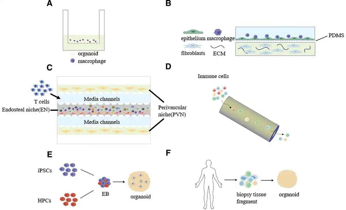
Static coculture platforms remain a foundational strategy for modeling immune cell behavior in vitro. These systems typically rely on physical proximity rather than fluid dynamics to support cell-cell interaction, and are often used due to their simplicity and ease of implementation. Zhu et al.[57] employed a static contact coculture approach to introduce immune cells into a lung organoid system. Utilizing a “spreading Matrigel” strategy, organoids were cultured within a thin layer of extracellular matrix (ECM) on the upper chamber of a Transwell insert, with part of their surface exposed to allow direct contact and attachment of immune cells (Figure 1A). Alveolar macrophages (AMs) and interstitial macrophages (IMs), isolated from lung tissue, were directly added to the upper chamber and placed in physical proximity to the organoids embedded in the Matrigel, without the need for any microfluidic injection. Although hydrodynamic forces such as micropumps were not applied, the Transwell structure allowed for paracrine signaling from mesenchymal stem cells (MSCs) seeded in the lower chamber, enabling the modeling of inflammatory stimuli and simulating structural and signaling interactions in a “pulmonary immune unit.” This simple yet well-controlled model preserved natural cell-cell contact and environmental cues and proved useful in studying macrophage chemotaxis, activation, and inflammatory modulation during acute lung injury.
Figure 1.
Immune cell integration strategies. (A) Static coculture of immune cells in a Transwell system. (B) Static coculture of immune cells within a microscale lung OoC. (C) Immune cell perfusion using a 5-channel microfluidic architecture. (D) Immune cell perfusion in a tubular organoid chip device. (E) Endogenous differentiation-based integration of immune cells. (F) In situ immune cell incorporation from biopsy tissue fragments. (These diagrams were independently created by our team using the BioGDP.com platform).
Unlike the standard Transwell design, Xia et al.[58] implemented a 3-layer microfluidic chip to spatially separate alveolar epithelium and stromal components. In this system, THP-1-derived macrophages were embedded within the epithelial compartment to reconstruct a localized pulmonary immune microenvironment (Figure 1B). Using static culture conditions, the system mimicked the alveolar epithelial–interstitial tissue interface. By coculturing alveolar epithelial cells, lung fibroblasts, and macrophages and treating them with varying doses of bleomycin (BLM), the model enabled systematic analysis of immune cell responses during the development of pulmonary fibrosis. Similar immune coculture strategies have been widely applied to organ models of the gut, liver, and lymphoid tissues to investigate organ-specific immune interactions.
For instance, in a gut-on-chip model, neutrophils derived from human peripheral blood were manually injected into the inlet channel, and the chip was subsequently incubated under static conditions. Neutrophils were able to migrate across a collagen gel containing macrophages and reach the polarized intestinal epithelial barrier[63]. Despite lacking active perfusion, this model effectively simulated neutrophil chemotaxis, adhesion, and tissue disruption under inflammatory stimuli. The results showed that under lipopolysaccharide (LPS) and N-formylmethionyl-leucyl-phenylalanine (fMLP) stimulation, neutrophils triggered epithelial disintegration and upregulation of inflammatory mediators, demonstrating that static systems can still achieve robust immune infiltration modeling and offer a controllable platform for studying acute inflammation mechanisms.
2.1.2 Dynamic perfusion models
To more closely mimic physiological immune circulation, dynamic perfusion-based models have gained increasing attention. These systems leverage microfluidic flow to enhance immune cell delivery, migration, and interaction with organoid structures. Ghoshal et al.[59] implemented a dynamic perfusion strategy to deliver chimeric antigen receptor T (CAR-T) cells into a preconstructed human multiple myeloma bone marrow-on-a-chip (hMM-on-a-chip) model, where CAR-T cells were introduced on the second day (48 h), monitored from 60 h onward, and maintained their activity for up to approximately 6 days (140 h). This 5-channel microfluidic system mimicked the endosteal and perivascular niches of the bone marrow. The central channel was lined with osteoblasts derived from bone marrow stromal cells, while the peripheral channels were engineered to form perfusable vascular endothelial networks[59] (Figure 1C). CAR-T cells were introduced through medium channels and actively migrated toward perivascular tumor regions containing multiple myeloma cells, achieving spatial colocalization and initiating cytotoxic immune responses. This flow-based delivery mimicked in vivo T-cell extravasation and infiltration and enabled real-time monitoring of cell migration, proliferation, and immune cytotoxicity. Using layer-resolved imaging and flow cytometry, the study comprehensively assessed CAR-T activation, antigen-specific killing, and cytokine release, highlighting the chip’s high potential for modeling cell-infusion immunotherapies.
In brain organoid studies, microglia introduction also followed a dynamic perfusion route. Researchers designed a 3D-printed, hollow tubular organoid-on-a-chip device in which the central channel and peripheral media chambers were simultaneously perfused with nutrients, oxygen, and immune cells[60] (Figure 1D). Microglia were loaded into the media reservoir at the chip center and cultured on a rocking platform (1 rpm), facilitating gradual and homogeneous infiltration of microglia into the brain organoid matrix via fluid flow. This noninvasive perfusion approach enabled immune cell integration without physical injection, minimizing structural damage and avoiding central hypoxia-associated immune inactivation often seen in large organoids. The perfusion system ensured homogeneous oxygenation and continuous chemotactic stimulation, providing microglia with conditions to remain functional and mobile, thereby significantly enhancing the physiological relevance and reproducibility of the organoid-based immune model.
2.1.3 Endogenous differentiation-based integration
Beyond exogenous introduction, immune cells can also be integrated into organoid systems through endogenous differentiation. This approach allows immune cells to codevelop alongside tissue lineages, more faithfully recapitulating ontogenetic processes. In the brain organoid model developed by Narasipura et al.[61], Microglia were not introduced exogenously or at a later developmental stage. Instead, they were integrated through an endogenous codevelopment strategy (Figure 1E). Compared with conventional cerebral organoids (COs), cerebral organoids with microglia (CO-iMs) were more efficient at generating CD45/CD11b/Iba-1 microglia and presented a physiologically relevant proportion of microglia (~7%). During embryoid body (EB) formation, researchers mixed human iPSCs with iPSC-derived hematopoietic progenitor cells (HPCs) at a defined ratio. Throughout subsequent organoid development (formation, expansion, and maturation), cultures were supplemented with macrophage colony-stimulating factor and interleukin-3 (IL-3) to synchronously induce the in situ differentiation, proliferation, and maturation of microglia-like cells from HPCs within the organoid matrix. This approach closely mimics the ontogeny of microglia during embryogenesis, wherein yolk sac–derived progenitors migrate into the developing brain and differentiate into mature microglia. By avoiding asynchronous cell injection and artificial implantation, this method reduces heterogeneity and enhances immune–neural integration. Moreover, the use of red fluorescent protein (RFP)-tagged HPCs allowed for genetic manipulation and lineage tracing of the microglial population, offering valuable insights into cell fate and function. Compared to exogenous methods, this endogenous differentiation strategy significantly improved microglial localization, viability, and immunocompetence while enabling coordinated development alongside neurons and astrocytes, establishing a more physiologically relevant immune-competent brain organoid model.
In parallel, Santos et al.[62] proposed an endogenous modeling method for the gut immune environment using an air–liquid interface (ALI) intestinal organoid system, directly derived from human small intestinal biopsy fragments (Figure 1F). These unmanipulated tissue segments retained their native epithelial, mesenchymal, and tissue-resident immune cell components and were embedded in collagen matrices for long-term culture in a Transwell system under ALI conditions. Notably, this system supported stable growth for over 1 year without disrupting the native mucosal structure or immune cell spatial organization. By preserving the intrinsic architecture and immune composition of the lamina propria, this model achieved high-fidelity reconstruction of the intestinal mucosal immune microenvironment. It offers a robust and physiologically relevant platform for studying diseases requiring in situ immune activation, such as celiac disease, where accurate simulation of localized immune interactions is essential. These endogenously maintained organoid models represent a significant advancement in modeling immune-tissue interactions without the need for exogenous manipulation, providing enhanced reproducibility and translational potential for autoimmune, infectious, and neuroinflammatory disease research.
2.2 Sources of immune cells
A primary challenge in constructing in vitro models with immune functionality lies in the appropriate selection of immune cell sources, which is essential to effectively reconstruct a realistic and complex immune microenvironment. Currently, commonly used immune cells can be broadly classified into 3 categories: PBMCs[59], differentiated immune cells derived from iPSCs or HPCs[73], and standardized experimental cell lines (e.g., Jurkat, NK-92)[74]. These 3 sources differ in terms of accessibility, retention of immune function, individual specificity, and expansion potential, thereby providing distinct foundations for various modeling strategies.
2.2.1 Cell lines: high standardization and reproducibility
In the development of in vitro immune models, commercially available immune cell lines (such as the Jurkat T-cell line, THP-1 monocyte cell line, NK-92 natural killer cell line, and neutrophils) are frequently employed as the initial platform due to their high degree of controllability, standardization, and reproducibility. These lines are commonly used for immune mechanism validation and preliminary drug screening studies[75,76]. For example, Qiang et al.[64] utilized macrophages differentiated from the THP-1 cell line to mimic red pulp-resident macrophages of the spleen, constructing a spleen-mimicking organ-on-chip system to investigate the role of macrophages in clearing abnormal red blood cells in sickle cell disease (SCD) (Figure 2A). This study revealed enhanced adhesion and phagocytosis dynamics between macrophages and SCD red blood cells under hypoxic conditions, offering a novel perspective on the functional roles of myeloid cells in tissue immunity.
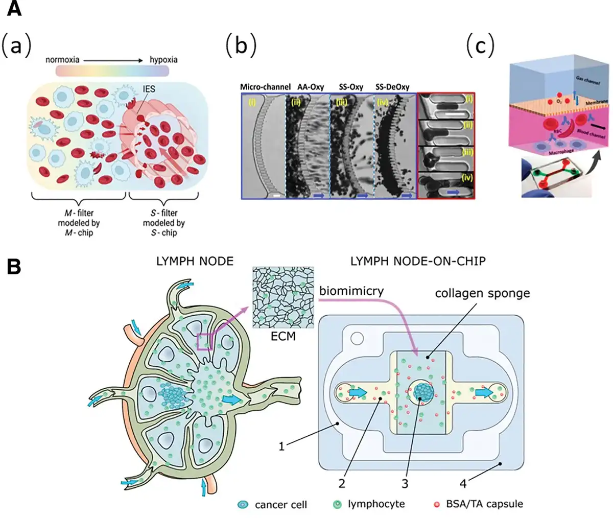
Figure 2.
Schematic representations of spleen-on-a-chip and lymph node-on-a-chip systems. (A) Spleen-on-a-chip: (a) Schematic of the oxygen gradient near the splenic sinus. Two structural and functional filters—the S-filter and M-filter—are biomimetically modeled in vitro using the S-chip and M-chip, respectively. (b) Cross-sectional view of the S-chip, showing the process and obstruction of red blood cells (RBCs) traversing the biomimetic splenic slits:(i) microfluidic channels; (ii) AA-type oxygenated RBC filtration; (iii) SS-type oxygenated RBC filtration; (iv) SS-type deoxygenated RBC filtration. (c) Schematic of the M-chip: blood flows through channels lined with adherent macrophages, while oxygen concentration is regulated via an overlying gas channel[64]. Copyright 2023, The Authors. (B) Lymph node-on-a-chip: schematic of a natural human lymph node and the structural design of the LNOC[38]. Copyright 2019, The Authors.
Similarly, German et al. employed the Jurkat cell line, derived from human T-cell leukemia, as a model T lymphocyte and introduced it into a lymph-node-on-chip system to simulate the behavior of circulating T cells within lymphatic fluid[38] (Figure 2B). Owing to its remarkable stability and scalability, this immortalized commercial cell line is widely utilized in studies of T-cell signaling and immune regulation. In their experiments, Jurkat cells were coinjected with bovine serum albumin/tannic acid (BSA/TA) nanoparticles of varying diameters into the chip to assess their distribution, penetration, and delivery performance around 4T1 breast cancer organoids. The findings demonstrated that such standardized cell lines not only emulate fundamental immune behaviors but also serve as effective models for evaluating drug delivery and immune infiltration capacities.
2.2.2 PBMCs: conventional and efficient
PBMCs have emerged as a critical component in constructing individualized immune response models due to their abundant immune subpopulations and preserved innate immune activity. PBMCs are widely accessible and easily isolated, encompassing key immune subsets such as T cells, B cells, natural killer (NK) cells, and monocytes. At the same time, the use of patient-derived PBMCs requires careful ethical oversight, particularly with respect to informed consent and donor privacy when genetic or clinical data are involved. Their full immunocompetence makes them particularly suitable for modeling patient-specific tumor organoids or organ-on-chip systems aimed at evaluating personalized immune responses and immunotherapeutic drug efficacy[77–78–79].
For instance, Gao et al.[65] developed a microfluidic platform to evaluate the interactions between patient-derived tumor organoids (PDOs) and immune cells. PBMCs were isolated from lung cancer patients using density gradient centrifugation, and their T-cell subtypes were quantified via flow cytometry. These cells were then statically seeded onto the chip and cocultured with PDOs to simulate the in situ immune microenvironment. Under immune checkpoint blockade (e.g., anti-programmed cell death protein 1 [PD1]), CD8⁺ T cells within the PBMCs demonstrated significant chemotaxis into the organoids, inducing tumor cell apoptosis. The use of autologously paired PBMCs and PDOs allowed the coculture system to effectively mimic personalized immune landscapes, offering a high-fidelity experimental platform for assessing immunotherapeutic responses.
In a similar study, researchers investigated immune interactions with human kidney organoids by isolating T cells from PBMCs and coculturing them with kidney organoids derived from iPSCs. The PBMC-derived T cells successfully infiltrated the organoid tubules and interacted with internal structures, leading to significant structural damage. This immune-mediated injury was accompanied by the upregulation of immune response-related genes in both epithelial and stromal cells within the organoids, including heightened expression of human leukocyteantigen (HLA) genes[66]. Despite their robust native immune function and patient specificity, PBMCs suffer from limited scalability and standardization, making cell lines a more controlled alternative for high-throughput or highly reproducible studies.
2.2.3 iPSCs/HPCs: controllability and scalability
Although PBMCs excel in individualized modeling, their substantial batch-to-batch variability and poor scalability restrict their use in high-throughput applications. In contrast, advances in stem cell technology have introduced a promising alternative—immune cells derived from iPSCs or HPCs. These sources offer both controllability and expandability. Nonetheless, ethical considerations remain, particularly regarding genome editing procedures and the long-term storage of stem cell lines. Together, these features position them as viable replacements for PBMCs in standardized immunological studies.
For example, Narasipura et al.[61] developed a brain organoid model containing microglia-like cells, in which the immune components originated from iPSC-derived HPCs. iPSCs were cocultured with HPCs during early EB formation, where HPCs spontaneously differentiated into microglia-like cells within a neurodevelopmental context. These cells expressed canonical markers such as CD45⁺, CD11b⁺, and Iba1⁺, and retained microglial homeostatic genes like TMEM119, CX3CR1, and CSF1-R, indicating functional maturity. Compared to exogenously injected cells or immortalized lines, these stem cell-derived microglia not only exhibited higher abundance (~7%) but also integrated naturally within the developing brain organoid, interacting physiologically with neurons and astrocytes. Under HIV-infection conditions, the model displayed typical inflammatory responses and viral reactivity, validating its relevance in studying neuroimmune-viral interactions.
In another study, researchers established a human brain organoid-on-a-chip system incorporating microglia-like cells (iMGs) derived from embryonic stem cells (hESCs, WA01 line) via HPC differentiation[60]. These iMGs exhibited lower baseline activation and heightened responsiveness to inflammatory stimuli (e.g., LPS/adenosine triphosphate [ATP]), making them more reflective of native human microglial phenotypes compared to commercialized microglial lines like HMC3. Moreover, iMGs demonstrated strong integration capability and morphological consistency, maintaining a stable, ramified “resting” state within the cerebral microenvironment and transforming into an activated phenotype upon stimulation. This enhanced fidelity and controllability greatly improve the system’s reliability in modeling human neuroinflammation.
In Conclusion, from the high standardization of commercial cell lines to the innate immunological complexity of PBMCs and the controllability and plasticity of iPSC/HPC-derived cells, these 3 immune cell sources offer distinct yet complementary strategies for constructing functional in vitro immune models. Appropriately aligning the modeling objectives with the choice of immune cell source is a central principle in the successful reconstruction of immune functionality in organoid and organ-on-chip systems.
To better highlight the functional trade-offs among different immune cell sources, Table 3 provides a comparative summary across functional fidelity, patient specificity, and scalability, along with representative applications.
Table 3
Comparative evaluation of immune cell sources.
| Source | Functional fidelity (e.g., cytokine secretion, antigen presentation, plasticity) | Patient specificity (genomic stability, donor variability) | Scalability and reproducibility | Representative applications |
|---|---|---|---|---|
| Cell lines | High reproducibility; limited functional plasticity (e.g., poor M1/M2 polarization[80]) | Not patient-specific | High scalability | Mechanistic studies, early drug evaluation[64,75,76] |
| PBMCs | Retain key immune functions such as cytokine secretion, TCR signaling, and NK cell cytotoxicity[81] | Allows for personalized immunological readouts, high interindividual variability | Limited scalability | Widely used in tumor immunology[65] |
| iPSC/HPC-derived immune cells | High physiological fidelity | Genomic stability depends on reprogramming process[73] | Expansible | Modeling neuroimmune interactions[61] |
NK, natural killer; TCR, T-cell receptor.
2.3 Immune cell targeting
Immune cells, in fulfilling their defensive and regulatory roles, must rely on precise spatial positioning and dynamic response mechanisms to perform targeted recognition of specific tissue microenvironments and subsequently execute a cascade of effector functions. This process involves not only direct recognition of pathogenic agents but also the ability to traverse physiological barriers, infiltrate tissues, sense ECM signals, and coordinate responses with other cell types[82]. The rapid advancement of organ-on-chip and 3D organoid technologies has enabled researchers to simulate critical immunological events—such as barrier disruption, immune cell migration, and activation—within highly biomimetic environments, facilitating deeper insights into tissue-specific immune responses.
This section analyzes immune targeting behaviors through 2 main model categories (Figure 3):
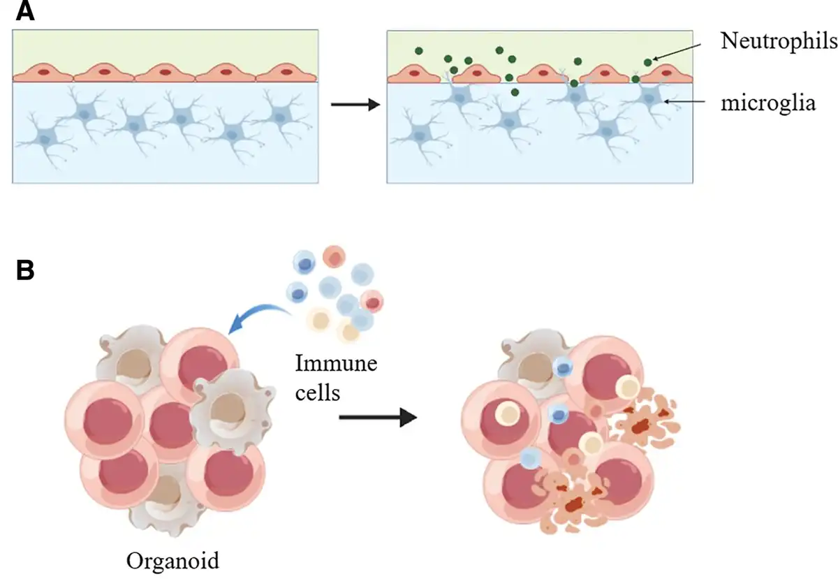
Figure 3.
Targeted recognition by immune cells. (A) Schematic representation of a barrier-crossing model. (B) Immune cell targeting and infiltration within a 3D organoid model. (These diagrams were independently created by our team using the BioGDP.com platform).
Barrier-on-chip models, which replicate physiological interfaces like epithelia or the blood–brain barrier (BBB), focusing on how immune cells traverse physical barriers and modulate tissue homeostasis;
3D organoid models, including tumor spheroids and PDOs, which emphasize the chemotaxis, recognition, and cytotoxic responses of immune cells in complex 3D tissue microenvironments.
2.3.1 Barrier-on-chip models
Gjorevski et al.[63] developed a fully functional intestinal immune organ-on-chip to recapitulate the entire process of neutrophil targeting and translocation across epithelial barriers. The model emulates typical inflammatory conditions in the gut, where neutrophils are recruited to mucosal interfaces and exhibit enhanced migratory and transmigration capacities when cocultured with activated THP-1-derived macrophages. The barrier was formed by perfusable Caco-2 epithelial tubules, while THP-1 macrophages were embedded in lateral collagen matrices to simulate lamina propria immune cells. Under stimulation with LPS and fMLP, and in conditions of epithelial barrier disruption, neutrophils were able to effectively traverse the ECM, reach sub-barrier regions, and cause epithelial structural disintegration, cell detachment, and death, accompanied by the release of tumor necrosis factor alpha (TNF-α) and matrix metalloproteinases (MMP-8/9). This model highlights the dual role of neutrophils: as frontline responders in host defense and as contributors to chronic inflammation and tissue damage, underscoring their importance in inflammatory bowel disease pathogenesis.
Similarly, Zeng et al. constructed a biomimetic BBB-on-chip model that integrates endothelial cells, astrocytes, and HMC3 microglia to simulate both physiological and pathological neurovascular environments[67]. The chip features a 3-channel parallel design: the central channel is filled with either Matrigel or fibrin to represent normal basement membrane or fibrin deposition in BBB dysfunction, while adjacent channels are seeded with hCMEC/D3 brain endothelial cells and HMC3 microglia. Upon LPS stimulation in the healthy BBB model, astrocyte surface area increased, microglial migration speed rose significantly to 0.58 μm/min, and CD68 expression was upregulated 2.3-fold. In contrast, microglia in the pathological fibrin-laden environment exhibited attenuated responses—migration slowed to 0.37 μm/min, and CD68 levels remained unchanged—indicating immune hypo-responsiveness in diseased microenvironments. These findings validate the model’s utility in studying immune dysfunctions and screening neuroinflammatory therapies.
2.3.2 Targeted 3D organoid models
In recent years, coculture systems integrating PDOs and immune cells—particularly T cells—have emerged as powerful tools for dissecting immune cell behavior within the 3D tumor microenvironment (TME)[83,84]. Jeong et al.[68] systematically reviewed immune-PDO coculture strategies involving PBMCs, tumor-infiltrating lymphocytes (TILs), and engineered T-cell receptor (TCR)/CAR-T cells, highlighting their chemotactic migration, immune synapse formation, and antigen-specific cytotoxicity within organoid structures. T cells were shown to actively migrate through Matrigel toward PDO surfaces, establish immune synapses, and induce interferon-γ (IFN-γ) release and tumor cell apoptosis. Moreover, the study discussed enhancing immune cell efficacy through interventions like CD39 inhibition and PD-1 blockade, demonstrating improved infiltration and cytotoxicity within 3D organoid systems. These results confirm that immune cells exhibit spatial targeting and functional specificity in organoid environments, making these platforms highly faithful models for simulating the tumor-immune landscape.
Similarly, Li et al.[69] constructed a 3D coculture model based on a gel-liquid interface, combining PBMCs with lung cancer organoids (LCOs), to replicate immune cell-directed infiltration and antitumor effects. In this system, PBMCs were seeded on top of the Matrigel, while LCOs were embedded at the gel base, creating a defined spatial path for immune cell chemotaxis and infiltration. Under PD-1 blockade, peripheral CD8⁺ T cells successfully traversed the matrix, deeply infiltrated organoid structures, and exhibited potent cytotoxicity, marked by increased IFN-γ and CD107a expression. This model demonstrated robust immune infiltration and tumor cell killing, providing a physiologically relevant platform for modeling the tumor-targeting capabilities of peripheral immune cells in 3D tumor organoids.
To achieve high-fidelity modeling of immune cell targeting under viral infection, Natarajan et al.[70] developed a microfluidic coculture system incorporating HLA-matched exogenous CD8⁺ T cells and liver organoids, aiming to investigate antigen-specific cytotoxic responses against hepatitis C virus (HCV). Researchers isolated and clonally expanded T cells specific to the NS3 peptide epitope (KLVALGINAV) from donors who had naturally cleared HCV infection. These T cells were then cocultured within a microfluidic chip with adult stem cell-derived liver organoids expressing HLA-A0201. Upon peptide pulsing of the organoids, the antigen-specific CD8⁺ T cells migrated through the chip’s microchannels and infiltrated the organoid compartment, executing antigen-dependent cytotoxicity. Within 60 h, organoid viability dropped below 20%, demonstrating a robust and specific immune effect. This platform effectively recapitulated the entire cascade of viral antigen presentation, immune cell migration, recognition, and killing within a controlled setting, offering a powerful paradigm for constructing targeted immune microenvironments in virus-infected organoid systems.
Beyond the use of exogenous immune interventions, other studies have leveraged endogenous tissue-resident immune cells to model viral immunity. Choi et al.[71] established a 3D ALI lung organoid model derived from unenzymatically processed adult human lung tissue, thereby retaining the native epithelial, stromal, and immune components—including T cells, B cells, NK cells, and macrophages—with a notable presence of CD69⁺CD103⁺ tissue resident memory T cells (TRM). Upon SARS-CoV-2 infection, the organoids accurately reflected viral tropism, predominantly targeting alveolar type II cells. Importantly, the model also activated both innate and adaptive immune responses intrinsic to the lung tissue, including cytokine production, antigen presentation, TCR clonal expansion, and activation of virus-specific CD8⁺ T cells. These processes occurred independently of peripheral lymphoid organs, faithfully mirroring the immunological dynamics of lung-resident responses. This study provides an important model for exploring tissue-specific antiviral immunity and immune memory under physiologically relevant conditions.
Collectively, these findings highlight that immune cells’ target recognition and effector functions are intricately dependent on the structural and dynamic signals within their microenvironment. Barrier-on-chip models, by simulating tissue permeability and inflammatory states, enable precise dissection of how immune cells traverse physical obstacles and modulate tissue homeostasis or injury. Meanwhile, 3D organoid coculture systems reveal how immune cells execute chemotaxis, antigen recognition, and cytotoxicity within complex tissue architectures. Whether modeling responses to tumors, viral infections, or autoimmune triggers, these advanced platforms provide high-fidelity, physiologically relevant environments that overcome the spatial, cellular, and functional limitations of traditional 2D cultures. A concise comparison of these 2 platforms is provided in Table 4 to highlight their respective features and applications.
Table 4
Comparative characteristics of immune cell targeting in barrier-on-chip and 3D organoid platforms.
| Feature | Barrier-on-chip models | 3D organoid models |
|---|---|---|
| Simulated structure | Physiological barriers (e.g., intestinal epithelium[63], BBB[67]) | 3D tissue architecture (e.g., tumor spheroids[83,84], viral organoids[70]) |
| Immune behavior | Translocation, barrier disruption, and adhesion by neutrophils/microglia | Infiltration, antigen recognition, and cytotoxicity by PBMCs or T cells |
| Disease applications | Inflammatory bowel disease, neuroinflammation, barrier dysfunction | Tumor immunity, antiviral responses (e.g., HCV[70], SARS-CoV-2[71]) |
BBB, blood–brain barrier; HCV, hepatitis C virus.
Such models offer critical support for decoding the spatial-temporal dynamics of immune interactions and developing next-generation immunotherapies. By bridging immunological function with microengineering and tissue biology, they mark a transformative shift toward precision modeling of immune responses in human disease.
Although organoid and chip-based systems have successfully recapitulated immune cell infiltration phenomena, a deeper understanding still requires elucidation of the molecular pathways driving these processes. In our framework, these regulatory mechanisms can be broadly categorized into 5 interconnected layers, which are illustrated in Figure 4 of this work. At the foundation lie chemokine gradients and adhesion molecule interactions, which directly govern the directionality and localization of immune cell migration. Building on this, major signaling pathways such as JAK/STAT[85], NF-κB[86], and PI3K/AKT[87,88] modulate transcriptional programs and metabolic states to fine-tune cellular responses. In parallel, the microenvironment exerts profound modulatory effects through factors like TGF-β/SMAD[89] signaling, hypoxia-driven hypoxia-inducible factor 1 alpha (HIF-1α) activation[90], lactate metabolism[91], and ECM remodeling[92], each of which reshapes immune infiltration capacity and functional states. Ultimately, these layered regulatory inputs converge on tissue effector functions, particularly antigen presentation and immune checkpoint signaling, thereby determining the efficacy and durability of immune responses. Among these dimensions, the present text focuses primarily on the first 2—chemokine-driven migratory cues and adhesion molecule-mediated interactions—as central regulators of immune cell migration and targeting. Their integration into organoid and chip-based systems not only reproduces physiologically relevant infiltration dynamics but also establishes a foundation for incorporating more complex regulatory layers in future immune modeling.
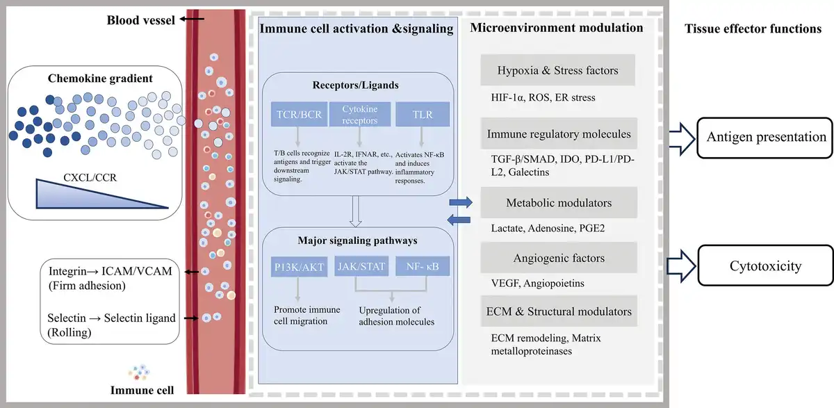
Figure 4.
Molecular drivers of immune cell infiltration (These diagrams were independently created by our team using the BioGDP.com platform).
2.3.3 Molecular pathways guiding immune cell infiltration
Chemokines play a central role in directing immune cell migration and tissue infiltration within organoid and OoC systems. Among the best-characterized pathways, the CXCL9/10/11-CXCR3 axis plays a pivotal role in the recruitment of immune cells and the formation of cancer-specific immunity in various cancers[92,93]. Elevated expression of CXCL9/10 by tumor-associated stromal cells has been shown to enhance T-cell chemotaxis, thereby promoting immune surveillance[94,95]. Similarly, the chemokine receptor CCR7 and its ligands CCL19 and CCL21 control a diverse array of migratory events in adaptive immune function[96]. Most prominently, CCR7 promotes homing of T cells and dendritic cells (DCs) to T cell areas of lymphoid tissues where T cell priming occurs, a process that is highly relevant for recreating immune priming niches in vitro. Conversely, the CXCL12–CXCR4 pathway often facilitates tumor progression by attracting immunosuppressive populations such as regulatory T cells and myeloid-derived suppressor cells (MDSCs)[97]. These opposing roles underscore the importance of chemokine gradients as critical regulators of immune cell targeting, and highlight opportunities to engineer microfluidic platforms capable of generating precise, controllable chemokine fields to modulate immune infiltration.
In addition to chemokine signaling, adhesion molecules are indispensable for guiding immune cells across endothelial or stromal barriers and for stabilizing immune cell interactions. A prototypical mechanism involves LFA-1 (integrin αLβ2) on T cells binding to intercellular adhesion molecule-1 (ICAM-1), a process essential for firm arrest[98]. Similarly, the interaction between very late antigen-4 (VLA-4) and its ligand vascular cell adhesion molecule-1 (VCAM-1) plays a critical role in immune cell adhesion and localization. This pathway facilitates immune cell retention and infiltration within inflamed tissues or TMEs, with its effectiveness strongly influenced by cellular activation status and local shear stress[99]. Together, these adhesion cascades act in concert with chemokine gradients to enable stepwise immune cell extravasation and tissue infiltration. Reproducing such interactions in vitro is therefore essential for physiologically relevant immune-on-a-chip models, where coating channels with ICAM-1 or VCAM-1 or modulating shear stress can be leveraged to better mimic immune cell trafficking dynamics.
2.4 Immune cell compositional strategies
2.4.1 Single-type immune cell systems: platforms for modeling specific immune responses
To simulate the dynamic progression of BLM-induced idiopathic pulmonary fibrosis (IPF), researchers developed a biomimetic lung organ-on-chip integrating macrophages. The chip design features a polydimethylsiloxane (PDMS) porous membrane that structurally separates the epithelial and stromal compartments and introduces THP-1-derived macrophages, enabling the in vitro construction of a controllable single-immune microenvironment[58] (Figure 5A). Upon BLM stimulation, macrophages initially polarize toward the M1 phenotype (expressing IL-1β, TNF-α), mediating inflammatory responses. With prolonged exposure, they shift toward the M2 phenotype, promoting TGF-β1 secretion and collagen deposition via activation of the PI3K-AKT pathway. Integrating transcriptomics, immunofluorescence staining, qPCR, and ELISA, the study systematically characterized macrophage functional transitions in IPF from molecular to tissue levels. This work offers both mechanistic insights and an experimental platform supporting antifibrotic strategies targeting M2 polarization.
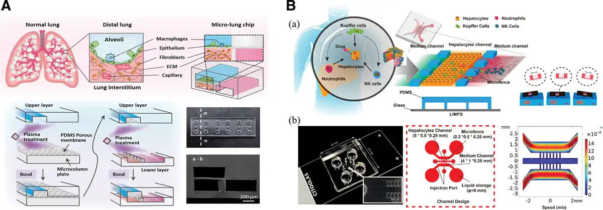
Figure 5.
Representative immune cell compositional strategies in OoC models. (A) Lung-on-a-chip: schematic illustration of the chip design, assembly process, final fabricated device, and scanning electron microscopy (SEM) image of the cross-section along line a–b[58]. Copyright 2025, The Authors (B) Liver-on-a-chip: (a) the illustration of the immune-mediated drug-induced liver injury (left), the design of the LIMPS (middle), and the gravity-based pumping (right); (b) structural overview of the liver microphysiological system (LIMPS) chip and a magnified view of the microfluidic channels[72]. Copyright 2024, The Authors.
In the nervous system, a similar approach incorporated microglia into a neuroimmune chip. The model integrates brain microvascular endothelial cells, astrocytes, and microglia to construct a 3D physiologically functional BBB organ-on-chip[67]. Upon LPS stimulation, the model demonstrated increased BBB permeability, microglial activation, and inflammatory cytokine release, effectively recapitulating hallmark features of neuroinflammation. This model underscores the utility of single-immune-cell systems for dissecting the relationship between immune activation and barrier integrity.
While single-immune-cell models offer the advantage of simplicity and precise control, facilitating focused investigations on cell-specific mechanisms, they inherently lack the complexity required to simulate coordinated immune cascades and intercellular synergy found in physiological immune responses.
2.4.2 Multi-immune-cell systems: reconstruction strategies approaching physiological complexity
In contrast, emerging multi-immune-cell coculture systems offer greater physiological relevance by enabling investigation of intricate cellular interactions and immune feedback mechanisms. Deng et al.[72] developed a liver-immune microphysiological system (MPS) integrating microfluidics and diverse immune cells to simulate drug-induced immune-mediated liver injury. The system features a 3-channel microfluidic design: HepG2 hepatocytes are seeded in the central channel, while the flanking channels introduce drug-containing medium and 3 immune cell types (THP-1-derived macrophages, HL-60-derived neutrophils, and NK-92 natural killer cells). Gravity-driven flow facilitates dynamic nutrient exchange and signal transduction (Figure 5B). When exposed to the clinically relevant dose (15 μM) of troglitazone (TGZ), the system captured hallmark responses: enhanced neutrophil-hepatocyte adhesion, increased M1 macrophage ratio, and elevated inflammatory cytokine expression, culminating in hepatocellular injury and death. Enabled by real-time imaging, the platform revealed intercellular interactions difficult to observe in traditional static cultures, offering a sensitive and predictive tool for preclinical assessment of immune-mediated hepatotoxicity.
In pulmonary organoid research, Zhu et al.[57] developed a more complex immune coculture model involving 2 primary macrophage subtypes (AMs and IMs), isolated respectively from bronchoalveolar lavage fluid and lung parenchyma. These were incorporated into a lung organoid model. AMs, located in the alveolar lumen, primarily handle phagocytosis and early immune responses; in contrast, IMs reside near blood vessels and basement membranes, specializing in antigen presentation and immune regulation. Upon LPS stimulation, the model successfully captured differences in migration, aggregation, cytokine secretion (e.g., IL-6, TNF-α), reactive oxygen species (ROS) production, and nod-, lrr-, and pyrin domain-containing protein 3 (NLRP3) inflammasome activation between AMs and IMs. The study highlights how coordinated actions between immune cell subsets enhance the physiological fidelity of complex immune microenvironment models.
In summary, single-immune-cell systems offer structural simplicity and tightly controlled variables, making them ideal for studying the phenotypic transitions and signaling pathways of specific immune cell types, such as M1/M2 polarization in macrophages or microglial activation. However, they fall short in replicating the multifaceted pathophysiological landscape of diseases, particularly in simulating cascade responses and functional complementarity among immune cells.
Conversely, multi-immune-cell systems enable a higher-order coupling of spatial architecture, signal transduction, and cell behavior by incorporating diverse immune subtypes. These systems more accurately mimic the complexity of native immune microenvironments. With advances in microfluidics, cellular labeling, and primary cell isolation, future in vitro disease models are expected to evolve toward greater complexity and dynamic multicellular interactions, ultimately offering more precise and reliable platforms for studying immunopathology and predicting therapeutic responses.
To achieve a comprehensive understanding of immune microenvironment reconstruction within organ-on-chip and organoid platforms, we categorized and synthesized current strategies along 4 primary dimensions.
First, we categorized immune cell integration approaches, highlighting how immune cells are introduced into the system through static coculture, perfusion-based delivery, or endogenous differentiation. These methods offer different advantages in spatial-temporal coordination with tissue structures and influence subsequent immune activity. Second, we examined the diversity of immune cell sources, encompassing a spectrum from standardized cell lines to immune cells derived from iPSCs and HPCs. Each source presents unique advantages in terms of accessibility, standardization, and physiological relevance. Third, we explored the target recognition and effector specificity of immune cells, highlighting how immune cells identify, migrate toward, and exert functions in response to specific tissue contexts. This dimension elucidates key mechanisms of cell-cell communication, barrier crossing, and antigen-specific responses. Fourth, we distinguished between single-immune-cell systems and multi-immune-cell systems, comparing their respective strengths and limitations. While single-cell systems offer controllability and mechanistic clarity, multicellular systems better recapitulate the complexity and feedback of in vivo immune responses.
This classification framework not only systematizes the current landscape of immune microenvironment modeling technologies but also provides theoretical guidance for establishing generalizable principles and tailored design strategies in future immune-integrated organoid and organ-on-chip platforms.
3. Applications of immune organoid coculture systems
3.1 The role of immune organoid-on-a-chip models in tumor therapy
Traditional animal models and 2D cell culture systems have significant limitations in simulating tumor biology and drug responsiveness, as they cannot fully replicate the complex TME of human tumors[100]. Organoids, capable of replicating the physiological and pathological characteristics of the original tissues in vivo, play a crucial role in accurately modeling tumor heterogeneity and the TME. The TME, composed of cancer cells, immune cell populations, stromal cells, and soluble molecules such as cytokines and growth factors, exerts a decisive influence on tumor initiation and progression by affecting multiple aspects of tissues, including metabolism, angiogenesis, and the immune system. Microfluidic OoC devices, featuring various micrometer-scale structures, support the coculture of cells or tissues, enabling the study of physiological and pathological systems in a highly controlled manner[101]. Utilizing organoid-on-a-chip models to investigate the killing effects of immune cell therapy on tumors allows for the study of key physiological aspects of tissues or organs, the interactions between cells (parenchymal cells, vascular cells, and immune cells) and their ECM molecules, the impact of native tissue architecture (geometry, dynamic flow, or mechanical forces) on tissue function, as well as the elucidation of mechanisms underlying tissue-specific diseases and drug testing[69]. Meanwhile, employing personalized tumor models that resemble the characteristics of the original tumor, preserving genomic, phenotypic features, and drug response profiles, enables more accurate prediction of patient responses to drugs. Tumor organoid models thus facilitate personalized cancer treatment approaches tailored to each patient[102]. By dynamically reconstructing the heterogeneous TME, organoid-on-a-chip models serve the following roles in the field of tumor therapy: (1) evaluation of immune cell therapy; (2) accelerating the translation process of antitumor drugs from in vitro validation to clinical trials, providing a highly biomimetic research platform for breaking through the bottlenecks in tumor therapy; and (3) in-depth analysis of cancer molecular mechanisms such as drug resistance. Figure 6 presents a summary diagram of the applications of organoids and OoC technologies in various aspects.
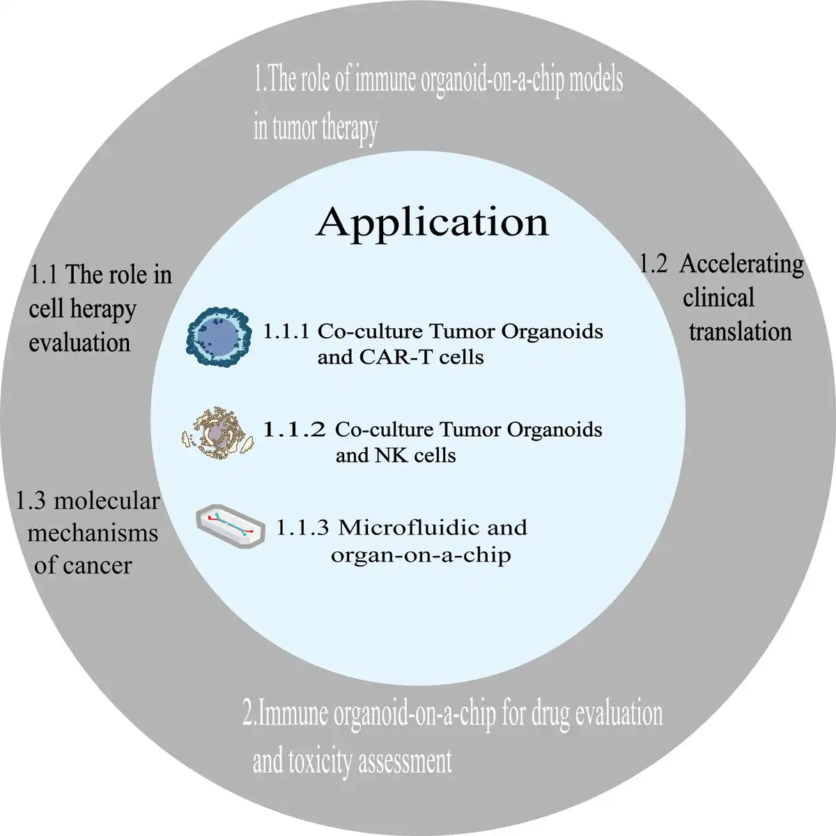
Figure 6.
Summary of applications of organoids and OoC technologies.
3.1.1 The role of tumor organoid-on-a-chip in the evaluation of immune cell therapy
The immune microenvironment plays a pivotal role in tumor therapy research, as it not only participates in tumor initiation and progression but also serves as a critical barrier to the efficacy of immunotherapy. Reshaping the immune microenvironment can significantly enhance the response rate to immunotherapy. Tumor organoid-on-a-chip technology integrates patient-derived tumor tissues with microfluidics to construct highly biomimetic 3D dynamic models, providing an in vitro research platform that closely approximates clinical reality for precise medication. By simulating the processes of drug penetration, metabolism, and cellular response within tumor tissues, this technology enables efficient screening of personalized treatment regimens (such as testing sensitivity to targeted drugs and optimizing combinations of immunotherapy and chemotherapy agents) (Figure 7A)[103]. Illustrates a schematic diagram of the application of tumor organoid-on-a-chip in immune cell therapy. Depending on the types of immune cells contained within the tumor organoids, they can be categorized into tumor organoids with an inherent immune microenvironment and tumor organoids cocultured with immune cells for cell therapy applications.
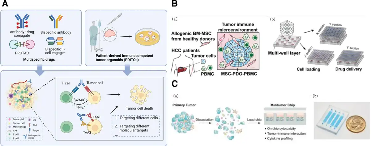
Figure 7.
Representative models illustrating the roles of immune organoid-on-a-chip models in tumor therapy. (A) Utilizing patient-derived immunocompetent tumor organoids as a model for evaluating multispecific drugs in cancer research[103]. Copyright 2025, The Authors. (B) Establishing a HCC TME on a high-throughput microfluidic chip through coculturing organoids derived from HCC specimens with MSCs and PBMCs. (a) Acquisition and coculturing of organoids. (b) Design of the microfluidic chip for high-throughput organoid culture and drug screening[28]. Copyright, The Authors. (C) Operating principle of the microtumor chip. (a) Design concept of the microtumor chip. (b) Physical appearance of the microtumor chip (featuring 4 parallel injection ports)[104]. Copyright 2022, The Authors.
CAR-T cell therapy is a novel form of adoptive immunotherapy that involves the genetic engineering of T lymphocytes using viral vectors carrying CARs. This represents a significant advancement in the field of cancer treatment[105]. Jiang et al. constructed 2 types of bladder cancer organoids (BCOs) expressing or not expressing B7H3, along with CAR-T cells targeting B7H3. They cocultured these to evaluate the antitumor function of CAR-T cells. When B7H3 CAR-T cells were cocultured with B7H3-expressing BCOs, the secretion of cytokines IFN-γ and IL-2 significantly increased. However, no significant differences were observed in ascites-derived organoids that did not express B7H3. The results indicated that specific antigen recognition and immune activation occurred during the coculture process, and the immune organoid coculture system could be used to determine drug sensitivity and immune-specific responses in vitro[106]. Song et al. established a glioblastoma (GBM) organoid model and observed the antitumor effects by coculturing it with CAR-T cells in combination with the inhibitor of apoptosis protein (IAP) antagonist birinapant. The results showed that the IAP antagonist made patient-derived primary GBM organoids more sensitive to apoptosis induced by cytokines (such as tumor necrosis factor) secreted by CAR-T cells, thereby preventing antigen escape and enhancing the efficacy of CAR-T cell therapy[107]. Wehrli et al. constructed a genetically engineered cellular product, mesoFAP CAR-TEAM cells, which target mesothelin and secrete fibroblast activation protein (FAP). Using a PDAC organoid model derived from patient tumors along with immune/stromal cells, they evaluated the efficacy of mesoFAP CAR-TEAM cells. Compared with engineered T cells targeting only a single antigen, mesoFAP-treated organoid cocultures showed a significant reduction in cancer-associated fibroblasts (CAFs) on days 2 and 5, indicating that MesoFAP CAR-TEAM cells exhibited stronger and more targeted effects in eliminating PDAC and CAFs[108]. Porter et al. utilized a prostate cancer patient-derived organoid model to test the impact of Lewis Y CAR-T cells in combination with carboplatin, docetaxel, or the checkpoint inhibitor nivolumab on tumor growth. The study revealed that carboplatin could synergize with CAR-T therapy, promoting early and sustained infiltration and activation of CAR-T cells in specific prostate cancers[109].
NK cells are major effector cells in antitumor immunity. With the increasing number of clinical trials aimed at developing and improving chimeric antigen receptor-NK immunotherapies, there is a growing demand for a rapid and accurate detection system to screen for changes in the cytotoxic function of NK cells[110]. For instance, Oh et al. demonstrated the tumor-killing effect of NK cells by coculturing them with LCOs. Furthermore, pretreating NK cells with the TGF-β inhibitor A83-01 significantly enhanced NK cell-induced cell death in LCOs[111]. In addition to studying interactions between tumor organoids and NK cells, researchers have also explored the interactions between normal tissues and NK cells. For example, Ziegler et al. cocultured primary human liver organoids with CD56bright NK cells, NK cells can be divided into 2 subsets: CD56bright and CD56dim, which represent the major population of intrahepatic resident NK cells. Their study revealed that NK cells migrate into liver organoids and upregulate T cell immunoreceptor with Ig and ITIM domains (TIGIT) expression while downregulating danx accessory molecule-1 (DNAM-1) expression. TIGIT is a coinhibitory receptor that reduces NK cell cytotoxicity and is involved in NK cell exhaustion. Additionally, TIGIT is associated with liver regeneration[112]. The aforementioned studies indicate that the organoid-based NK cell coculture system is a reliable platform. The coculture model of tumor organoids and NK cells not only allows for the assessment of NK cell cytotoxicity but also serves as an in vitro tool for studying the interactions between human immune cells and tissue cells.
OoC technology plays a significant role in the research and development, optimization, and efficacy prediction of cell therapies by reconstituting the TME. Zou et al. utilized microfluidic OoC technology to establish a MSC-patient-derived organoid-peripheral blood mononuclear cell (MSC-PDO-PBMC) model (Figure 7B) to investigate the response of hepatocellular carcinoma (HCC) organoids to immunotherapy drugs. The study demonstrated that, compared to traditional organoid models cultured without an immune microenvironment, the MSC-PDO-PBMC model could more accurately predict the response of HCC patients to the immunotherapy drug atezolizumab. This model exhibits greater potential for precisely predicting patient responses to anti-PD-L1 drugs. Additionally, this platform significantly reduces the time required for high-throughput organoid culture and drug screening, particularly for predicting the efficacy of immunotherapy drugs such as immune checkpoint inhibitors. Ao et al. developed a microfluidic-based microtumor chip (Figure 7C) that mimics the in vivo tumor immune microenvironment, allowing for local cell-to-cell interactions and preserving autocrine and paracrine signaling. By constructing models of in situ breast cancer and renal cell carcinoma with constitutive differences in PD-L1 expression levels, they found that tumor cells in both models exhibited responses to anti-PD1 therapy 10 days after tumor inoculation[104].
Overall, immune cell therapies have proven effective in treating hematological malignancies in recent years. However, their application in solid tumor treatment remains in the exploratory stage, primarily due to the inadequacy of these therapies in addressing the TME. Retaining the biological characteristics of primary tumor tissues is crucial for research on immune cell therapies in early stage solid tumors[113]. Therefore, the adoption of organoid and OoC models addresses the deficiencies in existing models, particularly the lack of a tumor immune microenvironment. These advanced models play a significant role in gaining a deeper understanding of the interaction mechanisms between immune cells and the TME, evaluating the efficacy and safety of immune cell therapies, and subsequently developing more precise and effective therapeutic drugs for tumor treatment.
3.1.2 The role of tumor organoid-on-a-chip in clinical translation
Tumor organoids demonstrate significant advantages in the field of personalized therapy and clinical translation. Their core value lies in constructing “individualized disease models” through high-fidelity simulation of patient-specific pathological features and heterogeneity, thereby providing a quantifiable and iterable research platform for precision medicine.
In personalized therapy, tumor organoids can be directly derived from patient tumor or normal tissue biopsy samples, preserving the genomic and epigenetic characteristics of the original tissue, as well as its drug response properties. This enables high-throughput screening and efficacy prediction of individual drug sensitivity. For example, Chiriaco et al. utilized patient-derived mesenchymal epithelial transition factor (MET)-overexpressing tumor organoids to evaluate the targeted killing effect of MET-CAR-T cells on MET-overexpressing tumors. Clinical data indicate that MET overexpression is one of the molecular mechanisms underlying resistance to epidermal growth factor receptor (EGFR), human epidermal growth factorreceptor 2 (HER-2), and v-Raf murine sarcoma viral oncogene homolog B (BRAF) targeted therapies in various cancers. The results showed that MET-CAR-T cells could prevent the acquisition of resistance to MET-targeted agents by MET-amplified cancer cells carrying secondary mutations in downstream signaling pathways. MET-CAR-T cells overcame secondary resistance to anti-MET drugs, providing a new option for screening patient subgroups suitable for MET-targeted immunotherapy and offering hope for drug-resistant patients[114]. Logun et al. conducted autologous CAR-T cell therapy simultaneously on patient-derived and in vitro-derived glioblastoma organoids (GBOs) from the same patient. The results demonstrated that CAR-T cell therapy led to a reduction in target antigens and tumor cell lysis in GBOs, with the degree correlating with the chimerism rate of CAR-T cells detected in the patient’s cerebrospinal fluid (CSF). Additionally, the cytokine release patterns in GBOs over time were similar to those in the patient’s CSF samples, providing crucial information for clinical trials[115]. Maulana et al. developed a breast cancer organoid-on-a-chip model with an endothelial barrier, enabling real-time monitoring of cytokine release, CAR-T cell infiltration, and specific tumor cell lysis during an 8-day perfusion culture. This allowed for the real-time observation of CAR-T cell migration and killing, revealing the dependency of CAR-T cell response on target antigen density. Furthermore, compared to mouse models (which take months), the ability to directly obtain patient-derived organoids and rapidly establish tumor-on-a-chip models from patients enables this model to assist in clinical decision-making, which often needs to be completed within a short timeframe (Figure 8).
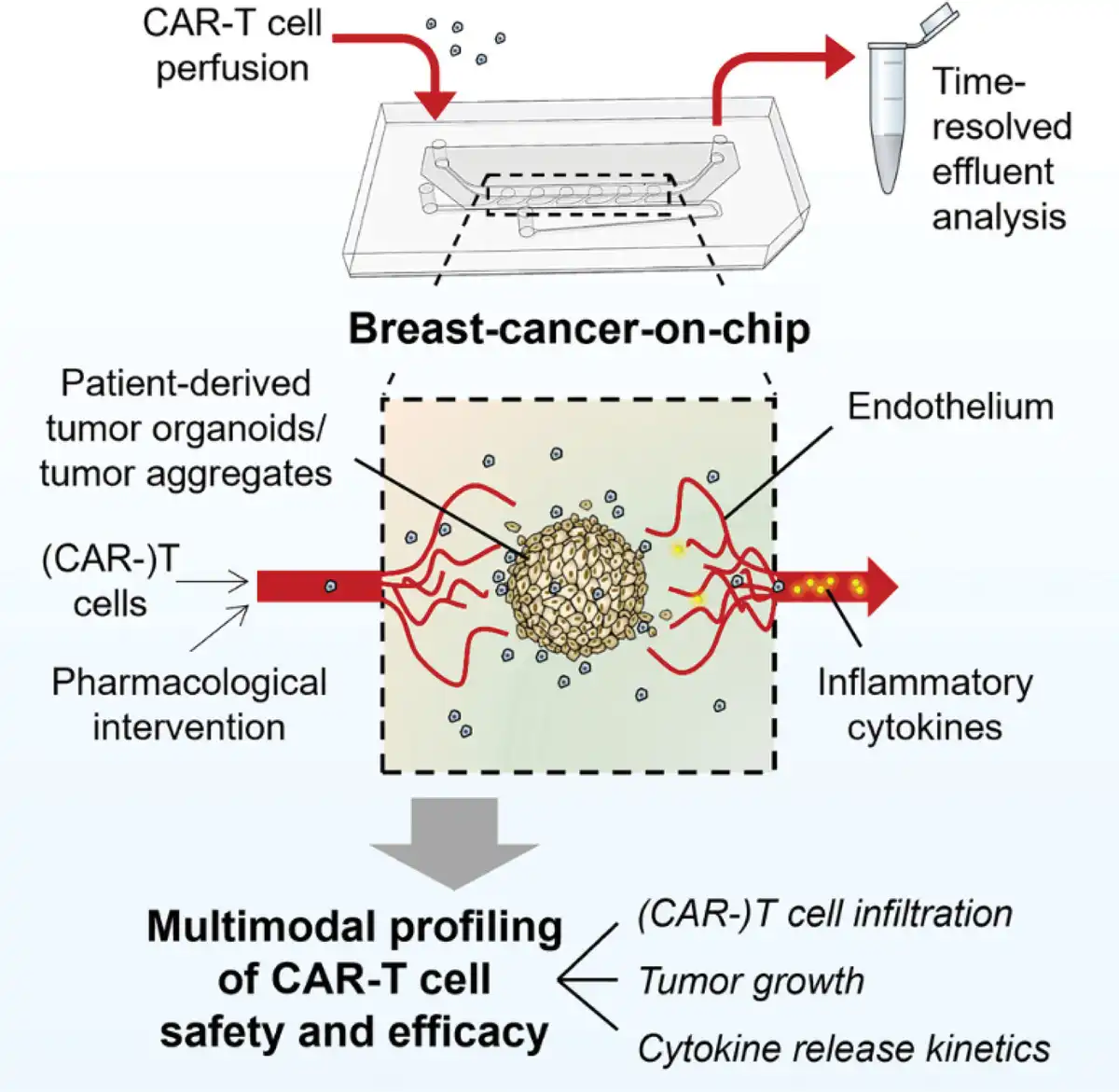
Figure 8.
Schematic diagram of the research on a breast cancer chip model integrating an endothelial barrier for investigating the therapeutic efficacy of CAR-T cells[115]. Copyright 2025, The Authors.
In terms of clinical translation, Vlachogiannis et al. established PDO models from 71 patients with metastatic gastrointestinal cancers and conducted tests using 55 drugs. The drug sensitivity tests demonstrated a sensitivity of 100%, a specificity of 93%, a positive predictive value of 88%, and a negative predictive value of 100% in predicting clinical treatment responses[116]. Ooft et al. utilized PDOs derived from metastatic lesions to identify patients with colorectal cancer (CRC) who would not respond to standard chemotherapy regimens. The results showed that for patients treated with irinotecan-based regimens, the PDO assay successfully predicted the response of biopsy-derived lesions in over 80% of cases, without misclassifying any patients who would have benefited from the treatment[117].
3.1.3 Applications of organoid-on-a-chip in studying cancer molecular mechanisms
In recent years, an increasing number of studies have discovered that certain tumor tissues exhibit resistance to immune checkpoints. Despite the promising therapeutic effects of immunotherapy, both intrinsic and acquired resistance have posed challenges[100]. The groundbreaking advancements in organoid technology have opened up new dimensions in cancer research: tumor organoids not only preserve the genetic heterogeneity and phenotypic diversity of the original tumors but also precisely reconstruct the dynamic interaction networks between tumors and their microenvironments. This provides a high-fidelity research platform for elucidating the mechanisms underlying tumor evolution, metastasis, and drug resistance.
Cuenca-Escalona et al. utilized a patient-derived CRC organoid model for 3D coculture with human monocyte-derived myeloid-derived suppressor cells (M-MDSCs). It is clinically known that tumor-derived prostaglandin E2 (PGE2) mediates the differentiation of MDSCs and confers them with tumor-promoting properties. The experimental results demonstrated that tumor-derived PGE2 signals through EP2 and EP4 receptors, and EP2/4 signaling contributes to the induction of a tumor-promoting phenotype and function in M-MDSCs. This supports the therapeutic value of targeting the PGE2-EP2/4 axis to alleviate the immunosuppressive effects of M-MDSCs and promote the development of antitumor immunity[118]. Choi et al. employed a LCO model containing blood vessels. Their study revealed that fibroblasts in fibrotic LCOs promoted cell proliferation and the expression of drug resistance-related genes. Fibrotic LCOs exhibited significantly greater changes in resistance to sensitizing targeted anticancer drugs. The expression of JAK2, FGFR1, and IL-6R genes, which are closely associated with the activation of pathways related to cancer cell proliferation, invasion, metastasis, and drug resistance, was generally enhanced in fibrotic LCOs[119]. Herpers et al. conducted high-throughput, large-scale screening on patient-derived organoid samples and identified a bispecific antibody, MCLA-158, which can specifically trigger the degradation of the epidermal growth factor receptor in leucine-rich repeat-containing G protein-coupled receptor 5-positive (LGR5+) cancer stem cells while exhibiting minimal toxicity to healthy LGR5+ colon stem cells[120].
3.2 Immuno-organoid-on-a-chip for drug evaluation and toxicity assessment
3.2.1 Drug evaluation
The immune microenvironment of tumor organoids can be derived not only from immune therapy cells but also from endogenous immune cells or externally added immune cells, thereby constructing a more physiologically relevant immune microenvironment that enables a more accurate evaluation of drug effects, especially for drugs that are difficult to assess using animal models. For instance, Crespo et al. proposed evaluating candidate drug combinations for oncolytic virus immunotherapy. Due to the limitations of animal models, their study relied on an organoid-based and immune cell cocultured explant patient-derived model system[121]. Zhu et al. utilized GBOs to demonstrate that the Zika virus can selectively replicate in glioblastoma stem cells (GSCs) but not in differentiated glioblastoma cells, leading to GSCs death and a subsequent loss of self-renewal and proliferative capacity. This model provides a platform closer to the human environment for assessing the efficacy of combining oncolytic viruses with immunotherapy, thus aiding in more accurate predictions of the clinical efficacy of combination therapies[122]. Teijeira et al. employed a tumor organoid system cocultured with T cells and autologous fibroblasts to test the costimulatory effects of a FAP-targeted 4-1BBL bispecific antibody fusion protein currently in clinical trials. This system not only releases IFN-γ but also achieves more effective tumor cell killing. It serves as a suitable model for testing the requirements for redirected killing of colon cancer induced by carcinoembryonicantigen (CEA)-targeted T cell-engaging antibodies undergoing clinical trials and allows for the testing of combination treatment regimens in relevant preclinical systems[123]. Ding et al. constructed microtumor organoid spheres (MOS) capable of capturing original stromal cells and permitting T cell infiltration. They demonstrated that immunotherapies such as PD-1 blockade and bispecific antibodies can activate TILs to attack tumor cells within MOS[124]. Zhao et al. utilized an ovarian cancer organoid system derived from high-grade serous subtype patient samples of ovarian cancer. These organoids recapitulated the histological and molecular heterogeneity of ovarian cancer while preserving key immune microenvironment components (including T cells, monocytes, macrophages, and B cells) and blood vessels. They showed potential in testing cisplatin sensitivity in patients resistant to carboplatin and paclitaxel, exhibiting significant responses in cancer proteoglycan and p53 (TP53) signaling pathways, as well as to PARP inhibitors, indicating the model’s potential in drug sensitivity testing for various types of drugs[125]. Maurer et al. constructed an intestinal model featuring tissue-resident innate immune cells with characteristics of mucosal macrophages and dendritic cells. They further demonstrated that precolonizing the model’s intestine with Lactobacillus rhamnosus can reduce tissue damage caused by Candida albicans, decrease its translocation, limit fungal burden, and effectively create a more physiological and immunocompetent microenvironment[126]. Zumwalde et al. utilized mammary ductal organoids and in vitro expanded T cells for coculture. Their study found that T cells can produce the antitumor cytokine IFN-γ and effectively kill bisphosphonate-treated breast cancer cells, inhibiting breast cancer growth. Using an in vitro tumor organoid model, they fully demonstrated the ability of these T cells to respond to bisphosphonate drugs approved by the US Food and Drug Administration, serving as a novel immunotherapeutic approach to inhibit breast cancer growth[127].
3.2.2 Toxicity assessment
Immuno-organoid-on-a-chip systems play a crucial role in predicting immunotoxicity. By accurately simulating the human immune microenvironment, these chips can integrate the interactions between various immune cells and target tissues, dynamically presenting the complex effects of drugs on the immune system. For example, Deng et al. constructed a hepatic immune MPS that effectively demonstrated the liver injury induced by TGZ, a drug withdrawn from the market due to hepatotoxicity. Their study revealed that in this model, the presence of TGZ enhanced the interactions between macrophages and neutrophils. At clinically relevant blood concentrations, TGZ could induce hepatocyte damage, a phenomenon not observed in other in vitro experiments[72]. Although immuno-organoids hold significant promise in drug toxicity assessment, research on toxicity and efficacy evaluation involving immune-engaged organoids is currently limited due to technical bottlenecks, such as the difficulty in integrating immune cells. However, as multicellular coculture technologies and microenvironment modulation strategies mature, these current technical limitations are expected to be overcome, enabling immuno-organoid-on-a-chip systems to play a pivotal role in more accurately predicting drug toxicity in the future.
4. Future perspectives
The rapidly evolving field of immune organoid-on-a-chip technology now stands at a critical inflection point. To transcend current limitations and achieve truly predictive human immunological models, future efforts must prioritize 4 interconnected frontiers:
4.1 Scalable and personalized immune cell sourcing
The scarcity and functional heterogeneity of autologous immune cells constitute a fundamental bottleneck in building personalized disease models, directly limiting accurate prediction of tumor immunotherapy responses. The lack of scalable immune cell sources reduces efficiency in patient-specific drug screening, impeding the implementation of precision medicine in solid tumors.
4.2 Multiorgan chips for systemic toxicity profiling of cellular therapeutics
Existing single-organ models fail to capture interorgan toxicity cascades of cellular drugs (e.g., CAR-T), resulting in undetected lethal side effects (e.g., cytokine release syndrome, PDACCRS) during preclinical studies. This disconnect significantly increases clinical trial failure risks and patient safety threats.
4.3 Bionic dual-vasculature chips for integrated immune microenvironments
While the lymphatic system mediates the majority of antigen presentation and immune cell activation in vivo, current chips only simulate intravascular immune microenvironments, causing distortion of critical physiological processes like immune cell trafficking and tolerance induction. Neglecting lymph-vascular synergy compromises physiological relevance in inflammation and cancer microenvironment research.
4.4 AI-driven closed-loop optimization of immune chips
The nonlinear interactions among dynamic variables within immune microenvironments exceed the capabilities of manual experimental design, leading to inefficient parameter optimization. Artificial intelligence (AI)-powered decision systems would not only enhance data utilization efficiency in immune organoid-on-a-chip platforms but also enable more comprehensive and efficient evaluation of immunologically relevant pharmacodynamic effects of therapeutics.
Acknowledgements
This work was financially supported by the National Natural Science Foundation of China (Grant No. 82373840) and the Jiangsu Key Laboratory of Neuropsychiatric Diseases (Grant Nos. BM2013003 and ZZ2009).
Conflicts of interests
The authors declare that they have no conflicts of interest.
References
- [1] Kim J, Koo BK, Knoblich JA. Human organoids: model systems for human biology and medicine. Nat Rev Mol Cell Biol. 2020;21(10):571–584.
- [2] Clevers H. Modeling development and disease with organoids. Cell. 2016;165(7):1586–1597.
- [3] Sasai Y. Next-generation regenerative medicine: organogenesis from stem cells in 3D culture. Cell Stem Cell. 2013;12(5):520–530.
- [4] Lancaster MA, Knoblich JA. Organogenesis in a dish: modeling development and disease using organoid technologies. Science. 2014;345(6194):1247125.
- [5] McCauley HA, Wells JM. Pluripotent stem cell-derived organoids: using principles of developmental biology to grow human tissues in a dish. Development. 2017;144(6):958–962.
- [6] Rossi G, Manfrin A, Lutolf MP. Progress and potential in organoid research. Nat Rev Genet. 2018;19(11):671–687.
- [7] Huh D, Hamilton GA, Ingber DE. From 3D cell culture to organs-on-chips. Trends Cell Biol. 2011;21(12):745–754.
- [8] Zhang B, Korolj A, Lai BFL, et al. Advances in organ-on-a-chip engineering. Nat Rev Mater. 2018;3(8):257–278.
- [9] Bhatia SN, Ingber DE. Microfluidic organs-on-chips. Nat Biotechnol. 2014;32(8):760–772.
- [10] Esch EW, Bahinski A, Huh D. Organs-on-chips at the frontiers of drug discovery. Nat Rev Drug Discov. 2015;14(4):248–260.
- [11] Takebe T, Zhang B, Radisic M. Synergistic engineering: organoids meet organs-on-a-chip. Cell Stem Cell. 2017;21(3):297–300.
- [12] Ingber DE. Is it time for reviewer 3 to request human organ chip experiments instead of animal validation studies. Adv Sci. 2020;7(22):2002030.
- [13] Park SE, Georgescu A, Huh D. Organoids-on-a-chip. Science. 2019;364(6444):960–965.
- [14] Man Y, Liu Y, Chen Q, et al. Organoids‐on‐a‐chip for personalized precision medicine. Adv Healthc Mater. 2024;13(30):2401843.
- [15] Yu F, Hunziker W, Choudhury D. Engineering microfluidic organoid-on-a-chip platforms. Micromachines (Basel). 2019;10(3):165.
- [16] Wang H, Ning X, Zhao F, et al. Human organoids-on-chips for biomedical research and applications. Theranostics. 2024;14(2):788–818.
- [17] Hofer M, Lutolf MP. Engineering organoids. Nat Rev Mater. 2021;6(5):402–420.
- [18] Xing Y, Liu J, Guo X, et al. Engineering organoid microfluidic system for biomedical and health engineering: a review. Chin J Chem Eng. 2021;30:244–254.
- [19] Ebrahimkhani MR, Young CL, Lauffenburger DA, et al. Approaches to in vitro tissue regeneration with application for human disease modeling and drug development. Drug Discov Today. 2014;19(6):754–762.
- [20] Wang YI, Carmona C, Hickman JJ, et al. Multiorgan microphysiological systems for drug development: strategies, advances, and challenges. Adv Healthc Mater. 2018;7(2):1701000.
- [21] Kim SB, Koo KI, Bae H, et al. A mini-microscope for in situ monitoring of cells. Lab Chip. 2012;12(20):3976–3982.
- [22] Sung JH, Wang YI, Sriram NN, et al. Recent advances in body-on-a-chip systems. Anal Chem. 2019;91(1):330–351.
- [23] Berthier E, Young EW, Beebe D. Engineers are from PDMS-land, Biologists are from Polystyrenia. Lab Chip. 2012;12(7):1224–1237.
- [24] Rogers KW, Schier AF. Morphogen gradients: from generation to interpretation. Annu Rev Cell Dev Biol. 2011;27:377–407.
- [25] Kretzschmar K, Clevers H. Organoids: modeling development and the stem cell niche in a dish. Dev Cell. 2016;38(6):590–600.
- [26] Clevers H. Advances in organoid technology: Hans Clevers, Madeline Lancaster, and Takanori Takebe. Cell Stem Cell. 2017;20(6):759–762.
- [27] Lou YR, Leung AW. Next generation organoids for biomedical research and applications. Biotechnol Adv. 2018;36(1):132–149.
- [28] Zou Z, Lin Z, Wu C, et al. Micro-engineered organoid-on-a-chip based on mesenchymal stromal cells to predict immunotherapy responses of HCC patients. Adv Sci (Weinh). 2023;10(27):e2302640.
- [29] Zhang T, Yang S, Ge Y, et al. Unveiling the heart’s hidden enemy: dynamic insights into polystyrene nanoplastic-induced cardiotoxicity based on cardiac organoid-on-a-chip. ACS Nano. 2024;18(45):31569–31585.
- [30] Abdulla A, Chen S, Chen Z, et al. Three-dimensional microfluidics with dynamic fluidic perturbation promotes viability and uniformity of human cerebral organoids. Biosens Bioelectron. 2023;240:115635.
- [31] Mitrofanova O, Nikolaev M, Xu Q, et al. Bioengineered human colon organoids with in vivo-like cellular complexity and function. Cell Stem Cell. 2024;31(8):1175–1186.e7.
- [32] Goluba K, Parfejevs V, Rostoka E, et al. Personalized PDAC chip with functional endothelial barrier for tumour biomarker detection: a platform for precision medicine applications. Mater Today Bio. 2024;29:101262.
- [33] Hu Y, Zhang H, Wang S, et al. Bone/cartilage organoid on-chip: construction strategy and application. Bioact Mater. 2023;25:29–41.
- [34] Yu J, Yin Y, Leng Y, et al. Emerging strategies of engineering retinal organoids and organoid-on-a-chip in modeling intraocular drug delivery: current progress and future perspectives. Adv Drug Deliv Rev. 2023;197:114842.
- [35] Yu T, Yang Q, Peng B, et al. Vascularized organoid-on-a-chip: design, imaging, and analysis. Angiogenesis. 2024;27(2):147–172.
- [36] Wang H, Zhu W, Xu C, et al. Engineering organoids-on-chips for drug testing and evaluation. Metabolism. 2025;162:156065.
- [37] Hu C, Yang S, Zhang T, et al. Organoids and organoids-on-a-chip as the new testing strategies for environmental toxicology-applications & advantages. Environ Int. 2024;184:108415.
- [38] Polini A, Del ML, Barra A, et al. Towards the development of human immune-system-on-a-chip platforms. Drug Discov Today. 2019;24(2):517–525.
- [39] Nanduri LS, Baanstra M, Faber H, et al. Purification and ex vivo expansion of fully functional salivary gland stem cells. Stem Cell Rep. 2014;3(6):957–964.
- [40] Zhou G, Lieshout R, van Tienderen GS, et al. Modelling immune cytotoxicity for cholangiocarcinoma with tumour-derived organoids and effector T cells. Br J Cancer. 2022;127(4):649–660.
- [41] Saxton SH, Stevens KR. 2D and 3D liver models. J Hepatol. 2023;78(4):873–875.
- [42] Horvath P, Aulner N, Bickle M, et al. Screening out irrelevant cell-based models of disease. Nat Rev Drug Discov. 2016;15(11):751–769.
- [43] Shah SB, Singh A. Creating artificial lymphoid tissues to study immunity and hematological malignancies. Curr Opin Hematol. 2017;24(4):377–383.
- [44] Maulana TI, Teufel C, Cipriano M, et al. Breast cancer-on-chip for patient-specific efficacy and safety testing of CAR-T cells. Cell Stem Cell. 2024;31(7):989–1002.e9.
- [45] Kroll KT, Mata MM, Homan KA, et al. Immune-infiltrated kidney organoid-on-chip model for assessing T cell bispecific antibodies. Proc Natl Acad Sci U S A. 2023;120(35):e1989645176.
- [46] Parlato S, De Ninno A, Molfetta R, et al. 3D Microfluidic model for evaluating immunotherapy efficacy by tracking dendritic cell behaviour toward tumor cells. Sci Rep. 2017;7(1):1093.
- [47] Ghoshal D, Petersen I, Ringquist R, et al. Multi-niche human bone marrow on-a-chip for studying the interactions of adoptive CAR-T cell therapies with multiple myeloma. BioRxiv. 2024;316:123016.
- [48] Zhang J, Tavakoli H, Ma L, et al. Immunotherapy discovery on tumor organoid-on-a-chip platforms that recapitulate the tumor microenvironment. Adv Drug Deliv Rev. 2022;187:114365.
- [49] Morrison AI, Sjoerds MJ, Vonk LA, et al. In vitro immunity: an overview of immunocompetent organ-on-chip models. Front Immunol. 2024;15:1373186.
- [50] Ye W, Luo C, Li C, et al. Organoids to study immune functions, immunological diseases and immunotherapy. Cancer Lett. 2020;477:31–40.
- [51] Morsink M, Willemen N, Leijten J, et al. Immune organs and immune cells on a chip: an overview of biomedical applications. Micromachines. 2020;11(9):849.
- [52] Sun W, Luo Z, Lee J, et al. Organ‐on‐a‐chip for cancer and immune organs modeling. Adv Healthc Mater. 2019;8(4):1801363.
- [53] Liu K, Chen X, Fan Z, et al. From organoids to organoids-on-a-chip: current applications and challenges in biomedical research. Chin Med J (Engl). 2025;138(7):792–807.
- [54] Van Os L, Engelhardt B, Guenat OT. Integration of immune cells in organs-on-chips: a tutorial. Front Bioeng Biotechnol. 2023;11:1191104.
- [55] Shanti A, Teo J, Stefanini C. In vitro immune organs-on-chip for drug development: a review. Pharmaceutics. 2018;10(4):278.
- [56] Yoon PS, Del Piccolo N, Shirure VS, et al. Advances in modeling the immune microenvironment of colorectal cancer. Front Immunol. 2021;11:614300.
- [57] Zhu J, Zhou J, Feng B, et al. MSCs alleviate LPS-induced acute lung injury by inhibiting the proinflammatory function of macrophages in mouse lung organoid–macrophage model. Cell Mol Life Sci. 2024;81(1):124.
- [58] Xia J, Dong R, Fang Y, et al. A micro-lung chip with macrophages for targeted anti-fibrotic therapy. Biofabrication. 2025;17(2):025020.
- [59] Ghoshal D, Petersen I, Ringquist R, et al. Multi-niche human bone marrow on-a-chip for studying the interactions of adoptive CAR-T cell therapies with multiple myeloma. Biomaterials. 2025;316:123016.
- [60] Ao Z, Cai H, Wu Z, et al. Tubular human brain organoids to model microglia-mediated neuroinflammation. Lab Chip. 2021;21(14):2751–2762.
- [61] Narasipura SD, Zayas JP, Ash MK, et al. Inflammatory responses revealed through HIV infection of microglia-containing cerebral organoids. J Neuroinflammation. 2025;22(1):36.
- [62] Santos A JM, van Unen V, Lin Z, et al. A human autoimmune organoid model reveals IL-7 function in coeliac disease. Nature. 2024;632(8024):401–410.
- [63] Gjorevski N, Avignon B, Gerard R, et al. Neutrophilic infiltration in organ-on-a-chip model of tissue inflammation. Lab Chip. 2020;20(18):3365–3374.
- [64] Qiang Y, Sissoko A, Liu ZL, et al. Microfluidic study of retention and elimination of abnormal red blood cells by human spleen with implications for sickle cell disease. Proc Natl Acad Sci U S A. 2023;120(6):e2217607120.
- [65] Gao M, Ding W, Wang Y, et al. Quantitatively evaluating interactions between patient-derived organoids and autologous immune cells by microfluidic chip. Anal Chem. 2024;96(32):13061–13069.
- [66] Kroll KT, Mata MM, Homan KA, et al. Immune-infiltrated kidney organoid-on-chip model for assessing T cell bispecific antibodies. Proc Natl Acad Sci U S A. 2023;120(35):e2305322120.
- [67] German SV, Abalymov AA, Kurochkin MA, et al. Plug-and-play lymph node-on-chip: secondary tumor modeling by the combination of cell spheroid, collagen sponge and T-cells. Int J Mol Sci. 2023;24(4):3183.
- [68] Jeong S, Kang M. Exploring tumor–immune interactions in co-culture models of T cells and tumor organoids derived from patients. Int J Mol Sci. 2023;24(19):14609.
- [69] Li K, Liu C, Sui X, et al. An organoid co-culture model for probing systemic anti-tumor immunity in lung cancer. Cell Stem Cell. 2025;32(8):1218–1234.e7.
- [70] Natarajan V, Simoneau CR, Erickson AL, et al. Modelling T-cell immunity against hepatitis C virus with liver organoids in a microfluidic coculture system. Open Biol. 2022;12(3):210320.
- [71] Choi SS, van Unen V, Zhang H, et al. Organoid modeling of lung-resident immune responses to SARS-CoV-2 infection. Res Sq [Preprint]. 2023:rs.3.rs-2870695.
- [72] Deng Q, Yang Y, Liu Y, et al. Assessing immune hepatotoxicity of troglitazone with a versatile liver-immune-microphysiological-system. Front Pharmacol. 2024;15:1335836.
- [73] Fang M, Allen A, Luo C, et al. Unlocking the potential of iPSC-derived immune cells: engineering iNK and iT cells for cutting-edge immunotherapy. Front Immunol. 2024;15:1457629.
- [74] Pandey R, Chiu C, Wang L. Immunotherapy study on non-small-cell lung cancer (NSCLC) combined with cytotoxic T cells and miRNA34a. Mol Pharm. 2024;21(3):1364–1381.
- [75] Ayuso JM, Truttschel R, Gong MM, et al. Evaluating natural killer cell cytotoxicity against solid tumors using a microfluidic model. Oncoimmunology. 2019;8(3):1553477.
- [76] Ode Boni BO, Lamboni L, Souho T, et al. Immunomodulation and cellular response to biomaterials: the overriding role of neutrophils in healing. Mater Horiz. 2019;6(6):1122–1137.
- [77] Polini A, Del Mercato LL, Barra A, et al. Towards the development of human immune-system-on-a-chip platforms. Drug Discov Today. 2019;24(2):517–525.
- [78] Boucherit N, Gorvel L, Olive D. 3D tumor models and their use for the testing of immunotherapies. Front Immunol. 2020;11:603640.
- [79] Chen X, Jiang L, Liu X. Natural killer cells: the next wave in cancer immunotherapy. Front Immunol. 2022;13:954804.
- [80] Mohd Yasin ZN, Mohd Idrus FN, Hoe CH, et al. Macrophage polarization in THP-1 cell line and primary monocytes: a systematic review. Differentiation. 2022;128:67–82.
- [81] Chanput W, Mes JJ, Wichers HJ. THP-1 cell line: an in vitro cell model for immune modulation approach. Int Immunopharmacol. 2014;23(1):37–45.
- [82] Miller CP, Shin W, Ahn EH, et al. Engineering microphysiological immune system responses on chips. Trends Biotechnol. 2020;38(8):857–872.
- [83] Lv B, Wang Y, Ma D, et al. Immunotherapy: reshape the tumor immune microenvironment. Front Immunol. 2022;13:844142.
- [84] Yuki K, Cheng N, Nakano M, et al. Organoid models of tumor immunology. Trends Immunol. 2020;41(8):652–664.
- [85] O’Shea JJ, Schwartz DM, Villarino AV, et al. The JAK-STAT pathway: impact on human disease and therapeutic intervention. Annu Rev Med. 2015;66:311–328.
- [86] Pandey P, Lakhanpal S, Mahmood D, et al. An updated review summarizing the anticancer potential of flavonoids via targeting NF-kB pathway. Front Pharmacol. 2025;15:1513422.
- [87] Zeng Y, Du Y, Xu X, et al. On-chip modeling of physiological and pathological blood-brain barrier microenvironment for studying glial responses to neuroinflammation. Nano Today. 2023;52:101947.
- [88] Abu-Eid R, Ward FJ. Targeting the PI3K/Akt/mTOR pathway: a therapeutic strategy in COVID-19 patients. Immunol Lett. 2021;240:1–8.
- [89] Batlle E, Massagué J. Transforming growth factor-b signaling in immunity and cancer. Immunity. 2019;50(4):924–940.
- [90] Zhan Y, Xu J, Zhang Z, et al. Targeting SPHK1 in macrophages remodels the tumor microenvironment and enhances anti‐PD‐1 immunotherapy efficacy in colorectal cancer liver metastasis. Cancer Commun. 2025;45(10):1203–1228.
- [91] Colegio OR, Chu N, Szabo AL, et al. Functional polarization of tumour-associated macrophages by tumour-derived lactic acid. Nature. 2014;513(7519):559–563.
- [92] Grandhi TSP, Mebrahtu M, Musso R, et al. A microphysiological assay for studying T-cell chemotaxis, trafficking and tumor killing. Biofabrication. 2024;17(1):015004.
- [93] Wang X, Zhang J, Zhou G. The CXCL11‐CXCR3A axis influences the infiltration of CD274 and IDO1 in oral squamous cell carcinoma. J Oral Pathol Med. 2021;50(4):362–370.
- [94] Feodoroff M, Hamdan F, Antignani G, et al. Enhancing T-cell recruitment in renal cell carcinoma with cytokine-armed adenoviruses. Oncoimmunology. 2024;13(1):2407532.
- [95] Cui Y, Miao Y, Cao L, et al. Activation of melanocortin-1 receptor signaling in melanoma cells impairs T cell infiltration to dampen antitumor immunity. Nat Commun. 2023;14(1):5740.
- [96] Comerford I, Harata-Lee Y, Bunting MD, et al. A myriad of functions and complex regulation of the CCR7/CCL19/CCL21 chemokine axis in the adaptive immune system. Cytokine Growth Factor Rev. 2013;24(3):269–283.
- [97] Smit MJ, Schlecht-Louf G, Neves M, et al. The CXCL12 CXCR4 ACKR3 axis in the tumor microenvironment: signaling, crosstalk, and therapeutic targeting. Annu Rev Pharmacol Toxicol. 2021;61(1):541–563.
- [98] Ma VP, Hu Y, Kellner AV, et al. The magnitude of LFA-1/ICAM-1 forces fine-tune TCR-triggered T cell activation. Sci Adv. 2022;8(8):eabg4485.
- [99] Reinhardt PH, Elliott JF, Kubes P. Neutrophils can adhere via a4b1 -integrin under flow conditions. Blood. 1997;89(10):3837–3846.
- [100] Yang Y, Cui J, Kong Y, et al. Organoids: new frontiers in tumor immune microenvironment research. Front Immunol. 2024;15:1422031.
- [101] Gharib G, Bütün I, Muganli Z, et al. Biomedical applications of microfluidic devices: a review. Biosensors (Basel). 2022;12(11):1023.
- [102] Aboulkheyr Es H, Montazeri L, Aref AR, et al. Personalized cancer medicine: an organoid approach. Trends Biotechnol. 2018;36(4):358–371.
- [103] Zhao Z, Wu X, Zhang T, et al. Evaluation of multispecific drugs based on patient-derived immunocompetent tumor organoids. ChemBioChem. 2025;26(3):e202400731.
- [104] Ao Z, Cai H, Wu Z, et al. Evaluation of cancer immunotherapy using mini-tumor chips. Theranostics. 2022;12(8):3628–3636.
- [105] Bupha-Intr O, Haeusler G, Chee L, et al. CAR-T cell therapy and infection: a review. Expert Rev Anti Infect Ther. 2021;19(6):749–758.
- [106] Jiang Y, Sun X, Song X, et al. Patient-derived bladder cancer organoid model to predict sensitivity and feasibility of tailored precision therapy. Curr Urol. 2023;17(4):221–228.
- [107] Song EZ, Wang X, Philipson BI, et al. The IAP antagonist birinapant enhances chimeric antigen receptor T cell therapy for glioblastoma by overcoming antigen heterogeneity. Mol Ther Oncolytics. 2022;27:288–304.
- [108] Wehrli M, Guinn S, Birocchi F, et al. Mesothelin CAR T cells secreting anti-FAP/Anti-CD3 molecules efficiently target pancreatic adenocarcinoma and its stroma. Clin Cancer Res. 2024;30(9):859–1877.
- [109] Porter LH, Zhu JJ, Lister NL, et al. Low-dose carboplatin modifies the tumor microenvironment to augment CAR T cell efficacy in human prostate cancer models. Nat Commun. 2023;14(1):5346.
- [110] Paul S, Lal G. The molecular mechanism of natural killer cells function and its importance in cancer immunotherapy. Front Immunol. 2017;8:1124.
- [111] Oh B, Kim J, Kim N, et al. Lung cancer organoid system to evaluate the cytotoxicity of natural killer cells. Int J Stem Cells. 2024;18(1):99–106.
- [112] Ziegler AE, Fittje P, Müller LM, et al. The co-inhibitory receptor TIGIT regulates NK cell function and is upregulated in human intrahepatic CD56(bright) NK cells. Front Immunol. 2023;14:1117320.
- [113] Ning RX, Liu CY, Wang SQ, et al. Application status and optimization suggestions of tumor organoids and CAR-T cell co-culture models. Cancer Cell Int. 2024;24(1):98.
- [114] Chiriaco C, Donini C, Cortese M, et al. Efficacy of CAR-T immunotherapy in MET overexpressing tumors not eligible for anti-MET targeted therapy. J Exp Clin Cancer Res. 2022;41(1):309.
- [115] Logun M, Wang X, Sun Y, et al. Patient-derived glioblastoma organoids as real-time avatars for assessing responses to clinical CAR-T cell therapy. Cell Stem Cell. 2025;32(2):181–190.e4.
- [116] Vlachogiannis G, Hedayat S, Vatsiou A, et al. Patient-derived organoids model treatment response of metastatic gastrointestinal cancers. Science. 2018;359(6378):920–926.
- [117] Ooft SN, Weeber F, Dijkstra KK, et al. Patient-derived organoids can predict response to chemotherapy in metastatic colorectal cancer patients. Sci Transl Med. 2019;11(513):eaay2574.
- [118] Cuenca-Escalona J, Subtil B, Garcia-Perez A, et al. EP2 and EP4 blockade prevents tumor-induced suppressive features in human monocytic myeloid-derived suppressor cells. Front Immunol. 2024;15:1355769.
- [119] Choi YM, Lee H, Ann M, et al. 3D bioprinted vascularized lung cancer organoid models with underlying disease capable of more precise drug evaluation. Biofabrication. 2023;15(3):034104.
- [120] Herpers B, Eppink B, James MI, et al. Functional patient-derived organoid screenings identify MCLA-158 as a therapeutic EGFR × LGR5 bispecific antibody with efficacy in epithelial tumors. Nat Cancer. 2022;3(4):418–436.
- [121] Teijeira Crespo A, Burnell S, Capitani L, et al. Pouring petrol on the flames: using oncolytic virotherapies to enhance tumour immunogenicity. Immunology. 2021;163(4):389–398.
- [122] Zhu Z, Gorman MJ, McKenzie LD, et al. Zika virus has oncolytic activity against glioblastoma stem cells. J Exp Med. 2017;214(10):2843–2857.
- [123] Teijeira A, Migueliz I, Garasa S, et al. Three-dimensional colon cancer organoids model the response to CEA-CD3 T-cell engagers. Theranostics. 2022;12(3):1373–1387.
- [124] Ding S, Hsu C, Wang Z, et al. Patient-derived micro-organospheres enable clinical precision oncology. Cell Stem Cell. 2022;29(6):905–917.e6.
- [125] Zhao Y, Wang C, Deng W, et al. Patient-derived ovarian cancer organoid carries immune microenvironment and blood vessel keeping high response to cisplatin. MedComm (2020). 2024;5(9):e697.
- [126] Maurer M, Gresnigt MS, Last A, et al. A three-dimensional immunocompetent intestine-on-chip model as in vitro platform for functional and microbial interaction studies. Biomaterials. 2019;220:119396.
- [127] Zumwalde NA, Haag JD, Sharma D, et al. Analysis of immune cells from human mammary ductal epithelial organoids reveals Vδ2+ T cells that efficiently target breast carcinoma cells in the presence of bisphosphonate. Cancer Prev Res (Phila). 2016;9(4):305–316.
This article is available under Open Access.
© 2026 The Authors