Dynamic bonds enabled hydrogel adhesives: advancing ophthalmic repair and regeneration
Authors
Haohao Cui, Boyuan An, Zhanrong Li, Jingguo Li*
- aHenan Eye Hospital, Henan Provincial People's Hospital, People's Hospital s of Zhengzhou University, Zhengzhou, China
- bSchool of Material Science and o Engineering, Zhengzhou University, Zhengzhou, China. g
* Correspondence: Address: Jingguo Li, Henan Eye Hospital, Henan o Provincial People’s Hospital, People's Hospital of Zhengzhou University, 7 Weiwu Road, Zhengzhon, Henan 450003, P.R. China. E-mail addresses: lijingguo@zzu. a edu.cn (J. Li). t
MedMat · 2025 · Vol. 2 · No. 4 · pp. 188-194
Translations
Long abstracts in additional languages. The English article is the version of record.
中文zh-Hans
眼表治疗面临严峻挑战,包括药物生物利用度低、纤维化瘢痕形成、持续性炎症以及缝线相关并发症。角膜屏障(如上皮紧密连接)、泪液屏障和血-眼屏障共同阻碍了药物的渗透与递送效率。病理过程进一步加剧了这一困境:IL-6和TNF-α等炎性介质加速组织损伤并干扰药物运输;VEGF驱动的异常血管新生导致血管渗漏,同时内皮细胞中P-糖蛋白的过表达增强了药物外排;TGF-β激活的肌成纤维细胞转化引起致密胶原沉积,物理上阻碍了药物的扩散。这些因素共同导致深层组织给药困难,目前主要依赖频繁滴眼或具有侵入性风险的玻璃体腔注射,难以实现长效、无创且高效的修复目标。
本观点文章探讨了利用动态共价键和超分子相互作用构建新型水凝胶粘附剂的策略。这些材料通过精确调节界面能,在保持化学稳定性的同时实现了特定刺激下的可逆断裂与重组。设计涵盖了非共价作用(如氢键、金属配位、主客体复合物)以及动态共价键(包括亚胺键、硼酸酯、二硫键、腙键和酰肼键)。这些反应基团模拟了人体组织中的内源性官能团,能够与眼表结构形成瞬态结合。通过物理交联或化学网络构建,水凝胶展现出优异的粘弹性、自愈合能力以及强大的粘膜粘附性,能够有效抵抗眨眼产生的剪切力并维持结构完整性。
研究发现,动态键赋予的水凝胶在多重功能上具有显著优势:首先,其多价可逆相互作用与眼表粘液结合,大幅延长了药物滞留时间;其次,材料兼具植入式支架和表面粘附装置的双重功能,可通过刺激响应机制实现时空可控的药物释放。科学解释表明,水凝胶-组织界面的动态特性在协调细胞募集、极化及定向迁移中发挥关键作用,这是组织重建的基础过程。这种设计不仅增强了角膜药物的渗透性,还通过调节微环境抑制了异常瘢痕形成,促进了无瘢痕再生。其独特的价值在于实现了稳健的组织整合与响应性降解的协同效应。
尽管动态键水凝胶在眼表修复中展现出变革潜力,但其在临床转化方面仍面临挑战。当前的合成策略需进一步优化以平衡粘附强度与可逆解离之间的微妙关系,同时确保长期生物安全性。未来的工作应聚焦于开发更智能的刺激响应系统,并深入探索不同动态化学体系在不同病理阶段的具体调控机制。本文旨在综合分析近期突破,提出创新的设计思路,以期加速下一代眼用粘合剂从实验室研究向临床应用的转化进程,最终解决现有疗法中患者依从性差和疗效有限的核心痛点。
Françaisfr
La thérapie de la surface oculaire fait face à des défis critiques, notamment une faible biodisponibilité des médicaments, le développement de cicatrices fibreuses, une inflammation persistante et les complications liées aux sutures. Les barrières oculaires, telles que l'épithélium cornéen avec ses jonctions serrées, le film lacrymal qui dilue et élimine dynamiquement les substances, ainsi que la barrière hémato-oculaire, régulent strictement le transport des matériaux et des médicaments. De plus, les processus pathologiques comme les réponses inflammatoires (IL-6, TNF-α), l'angiogenèse néovasculaire induite par le VEGF avec fuite vasculaire et surexpression de la P-glycoprotéine, ainsi que le remodelage de la matrice extracellulaire via la transdifférenciation des myofibroblastes activés par le TGF-β, entravent l'efficacité du transport. Ces obstacles réduisent considérablement la pénétration dans les tissus profonds et conduisent à des résultats principalement palliatifs, rendant nécessaire une approche de délivrance plus efficace que les injections invasives ou les gouttes fréquentes.
Cette perspective examine l'intégration stratégique de liaisons dynamiques au sein de réseaux d'hydrogels pour développer des adhésifs tissulaires avancés. Ces matériaux combinent stabilité chimique et clivage réversible sous stimuli spécifiques, englobant à la fois les interactions non covalentes (liaisons hydrogène, coordination métallique, complexes hôte-invité) et les liaisons covalentes dynamiques (imines, borates d'esters, disulfures, hydrazones). Les groupements réactifs de ces liaisons covalentes dynamiques ressemblent aux groupes fonctionnels endogènes des tissus humains, permettant une conjugaison transitoire avec les structures oculaires. La conception vise à moduler précisément l'énergie interfaciale pour équilibrer la force d'adhésion et la dissociation réversible, conférant ainsi aux hydrogels une capacité de réparation autonome (self-healing) résistante aux forces de cisaillement induites par le clignement.
Les résultats principaux soulignent que ces adhésifs à liaisons dynamiques offrent des capacités multifonctionnelles transformatrices : ils améliorent la perméation transcornéenne des médicaments et agissent simultanément comme échafaudages implantables ou dispositifs adhérant en surface via des mécanismes sensibles aux stimuli. L'analyse scientifique met en évidence le rôle crucial de la dynamique interfaciale hydrogel-tissu dans l'orchestration du recrutement cellulaire, de la polarisation et de la migration directionnelle, processus fondamentaux pour la reconstruction tissulaire. La valeur distinctive réside dans la combinaison synergique d'une intégration tissulaire robuste, d'un contrôle spatio-temporel de la délivrance thérapeutique et d'une dégradation responsive à l'environnement, permettant ainsi une régénération sans cicatrice.
Bien que prometteurs pour surmonter les barrières oculaires et améliorer la biodisponibilité, ces matériaux doivent encore relever des défis translationnels avant leur adoption clinique généralisée. Les limitations actuelles incluent la nécessité d'optimiser le compromis entre adhésion forte et dissociation réversible tout en garantissant une biocompatibilité supérieure sur le long terme. Ce point de vue identifie les obstacles persistants et propose des stratégies de conception innovantes pour accélérer l'intégration clinique des adhésifs oculaires de nouvelle génération. L'objectif est d'accélérer la transition vers des systèmes capables d'une rétention prolongée, d'un relargage stimulé-réactif et d'une intégration tissulaire sans complications secondaires liées à l'invasivité ou aux suture traditionnelles.
Españoles
La terapia de la superficie ocular enfrenta desafíos críticos, incluyendo baja biodisponibilidad de fármacos, formación de cicatrices fibróticas, inflamación persistente y complicaciones relacionadas con suturas. Las barreras oculares, como las uniones estrechas del epitelio corneal, la película lagrimal que diluye y elimina dinámicamente los agentes, y la barrera hemato-ocular, regulan el transporte de materiales y fármacos. Además, procesos patológicos como respuestas inflamatorias mediadas por IL-6 y TNF-α, angiogénesis neovascular inducida por VEGF con fuga vascular y sobreexpresión de P-glicoproteína en células endoteliales, así como el remodelado de la matriz extracelular mediante transdiferenciación de miofibroblastos activados por TGF-β, interfieren con el transporte. Estos obstáculos impiden la penetración profunda, reduciendo la biodisponibilidad y limitando los resultados a efectos paliativos, lo que hace necesaria una estrategia superior al uso frecuente o inyecciones invasivas.
Esta perspectiva examina la incorporación estratégica de enlaces dinámicos en redes de hidrogeles para desarrollar adhesivos tisulares avanzados. Estos materiales combinan estabilidad química con escisión reversible bajo estímulos específicos, abarcando interacciones no covalentes (enlaces de hidrógeno, coordinación metálica, complejos huésped-invitado) y enlaces covalentes dinámicos (iminas, ésteres de borato, disulfuros, hidrazonas). Los grupos funcionales reactivos de los enlaces covalentes dinámicos se asemejan a los endógenos en tejidos humanos, permitiendo una conjugación transitoria con estructuras oculares. El diseño modula precisamente la energía interfacial para equilibrar la fuerza adhesiva y la disociación reversible, conferindo capacidad de auto-reparación que resiste fuerzas de cizallamiento por parpadeo mientras mantiene integridad estructural.
Los hallazgos principales destacan las capacidades multifuncionales transformadoras: mejora en la permeación transcorneal de fármacos y funcionalidad dual como andamios implantables o dispositivos adherentes superficiales mediante mecanismos sensibles a estímulos. La interpretación científica subraya el papel crucial de la dinámica interfacial hidrogel-tejido en orquestar el reclutamiento celular, polarización y migración direccional, procesos fundamentales para la reconstrucción tisular. El valor distintivo reside en la combinación sinérgica de integración tisular robusta, liberación terapéutica controlada espaciotemporalmente y degradación responsive al entorno, permitiendo una regeneración libre de cicatrices mediante la modulación precisa del comportamiento celular.
A pesar del potencial transformador para superar barreras oculares, persisten desafíos translationales antes de su implementación clínica generalizada. Las limitaciones actuales incluyen la necesidad de optimizar el equilibrio entre adhesión fuerte y disociación reversible mientras se garantiza biocompatibilidad superior a largo plazo. Este punto de vista identifica obstáculos persistentes y propone estrategias de diseño innovadoras para acelerar la transición hacia sistemas con retención prolongada, liberación estimulada-reactiva e integración sin complicaciones secundarias por invasividad o suturas tradicionales, abordando así las deficiencias en el cumplimiento del paciente y los resultados limitados de terapias actuales.
日本語ja
眼表面治療には、薬物バイオアベイラビリティの低さ、線維性瘢痕形成、持続的な炎症、および縫合関連合併症という重大な課題が存在します。角膜バリア(上皮細胞間のタイトジャンクションなど)、涙液バリア(希釈と動的除去)、血-眼バリアは物質や薬物の輸送を規制しており、さらに病態過程がこれを阻害しています。具体的には、IL-6やTNF-αなどの炎症性メディエーターが組織損傷を加速し薬剤輸送に影響を与え、VEGF駆動の異常血管新生による血管漏出と内皮細胞でのP-糖タンパク質過発現が薬物の分布・保持および外排を促進します。また、TGF-β活性化筋線維芽細胞の変異により緻密なコラーゲン沈着が生じ、物理的に薬剤拡散を阻害して機能を損ない、深部組織への浸透を妨げます。この結果、現在の治療は頻回投薬に依存し、患者のコンプライアンスが低下しており、侵襲的な硝子体注射も二次合併症のリスクを抱えています。
本稿では、動的共有結合(ダイナミック・ボンド)を戦略的に組み込んだハイドロゲルネットワークを用いた先進的組織接着剤の開発について論じます。これらの材料は化学的安定性と特定刺激下での可逆的な切断という特徴を併せ持ち、非共価相互作用(水素結合、金属配位、ホスト-ゲスト複合体など)と共有動的結合(イミン結合、ボラートエステル、ジスルフィド結合、ヒドラゾン結合、アシルヒドラゾン結合)を含みます。動的共有結合の反応性官能基は生体組織内の内因性官能基に類似しており、眼構造との一時的な共役を可能にして薬物利用効率を高めます。設計上、界面エネルギーを精密に調節することで接着強度と可逆解離のバランスを取り、瞬発的な自己修復能力を持たせつつ、まばたきによるせん断力に対する耐性を確保しています。
主要な知見として、動的結合ハイドロゲルは多機能性において顕著な優位性を示します:第一に、角膜を介した薬物透過性が向上し、第二に刺激応答機構を通じて埋め込み型スキャフォールドと表面接着デバイスの二重機能を果たすことが確認されました。科学的解釈では、ハイドロゲル-組織界面の動態が細胞募集、極化、方向性移動という組織再構築の基本プロセスを調整する上で決定的な役割を果たしていることが強調されています。これらの材料は多価可逆相互作用により眼粘液との結合を強化し薬物滞留時間を延長するとともに、微環境を精密に調節して瘢痕形成を抑制し、無痕再生を促進します。その独自性は、堅牢な組織統合と時空間制御された治療薬放出、ならびに応答性分解の相乗効果にあります。
動的結合ハイドロゲルは眼表面修復において変革的な可能性を示していますが、臨床実装に向けた課題も残されています。現在の合成戦略では、接着強度と可逆解離の微妙なバランスを最適化し、長期にわたる生体適合性を確保する必要があります。本稿では最近のブレークスルーを分析し、永続的残留性、刺激応答放出、堅牢な組織接着・浸透、優れた生体適合性を備えた新規デリバリーシステムの必要性を指摘しています。今後の研究は、次世代眼用接着剤の臨床実装を加速させるための革新的設計戦略に焦点を当て、侵襲性のリスクや患者コンプライアンスの問題を解決し、より効果的な治療法の実現を目指すべきです。
العربيةar
تواجه علاجات سطح العين تحديات حرجة، تشمل انخفاض التوافر الحيوي للأدوية، وتكون ندبات ليفية، والالتهاب المستمر، والمضاعفات المرتبطة بالخيوط الجراحية. تنظم الحواجز البصرية مثل الحاجز القرني (مثل الوصلات الضيقة في الظهارة)، وحاجز الدموع الذي يخفف ويزيل المواد ديناميكياً، والحاجز الدموي-البصري نقل المواد والأدوية. بالإضافة إلى ذلك، تتداخل العمليات المرضية كاستجابات الالتهاب التي تسرعها وسائط مثل IL-6 وTNF-α، والangiogenesis غير الطبيعي المدفوع بـVEGF مع تسرب الأوعية الدموية وزيادة التعبير عن P-glycoprotein في الخلايا البطانية، وإعادة تشكيل المصفوفة خارج الخلوية عبر تحول miofibroblasts المحفز بواسطة TGF-beta. هذه العوامل مجتمعة تعيق اختراق الأدوية للأنسجة العميقة، مما يقلل من فعاليتها ويؤدي إلى نتائج علاجية مسكنة فقط، بينما تعتمد العلاجات الحالية على الجرعات المتكررة أو الحقن داخل الزجاجي الذي يحمل مخاطر ثانوية.
يستعرض هذا المنظور دمج الروابط الديناميكية بشكل استراتيجي في شبكات الهيدروجيل لتطوير مواد لاصقة متقدمة للأنسجة. تتميز هذه المواد بتركيب فريد يجمع بين الاستقرار الكيميائي والانشطار العكسي تحت محفزات محددة، وتشمل التفاعلات غير التساهمية (مثل الروابط الهيدروجينية والتنسيق المعدني) والروابط التساهمية الديناميكية (بما في ذلك روابط الإيمين والإسترات البورونية وثنائيات الكبريت). تشبه المجموعات الوظيفية النشطة للروابط التساهمية الديناميكية المجموعات الموجودة طبيعياً في أنسجة الإنسان، مما يتيح ارتباطاً مؤقتاً مع الهياكل العينية لتعزيز استخدام الدواء. يهدف التصميم إلى تعديل دقيق لطاقة الواجهة لتحقيق التوازن بين قوة الالتصاق والانشطار العكسي، مما يمنح الهيدروجيلات قدرة على الإصلاح الذاتي ومقاومة قوى القص الناتجة عن الرمش.
تُظهر النتائج الرئيسية أن المواد اللاصقة المعززة بالروابط الديناميكية تمتلك قدرات متعددة الوظائف ذات إمكانات تحويلية: فهي تعزز نفاذية الأدوية عبر القرنية، وتعمل كدعامات قابلة للزرع وأجهزة لاصقة سطحية من خلال آليات استجابة للمحفزات. يوضح التفسير العلمي الدور الحاسم لديناميكيات الواجهة بين الهيدروجيل والأنسجة في تنسيق تجنيد الخلايا واستقطابها وهجرتها الموجهة، وهي عمليات أساسية تحكم إعادة بناء الأنسجة. تكمن القيمة المميزة لهذه المواد في الجمع التآزري بين التكامل النسيجي القوي، والتوصيل العلاجي المتحكم به مكانياً وزمانياً، والتحلل الاستجابة للبيئة المحيطة، مما يعزز التجديد دون ندبات.
على الرغم من الإمكانات التحولية لمواد الهيدروجيل ذات الروابط الديناميكية في إصلاح العين، إلا أنها لا تزال تواجه تحديات أمام التطبيق السريري الواسع. تشمل القيود الحالية الحاجة إلى تحسين التوازن الدقيق بين قوة الالتصاق والانشطار العكسي مع ضمان توافق حيوي فائق على المدى الطويل. يحدد هذا المنظور العقبات المستمرة ويقترح استراتيجيات تصميم مبتكرة لتسريع التنفيذ السريري لجيل جديد من المواد اللاصقة العينية، بهدف تحقيق احتفاظ طويل الأمد وإطلاق محفز واستجابة للبيئة دون مخاطر التدخل الجراحي أو الاعتماد المفرط على الجرعات المتكررة التي تؤثر سلباً على التزام المرضى.
Full Text
1. Introduction
Dynamic covalent bonds, characterized by their unique combination of chemical stability and reversible cleavage under specific stimuli, have been strategically incorporated into hydrogel networks to develop advanced tissue adhesives. These dynamic-bonded hydrogels exhibit remarkable adaptability to various substrates, including hydrated and topologically complex biological surfaces, through precise modulation of interfacial energy that balances adhesion strength with reversible dissociation. In addressing critical challenges in ocular surface therapy, particularly poor drug bioavailability, fibrotic scarring, persistent inflammation, and suture-related complications, these materials have demonstrated transformative potential.[1–2–3] Their multifunctional capabilities span: (1) enhanced transcorneal drug permeation, (2) dual functionality as both implantable scaffolds and surface-adherent devices via stimulus-responsive mechanisms, and (3) precise modulation of cellular behavior to promote scar-free regeneration. Recent advances have particularly highlighted the crucial role of hydrogel-tissue interfacial dynamics in orchestrating cellular recruitment, polarization, and directional migration—fundamental processes governing tissue reconstruction. The distinctive value of these adhesives emerges from their synergistic combination of robust tissue integration, spatiotemporally controlled therapeutic delivery, and environmentally responsive degradation. This perspective examines recent breakthroughs in dynamic bond chemistry, analyzes their ophthalmic applications, identifies persistent translational challenges, and proposes innovative design strategies to accelerate clinical implementation of next-generation ocular adhesives.
2. Dynamic bonds in advancing ophthalmic therapy
2.1 Challenges in ophthalmic therapy
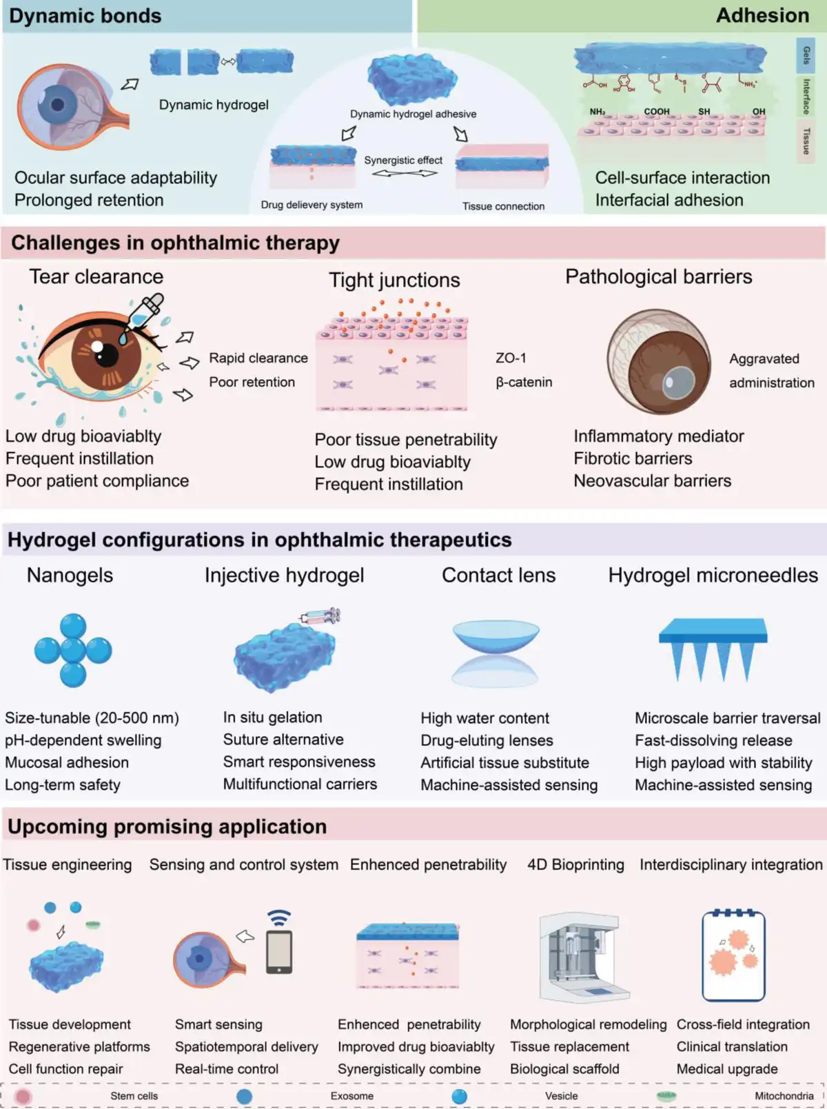
The ocular barriers, including corneal barrier (eg, corneal epithelial tight junctions and opposite polarity compared with corneal stromal cells), tear film barrier (eg, dilution and dynamic removal of tears), and blood-ocular barrier, regulate material or drug transport. In addition to this, the pathological processes, such as inflammatory responses, neovascularization, and extracellular matrix (ECM) remodeling during tissue injury progression, as barriers interfere with drug transport and efficacy.[4,5] Collectively, ocular barriers impede drug penetration into deeper tissues, reducing bioavailability and yielding primarily palliative outcomes. The main barrier-like components affect drug delivery during wound healing as follows. (1) Inflammatory mediator: IL-6 and TNF-α accelerate tissue damage and affect drug transport. (2) Neovascularization: VEGF-driven pathological angiogenesis promotes tissue repair, while vascular leakage affects the distribution and retention of drugs; P-glycoprotein overexpression in endothelial cells further potentiates drug efflux. (3) Extracellular matrix: TGF-β-activated myofibroblast transdifferentiation causes aberrant and dense collagen deposition during ocular wound healing, physically obstructing drug diffusion and distribution to compromise function (Figure 1).
Figure 1.
Dynamic bonds in hydrogel adhesives advancing ophthalmic repair and regeneration: challenges, configurations, and upcoming applications.
Consequently, current ophthalmic delivery relies heavily on frequent dosing, compromising patient compliance. Intravitreal injections enhance efficiency, while the invasiveness risks secondary complications. Novel delivery systems are thus imperative, requiring prolonged retention, stimuli-responsive release, robust tissue adhesion/penetration, and superior biocompatibility. Dynamic covalent bonds offer a potential platform for enabling these functionalities.
2.2 Dynamic bonds redefining ophthalmic therapeutics
Dynamic bonds encompass noncovalent interactions (hydrogen bonding, metal coordination, host–guest complexation, hydrophobic interactions, and ionic interactions) and covalent dynamic bonds (imine bonds, borate esters, disulfide linkages, hydrazone bond, and acylhydrazone bond), defined by reversible kinetics.[6,7] Noncovalent interactions exhibit rapid yet labile dynamics, whereas covalent dynamic bonds demonstrate slower but robust exchange. Crucially, the reactive moieties of dynamic covalent bonds resemble endogenous functional groups in human tissues, enabling transient conjugation with ocular structures to enhance drug utilization.
Hydrogel-based drug carriers significantly enhance bioavailability. Dynamic covalent bonds enable hydrogel fabrication with improved viscoelasticity and robust mucoadhesion. Their reversible breakage–reformation confers self-healing capacity, resisting blinking-induced shear forces while maintaining structural integrity. Multivalent reversible interactions between these hydrogels and ocular mucins markedly prolong drug retention. Physically cross-linked gels serve as carriers for sustained cargo delivery. Their network architectures, stabilized by noncovalent interactions, withstand blinking-induced mechanical stress without imposing a burden on ocular tissues.
Hydrogels for repair (e.g., in situ-forming bandage lenses) typically rely on Schiff base or disulfide bonds, as they offer moderate cross-linking kinetics and suitable mechanical strength under physiological pH, facilitating adherence to the wound. In contrast, nanogels for intravitreal therapy preferentially employ boronic ester bonds or host–guest interactions, owing to their responsiveness to specific intraocular biomarkers, reactive oxygen species (ROS), or matrix metalloproteinases (MMPs), enabling intelligent drug release. Dynamic bonds serve a dual purpose: acting within the internal network of the gel and mediating interactions at the tissue interface. Internally, they enhance drug-loading capacity or enable responsive dissociation, while at the interface, they strengthen adhesion to the tissue, thereby resisting clearance caused by blinking-induced stress.
Dynamic covalent gel networks facilitate stimuli-responsive drug release.[8,9] Drugs conjugated via dynamic covalent bonds or physically entrapped within such networks undergo precise liberation upon pathological microenvironment-triggered bond cleavage. For instance, imine bond-cross-linked hydrogels accelerate drug release in response to localized pH reduction at inflammatory/infectious sites; disulfide bond systems release cargo under elevated inflammatory conditions (eg, dry eye, keratitis, and uveitis); and light-triggered spatiotemporal control is achieved via photoisomerizable moieties (eg, azobenzene). This on-demand release enhances local drug concentration, minimizes off-target effects, and optimizes therapeutic efficacy.
Dynamic covalent bonds also enhance tissue penetration and targeting efficacy. For example, disulfide bond-containing nanoparticles undergo size-shedding via cleavage to penetrate dense barriers (eg, corneal epithelium and mucus layers), followed by postpenetration reconstitution; boronic ester-functionalized materials conjugate sialic acid moieties, enabling active targeting toward inflammatory cells.
3. The adhesion in ophthalmic therapeutics
3.1 The necessity of adhesion
Developing the adhesive properties of hydrogels aims to enhance their interaction with tissues. The ocular microenvironment imposes stringent demands on drug delivery and therapeutic materials. For surface therapies, hydrogels resist blinking-induced shear forces, enabling sustained retention on the dynamic ocular surface.[10,11] This extends treatment intervals from multiple daily doses to once every few days. Simultaneously, the robust yet reversible nature of dynamic covalent bonding minimizes structural compromise to corneal tissues, demonstrating exceptional biocompatibility. This achieves synergistic optimization of mucoadhesion and safety. For extensive tissue defects (e.g., corneal stromal lesions or transplantation), robust interfacial adhesion prevents dislodgement during therapeutic cycles, while balanced dynamic covalent bonding and mucoadhesion ensure modulated degradation kinetics at the injury site.
3.2 The mechanism of adhesion
The ocular tissue surface contains functional groups such as carboxyl, amino, hydroxyl, thiol, and fumarate moieties, exhibiting a net negative charge. Adhesion can thus be achieved via physicochemical interactions. Hydrogel surfaces may be functionalized with polyhydroxyl, polycarboxyl, polyphenolic, phenylboronic acid, aldehyde, or disulfide groups to achieve adhesion.
Alternative adhesion strategies include mechanical interlocking via microneedle arrays and topographic adhesion mediated by chitosan’s phase transition and interfacial diffusion under physiological versus acidic conditions. Current research extensively focuses on mussel-inspired adhesion strategies, which rely on polyphenol-enabled multivalent hydrogen bonding and covalent interactions to achieve interfacial tissue integration. Although direct evidence is lacking, the recruitment and migration-promoting effects of specific functional groups suggest that adhesive materials can modulate cellular behavior to some extent, thereby facilitating tissue repair.
3.3 Balance of adhesion and cohesion
The magnitude of adhesion arises from a balance between interfacial adhesive moieties and bulk cohesive strength, both of which must be optimized to achieve robust adhesion. For ocular surface modulation, cohesive contributions can be neglected; however, tissue integration requires careful consideration due to inherent persistence limitations at the tissue interface. The clinically prevalent mode of adhesive failure manifests as cohesive fracture within hydrogels, primarily governed by internal crack propagation mechanisms.
The synergistic integration of strong and weak bonds enables stable hydrogel adhesion, where robust bonds maintain structural integrity while labile bonds dissipate imposed stresses, thus ensuring cohesive stability. This fundamental principle has driven recent advances in dual-network and dual-cross-linking material strategies.
Adhesive failure represents another clinically relevant failure mode, primarily mediated by interfacial hydration layers that compromise tissue-adhesive interactions. Excessive tissue exudation impedes wound healing by inducing edema and facilitating microbial colonization. Hydrogels counter this through their superior hydration capacity, which simultaneously shields the wound bed and exposes engineered reactive sites to enable persistent interfacial integration. For internal tissue integration, hydrogel hydration capacity poses significant clinical risks: swelling-induced volumetric expansion may compress neural structures and elevate intraocular pressure. Thus, modulating hydrogel hydration capacity while preserving adhesive properties necessitates disease-specific trade-offs in clinical implementation.
A particularly distinctive scenario involves unilateral asymmetric adhesion, where anisotropic binding interfaces exclusively facilitate deep-tissue integration while preventing detrimental interfacial binding on nontargeted surfaces. This mechanism operates via 3 distinct pathways: passivation of surface adhesive moieties, integration of nonadhesive interfacial layers, or counter-charge neutralization combined with in situ network polymerization that internally confines adhesive functional groups. The diverse spectrum of adhesion modalities offers unprecedented therapeutic alternatives for ophthalmic applications, enabling disease-specific tailoring of distinctive adhesive hydrogels.
4. Biointegration of dynamic covalent bonds and adhesion
Biointegration leverages reversible, stimuli-responsive chemical bonds to engineer biomaterials capable of intelligent, robust, and enduring adhesion to living tissues. This approach targets the molecular design of stable, functional interfaces between materials and host tissues that resist fibrous encapsulation.[12]
Conventional materials, however, face significant challenges in achieving ideal biointegration, particularly concerning dynamic adaptation to tissue changes, long-term interfacial stability, and on-demand detachment. The integration of dynamic bonds overcomes these limitations by enhancing interfacial cross-linking dynamics and adaptability. Within adhered hydrogels, the dynamic chemical bond-enabled network undergoes adaptive remodeling in response to the local microenvironment (e.g., pH, enzymes, and redox state), accommodating tissue morphology and mechanical shifts while mitigating detrimental stress shielding or concentration. Furthermore, this internal dynamic network creates an ECM-like microenvironment conducive to cell infiltration, tissue integration, and the potential guidance of cell behavior and tissue ingrowth. Critically, dynamic bonds facilitate the immobilization of bioactive molecules (growth factors, antibodies, and cell-adhesive peptides), offering a platform for controllable immunomodulation.
At the material–tissue interface, dynamic covalent networks establish stronger, reversible covalent bonds with tissue surface molecules (collagen, fibrin, and cell surface receptors), surpassing the inherent weaknesses of traditional noncovalent interactions and enabling purposeful detachment. This dynamic nature also confers self-healing properties; upon damage (e.g., microfractures), the interface reversibly breaks and reorganizes, restoring structural integrity and adhesion strength. Importantly, the application of specific external stimuli (e.g., pH shift, light exposure, and reducing agents) allows for the on-demand, reversible disruption of the adhesive interface, facilitating noninvasive or minimally invasive device removal.[13,14]
Biointegration of dynamic covalent bonds and adhesion imparts dynamic mechanical properties, facilitates cellular infiltration and vascularization, and enables responsive release of bioactive factors to the tissue microenvironment. Concurrently, it achieves adaptive adhesion to soft tissues, rapidly establishing robust, reversible bonds on hydrated tissue surfaces. This holds significant promise for tissue engineering, implantable devices, adhesive sealing, and intelligent delivery systems.
5. Hydrogel configurations in ophthalmic therapeutics
Based on these core designs, hydrogels exhibit diverse ocular configurations, including nanogels, injectable hydrogels, hydrogel contact lenses, and hydrogel microneedles, designed to overcome conventional therapeutic limitations and enhance efficacy.
5.1 Nanogels
Nanogels refer not to the simplistic integration of nanoscale designs with hydrogels, but rather to nanoscale microstructures fabricated based on gel preparation principles. They possess not only the adhesive properties of hydrogels but also the nanoscale effects of nanomaterials, enabling penetration through biological barriers and prolonged retention at wound sites. This design concurrently integrates macroscopic and microscopic advantages, transcending traditional composite approaches. Such therapeutic strategies optimize treatment efficacy while facilitating scalable applications.
Compared with conventional nanoparticles (poor retention), metallic nanoparticles (weak penetration), lipid nanoparticles (prone to hydrolysis), and mesoporous silica (fibrosis induction), nanogels leverage biomimetic structural design and dynamic network modulation. They offer tunable elastic modulus, mucin-mimetic structures for prolonged retention, integrated multiresponsiveness (temperature/pH/ROS), high swelling ratios (10–50×), and high drug-loading capacity. Additionally, their high-water content and ECM-mimicking composition enhance biocompatibility—reducing inflammation, lubricating surfaces, and enabling controlled degradation—benefiting treatment of delicate ocular tissues. Also, nanogels exhibit superior coloading efficiency for both hydrophilic and hydrophobic drugs, with intelligent release enabled by dynamic bonds. In contrast, liposomes suffer from low drug-loading capacity and insufficient stability. Nanoparticles and micelles demonstrate poor hydrophilic drug encapsulation and are susceptible to premature drug release. Exosomes face challenges in isolation/purification and low drug-loading efficiency. Critically, while all these carriers can utilize thiol-mediated surface interactions for cellular internalization, potentially avoiding lysosomal clearance, nanogels uniquely retain tunable elasticity. This adjustable particle stiffness may better accommodate biomechanical stress transmission in biological systems.
5.2 Injectable hydrogel
Injectable hydrogels gel in situ at lesion sites, conforming to irregular defects and modulating both superficial and deep injuries. Typically, photo-cross-linked or dynamically bonded, they provide sustained hydration protection and controlled release of therapeutic agents for enhanced repair. Classified as surface dressings or internal fillers, their mechanical strength varies, exemplified by weak intravitreal gels versus low-modulus dry eye lubricants. Crucially, grafts and defect-filling designs require adhesion and optical transparency to mitigate detachment risks while preserving visual function.
Beyond superficial drug delivery, hydrogel-tissue engineering coordinates uniform cellular growth across tissue layers, preventing scar formation and functional impairment caused by heterogeneous cell proliferation. For transplants, injectable adhesive hydrogels achieve seamless donor–host integration to mitigate surgical trauma while enabling holistic wound modulation across superficial and deep strata for comprehensive recovery.
5.3 Hydrogel contact lens
Hydrogel contact lenses are fabricated into cornea-mimetic structures with specific curvature and exceptional transparency. They provide mechanical barrier protection for the cornea while enabling customized curvature correction for myopia, hyperopia, and astigmatism. Crucially, their high oxygen permeability prevents hypoxic edema.
Hydrogel contact lenses enable human–digital interfacing, facilitating digitally controlled release of encapsulated therapeutics and transmission of ocular surface pathology via electrical signals for expert computational analysis.
Hydrogel contact lenses may serve as biomimetic ocular prostheses. When suitable donors are unavailable, stable, biocompatible, customized hydrogels can replace ocular structures to preserve viable visual function.
5.4 Hydrogel microneedles
Hydrogel microneedles (400–800 μm in length) penetrate the corneal epithelium (50 μm thick), creating transient micropores (<1 μm in diameter) pain-free. This enables drug bypassing of tear clearance and mucus barriers while establishing adhesive anchoring via microneedle arrays.
Hydrogel microneedles can be engineered for sustained release via calcium chelation or burst release via pH responsiveness, tailored to specific therapeutic agents. Concurrently, the array penetrates corneal epithelial interstices, adsorbs tear inflammatory mediators (e.g., MMP-9, IL-6), enhances detection sensitivity through surface enhancement, continuously monitors intraocular pressure fluctuations, and wirelessly transmits data to mobile applications.
6. Emerging applications in ophthalmic therapeutics
6.1 Cascade regulation of ocular inflammation
In ocular diseases, inflammatory conditions (e.g., dry eye and alkali burns) constitute a major proportion and may induce pathological structural changes if untreated. While current medical systems manage such conditions, fewer yet more effective drugs remain critical due to concerns regarding ocular toxicity and drug resistance. Drug-adhesive material cascades may address this challenge. Persistent inflammation not only causes discomfort but also promotes scarring and ocular fragility, necessitating multistage biological cascade regulation (e.g., inflammatory and proliferative phases). Prolonged material–tissue interaction with controlled release is essential for such multiphase modulation. Dynamic covalent bonds with tunable kinetics can be designed to sequentially meet distinct repair phase demands, enabling cascade control of inflammatory diseases.
The existing strategy of coloading hydrophilic and hydrophobic drugs for sequential release confronts critical challenges: achieving sequential delivery of dual hydrophobic drugs, ensuring dual-drug carrier stability, and determining whether their therapeutic effects manifest synergy or antagonism—all demanding rigorous investigation.
6.2 Microinvasive therapeutic approaches for corneal disorders
Corneal transplantation presents complex clinical challenges requiring advanced surgical skill and management of potential immune rejection. Conventional suture fixation induces secondary trauma, scar formation, and risks of corneal neovascularization. Dynamic covalent-bonded hydrogel adhesives address these limitations by adaptively bridging donor and recipient tissues through self-adjusting interfacial bonding, eliminating suture-related complications while promoting seamless tissue integration. Critically, the adhesive process concurrently enables drug loading via dynamic covalent bonding, facilitating sustained release of immunosuppressants to circumvent side effects associated with repeated topical administration.
Compared with donor corneal or hydrogel transplantation, focal repair of damaged tissue for localized corneal defects offers greater clinical appeal by avoiding extensive replacement and invasive surgery. In situ-forming transparent hydrogels—combining tissue adhesion with pre-gel fluidity—provide a novel therapeutic alternative. This approach involves applying a pre-gel solution directly to the defect site, followed by gelation to stabilize the wound suture-free, thereby promoting epithelial reformation and stromal regeneration for vision recovery.
6.3 Hydrogel-mediated regulation in ocular tissue engineering
While pharmacotherapy carries inherent toxicity risks, biomaterial-mediated tissue modulation has emerged as a compelling alternative.[15,16] Magnetic materials direct cellular alignment, hydrogel stiffness governs proliferation, and surface properties regulate adhesion, with intravitreal hydrogel injection further maintaining ocular homeostasis. Hydrogels engineered from abundant biocompatible materials exert multiscale bioinfluence: from receptor-level signaling and migratory guidance to functional tissue restoration. Elucidating these mechanisms will pioneer multidimensional strategies for advancing ocular health.
7. Challenges and perspectives
The current research focus lies in unilaterally enhancing material properties such as adhesive capabilities, dynamic exchange capabilities, and intrinsic bioactivities—which can be optimized to exceptional levels—yet fails to consider the overall logic holistically. While dynamically bonded adhesive hydrogels have made significant progress in ocular drug delivery and tissue engineering, several challenges remain. The fundamental issue lies in the inability to achieve highly efficient treatment targeting of lesion sites. Addressing these will have profound implications for future innovations in ophthalmic materials.
A key challenge lies in achieving robust adhesion while maintaining gentle reversibility. The trade-off between adhesion strength and reversibility primarily stems from the kinetic mismatch of dynamic bonds: strong bonds (e.g., catechol-metal coordination) favor adhesion but hinder detachment, while weak bonds (e.g., hydrogen bonds) enable reversibility but compromise stability. Current cross-linking strategies lack spatiotemporal control to synchronize these opposing requirements. Moreover, for ocular applications, the degree of adhesion and adaptability requires quantitative gold standards, determining what levels ensure sufficient handling convenience and stability without compromising tissue interactions. Additionally, while hydrogels possess ECM-like properties, they are still foreign materials. Existing ISO 10993 standards focus on short-term cytotoxicity but neglect dynamic material–tissue interactions. For hydrogel adhesives, key gaps include: (1) no established protocols to evaluate chronic inflammatory responses induced by bond exchange and (2) insufficient correlation between in vitro degradation tests and actual in vivo behavior due to ocular microenvironment complexity. The incorporation of externally-triggered dynamic covalent bonds offers a pathway to reconcile the kinetic dichotomy between strong permanent anchors and weak reversible linkages. Precise spatiotemporal modulation of cross-link density enables optimization of the adhesion-reversibility trade-off through controlled bond lifetime tuning.
The next shortcoming lies in the long-term interactions of biocompatibility. The existing evaluation methods suffer from a short-term perspective in the temporal dimension, primarily assessing the acute cytotoxicity of materials over a few days, while failing to predict potential genotoxicity and ultra-long-term biological responses. These methods are static and incapable of simulating the complex dynamic physiological environment within the body, such as mechanical stresses and biochemical gradient changes. They lack the ability to monitor dynamic, continuously evolving processes, such as real-time fluctuations in inflammatory factors, dynamic variations in oxidative stress levels, and the evolution of the microenvironment at the tissue-material interface. Most critically, current approaches struggle to effectively integrate long-term changes across molecular levels (e.g., gene expression and proteomics), cellular levels, tissue levels, and organ/system levels, thereby hindering a holistic understanding of the long-term evolution of biocompatibility. Current detection devices can identify electrical signals and glucose changes and perform genetic analysis, but they lack sufficient sensitivity and specificity—specifically, the ability to reliably detect targets at low concentrations and accurately attribute signals to their precise sources. Integrating miniaturized, biocompatible biosensors into implantable devices or as standalone implants enables long-term, real-time, in situ monitoring of key biomarkers, such as inflammatory factors, tissue damage/repair biomarkers, metabolites, drug molecules, and tissue physical parameters. This provides an unprecedented perspective for understanding the dynamics of material–host interactions and offers early warnings of potential long-term complications. However, it requires multidisciplinary functional integration, and multidimensional, multitiered detection capabilities still demand further development.
Another barrier lies in the penetration capacity of hydrogels. Although current hydrogels can achieve nanoscale dimensions, their improved drug efficacy primarily stems from resisting blink-induced clearance. While the interactive nature of dynamic bonds enhances penetration potential, it still falls short of being satisfactory. For posterior segment diseases, microinvasive hydrogel therapies remain limited. Future strategies may involve functional integration to impart nanomotor functionality, thereby increasing penetration power, but this necessitates controllable environmental responsiveness. Externally-directed propulsion-enhanced targeting achieves both deep-tissue penetration and molecular specificity.
The exchange capacity across hydrogel adhesives presents another challenge. For the ocular surface, maintaining gas exchange (particularly oxygen) between the air and ocular tissues is essential to prevent potential hypoxic edema. Within connective tissues, definitive evidence demonstrating comprehensive exchange of growth factors, nutrients, and cellular components across the adhesive interface remains lacking. Machine learning-guided interfacial simulations may provide foundational mechanistic insights for designing hydrogels and optimizing mass transport across biointerfaces.
Translational hurdles present another significant challenge. Sterilization stability, scalable synthesis, spatiotemporally controlled applicability, and multidimensional in vivo efficacy validation all impact progress. Machine-assisted structural design will facilitate the development of novel functional hydrogels capable of meeting application demands within complex in vivo environments.
The 4D-printed hydrogel structures represent a potential future direction. Although 4D printing holds significant promise, its clinical translation primarily depends on resolving the biocompatibility of materials. Future research must focus on developing novel biocompatible cross-linking systems. For instance, exploring initiator-free click chemistry, enzyme-mediated cross-linking, or the use of endogenous cross-linkers could fundamentally eliminate the potential toxicity risks associated with exogenous photoinitiators. Furthermore, the long-term functional stability of 4D structures poses another major challenge. Future designs of smart hydrogels should ensure that their shape-memory effects remain stable under prolonged physiological conditions. Simultaneously, their stimulus-responsive mechanisms should exhibit greater targeting precision and specificity to enable truly on-demand therapeutic interventions. Shape-adaptive hydrogels offer enhanced compatibility with ocular motility. Furthermore, a comprehensive scientific understanding of the mechanical behavior of hydrogels on the ocular surface—including fatigue, stress relaxation, creep, and aging—remains limited. Future bioink designs must prioritize structural and functional congruence with native tissue architectures, actively emulating biological systems through biomimetic engineering.
Finally, hydrogel adhesives should incorporate rapid diagnostic capabilities. By specifically recognizing key pathological features at the lesion site and providing clinicians with detailed data, they could not only shorten diagnostic timelines and reduce costs but also facilitate future medical advancements.
Funding
This research was financially supported by the National Natural Science Foundation of China (Nos. 52173143 and 82371108), the Natural Science Foundation of Henan Province of China (Nos. 242300421018 and 252300421024), Henan Province Science and Technology Research and Development Plan Joint Fund Project (No. 242301420010), Henan Province Youth Health Science and Technology Innovation Talent Training Project (No. LJRC2024003), Henan Province Clinical Medical Scientist Training Program (No. HNCMS202402), and the Basic Science Key Project of Henan Eye Hospital (No. 24JCZD003).
Conflicts of interests
The authors declare that they have no conflicts of interest.
Author contributions
H.C.: conceived, organized, and wrote the manuscript. B.A.: conceptualization. Z.L.: conceptualization, funding acquisition, project administration, and supervision. J.L.: conceptualization, funding acquisition, investigation, project administration, resources, and supervision. All authors discussed and approved the final manuscript.
References
- [1] Li P, Sun W, Li J, et al. N-type semiconducting hydrogel. Science. 2024;384:557–563.
- [2] Shi J, Kim S, Li P, et al. Active biointegrated living electronics for managing inflammation. Science. 2024;384:1023–1030.
- [3] Pranantyo D, Yeo CK, Wu Y, et al. Hydrogel dressings with intrinsic antibiofilm and antioxidative dual functionalities accelerate infected diabetic wound healing. Nat Commun. 2024;15:954.
- [4] Huang J, Jiang T, Qie J, et al. Biologically inspired bioactive hydrogels for scarless corneal repair. Sci Adv. 2024;10:eadt1643.
- [5] Dang M, Slaughter KV, Cui H, et al. Colloid-forming prodrug-hydrogel composite prolongs lower intraocular pressure in rodent eyes after subconjunctival injection. Adv Mater. 2025;37:e2419306.
- [6] Soliman BG, Nguyen AK, Gooding JJ, et al. Advancing synthetic hydrogels through nature-inspired materials chemistry. Adv Mater. 2024;36:e2404235.
- [7] Chen M, Hu J, Gao H, et al. An immunotherapeutic artificial vitreous body hydrogel to control choroidal melanoma and preserve vision after vitrectomy. Sci Adv. 2023;9:eadh1582.
- [8] Yang B, Li Z, Yang Z, et al. Recapitulating hypoxic metabolism in cartilaginous organoids via adaptive cell-matrix interactions enhances histone lactylation and cartilage regeneration. Nat Commun. 2025;16:2711.
- [9] Duan S, Hua M, Zhang CW, et al. Noncovalent aggregation for diverse properties in hydrogels: a comprehensive review. Chem Rev. 2025;125:7918–7964.
- [10] Pal S, Shin J, DeFrates K, et al. Recyclable surgical, consumer, and industrial adhesives of poly(α-lipoic acid). Science. 2024;385:877–883.
- [11] Cui H, Li J. Hydrogel adhesives for tissue recovery. Adv Colloid Interface Sci. 2025;341:103496.
- [12] Ren H, Zhang Z, Cheng X, et al. Injectable, self-healing hydrogel adhesives with firm tissue adhesion and on-demand biodegradation for sutureless wound closure. Sci Adv. 2023;9:eadh4327.
- [13] Shi J, Lin Y, Li P, et al. Monolithic-to-focal evolving biointerfaces in tissue regeneration and bioelectronics. Nat Chem Eng. 2024;1:73–86.
- [14] Freedman BR, Kuttler A, Beckmann N, et al. Enhanced tendon healing by a tough hydrogel with an adhesive side and high drug-loading capacity. Nat Biomed Eng. 2022;6:1167–1179.
- [15] Wu J, Deng J, Theocharidis G, et al. Adhesive anti-fibrotic interfaces on diverse organs. Nature. 2024;630:360–367.
- [16] Chen L, Jin Z, Feng W, et al. A hyperelastic hydrogel with an ultralarge reversible biaxial strain. Science. 2024;383:1455–1461.
This article is available under Open Access.
© 2025 The Authors