Compelling standardized and high-throughput micro-/millifluidic plates for biomedical research: from laboratory to market
Authors
Longjun Gu, Peidi Liu, Wen Zhao, Yuhang Fan, Yuwen Wang, Pu Chen*
- aDepartment of Biomedical Engineering, Tissue Engineering and Organ Manufacturing (TEOM) Lab, Wuhan University TaiKang Medical School (School of Basic Medical Sciences), Wuhan, Hubei, China
- bDepartment of Intelligent Systems Engineering, Indiana University, Bloomington, Indiana, USA
- cTaiKang Center for Life and Medical Sciences, Wuhan University, Wuhan, Hubei, China.
* Correspondence: Address: Pu Chen, Department of Biomedical Engineering, Wuhan University, 115 Donghu Road, Room 305, #3 Building, Wuhan, Hubei 430071, China. Email: puchen@whu.edu.cn (P. Chen).
MedMat · 2025 · Vol. 2 · No. 2 · pp. 101-117
Abstract
Microfluidics has demonstrated significant potential for advancing biomedical research. However, the widespread adoption of current microfluidic devices within the biomedical community is hindered by 2 major challenges: standardization and throughput. To address these issues, emerging micro-/millifluidic plates based on microtiter plate format have been proposed. On the one hand, the established standards for microtiter plates facilitate clear communication between microfluidic engineers and end-users, enabling untrained users to manipulate micro-/millifluidic plates directly without the need to be proficient in understanding the mechanisms behind the platform. On the other hand, micro-/millifluidic plates inherit the high-throughput capabilities of microtiter plates, enhancing their utility in applications such as organ-on-a-chip and point-of-care testing. This review is intended to provide a timely and insightful overview of micro-/millifluidic plates, covering their design strategies, liquid-driven systems, applications, and commercialization status. Additionally, the review discusses the challenges facing micro-/millifluidic plates and highlights emerging positive trends in this field. We believe that our unique perspective on micro-/millifluidic plates can facilitate innovation and accelerate academic transformation by appealing to the microfluidic community to establish a consistent chip development plan to match end-user expectations in the biomedical field.
Translations
Long abstracts in additional languages. The English article is the version of record.
中文zh-Hans
微流控技术在推动生物医学研究方面展现出巨大潜力,然而其广泛应用仍面临两大核心挑战:标准化不足与通量有限。当前大多数微流控设备缺乏统一标准,导致工程师与普通用户之间存在沟通壁垒,非专业人员难以直接操作复杂的芯片系统,且传统设计往往牺牲了高通量筛选能力。本文旨在综述基于微量滴定板格式的新一代微/毫流体板的最新进展,通过整合现有文献与设计策略,为从实验室研发到市场化应用提供系统性见解,以解决上述瓶颈问题并促进该领域的标准化进程。
本综述深入分析了微/毫流体板的设计架构与工作原理,重点探讨其如何继承微量滴定板的国际标准格式(如96孔、384孔等)。这些新型平台采用液驱动系统作为核心机制,将复杂的流控功能集成于标准化的物理接口中,使得未受过专业训练的研究人员也能直接进行实验操作。文章详细梳理了从芯片设计策略到液体输送系统的技术细节,涵盖了器官芯片构建与即时检测(POCT)等多种应用场景下的具体实施方案,并评估了当前商业化产品的市场现状与技术成熟度。
研究指出,基于微量滴定板格式的微/毫流体平台通过标准化接口显著降低了使用门槛,实现了微流控技术与终端用户需求的无缝对接。这种设计不仅保留了传统微流控的高灵敏度与精确控制优势,更赋予了其高通量并行处理的能力,使其在器官芯片模型构建及大规模药物筛选中展现出独特价值。综述发现,当前的技术趋势正从单一功能向多功能集成转变,液驱动系统的优化进一步提升了实验的可重复性与操作便捷性,为生物医学研究提供了更为灵活且高效的工具平台。
尽管微/毫流体板在标准化与高通量方面取得了显著进展,但该领域仍面临芯片开发计划缺乏统一规范、商业化产品种类有限等挑战。本文强调,未来需要学术界与产业界共同努力,建立一致的芯片研发标准以匹配生物医学领域的实际需求,从而加速从实验室创新到市场转化的进程。通过呼吁微流控社区形成共识并制定统一的开发路线图,我们期望这一独特视角能推动技术创新,消除应用障碍,最终实现微流控技术在更广泛生物医学场景中的普及与深度整合。
Françaisfr
La microfluidique a démontré un potentiel significatif pour faire avancer la recherche biomédicale, mais son adoption généralisée au sein de la communauté est entravée par deux défis majeurs : le manque de standardisation et une faible capacité de débit. Les dispositifs actuels souffrent souvent d'une absence de normes claires, créant des barrières entre les ingénieurs en microfluidique et les utilisateurs finaux qui ne maîtrisent pas les mécanismes sous-jacents. Cet article vise à fournir un aperçu opportun et perspicace sur les plaques micro-/millifluidiques basées sur le format de plaque de microréacteurs, couvrant leurs stratégies de conception, systèmes liquides, applications et statut commercialisation pour adresser ces lacunes critiques.
L'approche présentée repose sur l'intégration des standards établis pour les plaques à micro-puits dans la conception de nouveaux dispositifs microfluidiques. Ces plateformes héritent du format standardisé permettant une communication claire entre ingénieurs et utilisateurs, facilitant ainsi la manipulation directe par des opérateurs non formés sans nécessiter d'une expertise approfondie en mécanismes fluidiques. Le texte examine les stratégies de conception détaillées, y compris les systèmes à entraînement liquide, tout en illustrant comment ces plaques sont adaptées pour des applications exigeantes telles que l'organe-sur-puce et les tests au point de soins, offrant une vue d'ensemble technique complète.
Les analyses révèlent que l'adoption du format plaque standardisée permet non seulement de surmonter la barrière de compétence mais aussi d'améliorer considérablement le débit des expériences. Les plaques micro-/millifluidiques combinent ainsi les avantages de la précision microfluidique avec une capacité de traitement à haut débit, rendant ces outils particulièrement utiles pour le criblage pharmaceutique et la modélisation tissulaire avancée. La revue met en lumière comment cette approche favorise l'innovation en alignant le développement des puces sur les attentes réelles du domaine biomédical, tout en identifiant les tendances émergentes positives qui redéfinissent les pratiques actuelles.
Malgré ces progrès, la communauté fait face à des défis persistants concernant la nécessité d'un plan de développement cohérent et l'accélération de la transformation académique vers le marché. L'article souligne que pour faciliter l'innovation future, il est impératif que la communauté microfluidique établisse une approche standardisée qui réponde aux besoins des utilisateurs finaux dans les applications biomédicales complexes. Les perspectives futures incluent le renforcement des collaborations entre ingénieurs et chercheurs cliniques pour surmonter les limitations actuelles de commercialisation et garantir que ces technologies avancées deviennent accessibles à un plus large éventail d'applications scientifiques et médicales.
Españoles
La microfluídica ha demostrado un potencial significativo para avanzar en la investigación biomédica; sin embargo, su adopción generalizada se ve obstaculizada por dos desafíos mayores: estandarización y capacidad de procesamiento. Los dispositivos actuales carecen a menudo de normas claras que faciliten la comunicación entre ingenieros y usuarios finales, lo que impide que personal no especializado manipule estos sistemas complejos sin un profundo conocimiento de sus mecanismos subyacentes. Esta revisión tiene como objetivo proporcionar una visión oportuna e insightful sobre las placas micro-/millifluídicas basadas en el formato de placa de micropocillos, cubriendo estrategias de diseño, sistemas impulsados por líquidos y su estado comercial para abordar estas limitaciones críticas.
El enfoque presentado se centra en cómo los nuevos dispositivos heredan la alta capacidad de procesamiento de las placas estándar mientras integran interfaces estandarizadas que permiten una comunicación clara entre ingenieros y usuarios. Estas plataformas utilizan sistemas de conducción líquida como mecanismo central, permitiendo que operadores no entrenados manipulen directamente las placas sin necesidad de comprender los detalles técnicos complejos del funcionamiento interno. El texto examina en detalle las estrategias de diseño, incluyendo la adaptación para aplicaciones específicas como órganos-en-chip y pruebas point-of-care, ofreciendo una descripción completa de cómo estas innovaciones técnicas están transformando el panorama experimental actual.
Los hallazgos principales indican que este formato estandarizado no solo reduce significativamente la barrera de entrada técnica sino que también mejora sustancialmente la capacidad de procesamiento paralelo en experimentos biológicos. Las placas micro-/millifluídicas combinan así las ventajas de precisión con una escalabilidad superior, haciéndolas particularmente útiles para cribado farmacológico y modelización tisular avanzada. La revisión destaca cómo esta perspectiva única puede facilitar la innovación al alinear el desarrollo de chips con las expectativas reales del campo biomédico, identificando tendencias emergentes positivas que están redefiniendo los estándares actuales en este sector tecnológico.
A pesar de estos avances, persisten desafíos importantes relacionados con la necesidad de un plan de desarrollo consistente y la aceleración de la transformación académica hacia el mercado. El artículo enfatiza que para facilitar la innovación futura es imperativo que la comunidad microfluídica establezca una estrategia común que responda a las necesidades específicas de los usuarios finales en aplicaciones biomédicas complejas. Las perspectivas futuras incluyen un llamado a establecer planes de desarrollo coherentes y colaboraciones más estrechas entre ingenieros e investigadores clínicos para superar las limitaciones actuales de comercialización, asegurando así la adopción generalizada de estas tecnologías avanzadas.
日本語ja
マイクロ流体技術は生物医学研究の進展に大きな可能性を示していますが、現在のマイクロ流体デバイスの普及には標準化と処理能力という2つの主要な課題が障壁となっています。多くの既存デバイスでは明確な基準が存在せず、エンドユーザーが専門知識を持たずに直接操作することが困難であり、また従来の設計では高スループット性が十分に活用されていません。本稿は、これらの問題を解決するために提案されたマイクロ/ミリ流体プレートに関する最新の知見を体系的にレビューし、実験室から市場への移行における標準化の重要性と、この分野の現状と将来展望について包括的な枠組みを提供することを目的としています。
本研究では、マイクロピペットプレートの既存規格に基づいた設計戦略と液駆動システムの詳細な分析を行います。これらの新規プラットフォームは、エンジニアとエンドユーザー間の明確なコミュニケーションを可能にする標準化されたフォーマットを採用しており、専門知識がなくても直接操作可能な点に特徴があります。レビューでは、器官オンチップやポイントオブケア検査などにおける具体的な応用例とともに、液体輸送メカニズムの技術的詳細、設計上の工夫、および現在の商業化状況について包括的に解説し、実用面での利点を明確に示しています。
分析の結果、マイクロ/ミリ流体プレートは標準化されたインターフェースを通じて使用ハードルを大幅に低下させるとともに、高スループット処理能力も維持・強化することが確認されました。これにより、複雑なメカニズムの理解が不要であっても実験が可能となり、生物医学研究における効率性と再現性が向上します。特に器官モデルや大規模スクリーニングにおいてその有用性が高く評価されており、技術的な進歩がエンドユーザーの期待に合致する形で発展していることが示されています。また、この分野でのポジティブな傾向として、標準化された開発アプローチへの移行が進んでいる点が強調されます。
しかしながら、マイクロ/ミリ流体プレートには依然として課題が残っており、特に一貫したチップ開発計画の欠如や市場普及までの道のりの長さなどが指摘されています。本稿は、この分野における革新を促進し学術的変革を加速させるためには、微流控コミュニティ全体で標準的な開発方針を確立し、エンドユーザーの期待に応えることが不可欠であると結論付けています。今後の展望として、技術者間の連携強化や商業化プロセスの最適化を通じて、これらの高度なプラットフォームがより広範な生物医学応用において普及し、研究と臨床現場での実装がさらに加速されることを目指しています。
العربيةar
أظهرت تقنية الميكروفلويديك إمكانات كبيرة في تقدم البحث الطبي الحيوي، إلا أن اعتمادها على نطاق واسع لا يزال يواجه تحديات رئيسية تتمثل في نقص التوحيد القياسي وقلة الإنتاجية. تواجه الأجهزة الحالية صعوبة في التواصل الواضح بين مهندسي الميكروفلويديك والمستخدمين النهائيين، حيث يتطلب التعامل معها فهمًا عميقًا للآليات المعقدة التي قد تكون غير متاحة للمستخدمين غير المدربين. يهدف هذا الاستعراض إلى تقديم نظرة شاملة ومحدثة حول ألواح المايكرو/ميلي فلويديك المستندة إلى تنسيق صواني العينات الدقيقة، تغطي استراتيجيات التصميم والأنظمة السائلة والتطبيقات والحالة التجارية لمعالجة هذه الثغرات الحرجة في المجال.
يرتكز النهج المعروض على دمج المعايير الراسخة لصواني العينات في تصميم الأجهزة الجديدة، مما يسمح بواجهة موحدة تسهل التواصل بين المهندسين والمستخدمين. تعتمد هذه المنصات الناشئة أنظمة دفع سائلة كآلية أساسية، وتمتلك القدرة على التعامل مع عمليات عالية الإنتاجية دون الحاجة إلى خبرة متخصصة في فهم الآليات الداخلية المعقدة. يغطي الاستعراض بالتفصيل استراتيجيات التصميم المختلفة والأنظمة السائلة المستخدمة، مع التركيز على تطبيقات محددة مثل الأجهزة العضوية-على-رقاقة واختبارات نقطة الرعاية، مقدماً تقييماً شاملاً للوضع التجاري الحالي لهذه التقنيات المتقدمة.
تُظهر النتائج الرئيسية أن استخدام تنسيق صواني العينات القياسي يقلل بشكل كبير من حواجز الاستخدام ويسمح بالتحكم الدقيق في العمليات البيولوجية المعقدة. توفر هذه الألواح الميكرو/ميلي فلويديك مزايا فريدة تجمع بين دقة التقنيات الدقيقة وقدرة الإنتاج العالي، مما يجعلها أدوات فعالة للغاية في تطبيقات مثل الفحص الدوائي واسع النطاق وبناء نماذج الأنسجة المتقدمة. يسلط الاستعراض الضوء على كيفية مساهمة هذا المنظور الفريد في تسريع الابتكار من خلال مواءمة خطط تطوير الرقاقات مع توقعات المستخدمين النهائيين في المجال الطبي الحيوي، مشيراً إلى الاتجاهات الإيجابية الناشئة التي تعيد تشكيل الممارسات الحالية.
على الرغم من التقدم المحرز، لا تزال هناك تحديات تواجه هذا المجال تتعلق بضرورة وضع خطة تطوير متسقة وتسريع التحول الأكاديمي نحو السوق. يؤكد الاستعراض أن تسهيل الابتكار المستقبلي يتطلب تعاوناً بين مجتمع الميكروفلويديك لإنشاء معايير موحدة تلبي احتياجات المستخدمين النهائيين في التطبيقات الطبية الحيوية المعقدة. تشمل الآفاق المستقبلية الدعوة إلى وضع خطط تطوير متسقة وتعاون أوثق بين المطورين والمستخدمين لتجاوز القيود الحالية، مما يضمن تبني هذه التقنيات المتقدمة على نطاق واسع ويسرع من تحولها من المختبرات إلى الأسواق التجارية.
Keywords
Full Text
1. Introduction
Over the past 3 decades, despite microfluidic practitioners making great efforts to expand the impact of microfluidics on biomedical applications, this innovative technology has not yet been widely embraced by end-users in the biomedical community. From an engineering standpoint, one primary reason is that microfluidic engineers, who are often nonexperts in the biomedical field, tend to overlook the experimental practices and workflows of end-users such as biologists and clinicians during the development of microfluidic chips. As a result, these microfluidic novices usually have to spend considerable time troubleshooting and adapting to these new tools rather than focusing on scientific advancements.[1] Another challenge is the delicate balance that objectively exists between the functional complexity and the throughput of microfluidic chips.[2] Complex, highly functional designs, along with customized operational processes, inherently limit the throughput of these chips, which in turn slows down sample production and data acquisition. Consequently, most existing microfluidic chips are generally suitable for small-scale laboratory research but are not easily scalable to meet the demands of applications in pharmaceutical and clinical diagnosis.[3]
Compared with microfluidic chips, the microtiter plate is widely recognized as a standard device in biomedical research due to its user-friendly operation and high-throughput testing capabilities. Since the establishment of microtiter plate standards (Table 1),[4–5–6–7–8–9–10–11–12] microtiter plates of various specifications have been mass-produced in industry and have become essential tools in a range of biomedical applications, including cell manipulation,[13] drug screening,[14] and clinical diagnosis.[15,16] Especially in COVID-19 testing, the microtiter plate significantly contributes to detecting the causative virus by using the reverse transcription polymerase chain reaction (RT-PCR) technique.[17] Alongside microtiter plates, a variety of commercial instruments such as robotic liquid handlers, plate readers, and high-content screening instruments have been specifically developed and employed for sample handling and analysis in microtiter plates. These advancements have made microtiter plates viable for workflow automation with minimal human intervention, thereby enhancing reproducibility and robustness in biomedical research.[18]
Table 1
Comparison of standards for microtiter plates.
| Standard | Scope | Design guidelines | Biocompatibility and safety | Industry adoption | Reference |
|---|---|---|---|---|---|
| ANSI/SLAS 1-2004 (R2012) | Footprint dimensions | Footprint, corner radius | NA | High-throughput testing industry | [4] |
| ANSI/SLAS 2-2004 (R2012) | Height dimensions | Plate height, top surface | NA | High-throughput testing industry | [5] |
| ANSI/SLAS 3-2004 (R2012) | Bottom outside flange dimensions | Flange height, flange width, chamfers (corner notches) | NA | High-throughput testing industry | [6] |
| ANSI/SLAS 4-2004 (R2012) | Well position | Well layout, well column position, well row position, positional tolerance, well markings | NA | High-throughput testing industry | [7] |
| ANSI/SLAS 6-2012 | Well bottom elevation | Well bottom elevation, well bottom elevation variation, intra-well bottom elevation variation, well depth, bottom thickness, well bottom width | NA | High-throughput testing industry | [8] |
| ISO 7581 | Bactericidal activity of a nonporous antimicrobial surface | NA | Bactericidal activity (Pseudomonas aeruginosa, Staphylococcus aureus, Enterococcus hirae, Escherichia coli) | Medical and pharmaceutical device industry | [9] |
| ISO 10993-5:2009 | In vitro cytotoxicity | NA | Cell damage, cell growth, cellular metabolism | Medical and pharmaceutical device industry | [10] |
| ASTM D6400-23 | Labeling of plastics designed to be aerobically composted in municipal or industrial facilities | Materials must meet compostability criteria | NA | Medical and pharmaceutical device industry | [11] |
| USP <1031> | Biocompatibility | NA | Biological reactivity | Medical and pharmaceutical device industry | [12] |
NA, not applicable.
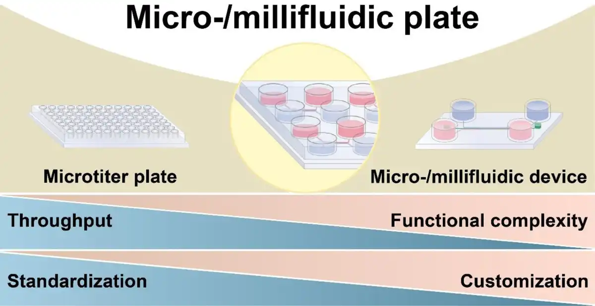
Chip design based on microtiter plate format represents a promising strategy to promote standardization and enhance the throughput of current micro-/millifluidic devices (Figure 1).[19–20–21] The platform that integrates micro-/millifluidic technology into a standard microtiter plate is defined as a micro-/millifluidic plate. Specifically, a microfluidic plate features micrometer-scale channels and handles fluid volumes in the nanoliter to microliter range, enabling precise fluid control, miniaturization, and integration of complex biological functions, making it well-suited for point-of-care testing (POCT). In contrast, a millifluidic plate has millimeter-scale channels and operates in the microliter to milliliter range, allowing for the culture and manipulation of larger biological models under controlled flow conditions, which is essential for organ-on-a-chip systems. On the one hand, the established international standards facilitate clear communication between microfluidic engineers and the end-users, enabling these inexperienced users to operate micro-/millifluidic plates directly without the need to be proficient in understanding the mechanisms behind the platform. Users are therefore allowed to conduct experiments using this emerging platform while maintaining their conventional practices. On the other hand, micro-/millifluidic plates inherit the benefits of microtiter plates in high-throughput testing and have a considerable capacity to fulfill the demands for the detection and analysis of multiple samples. Since the development of the microfluidic capillary electrophoresis array based on standardized microtiter plate footprint,[22,23] micro-/millifluidic plates have gained increasing attention and sustained fast development tendency in research laboratories (Table 2). Meanwhile, numerous start-up companies originating from academic laboratories are working to accelerate the translation of this proof-of-concept into practical applications, aiming to extend the influence of microfluidic technology within the biomedical field.
Table 2
Comparison of micro-/millifluidic plates, micro-/millifluidic devices, and microtiter plates.
| Feature | Micro-/millifluidic plates | Micro-/millifluidic devices | Microtiter plates |
|---|---|---|---|
| Definition | Hybrid platform combining micro-/millifluidic channels with the standardized format of microtiter plates | Miniaturized devices with precisely engineered channels for fluid manipulation | Standard well plates (eg, 48-, 96-, 384-well) used for liquid-based assays |
| Standardization | Partially standardized (ANSI/SLAS, ISO, and ASTM standards applied but still evolving) | Highly customized, lacks universal standards | Fully standardized with well-established formats |
| Throughput | High throughput, compatible with automation and parallel experiments | Low-to-medium throughput | High throughput, optimized for large-scale screening |
| Functional complexity | - Integrates microfluidic functionalities with high-throughput - Suitable for complex assays like cell culture, drug screening, and diagnostics - Supports automation and real-time monitoring | - Provides precise fluid control and microenvironment management - Suitable for organ-on-chip and lab-on-a-chip applications - Complex designs enabling multistep assays | - Basic fluid handling for static assays - Primarily used in drug screening, ELISA, and biochemical tests - Limited control over fluid dynamics and real-time monitoring |
| Customization | Highly customizable—designs can be tailored for specific applications | Highly customizable—chip designs can be tailored for specific applications | Minimal customization—fixed well sizes and layouts |
| Advantages | - Flexible for a wide range of applications - Compatible with high-throughput automation systems - Well-defined plate format for easy integration | - Highly customizable for specialized applications - Ideal for complex, multistep assays - Excellent fluidic control for precise experiments | - Economical and mass-produced - Universal compatibility with many laboratory instruments - Well-established for high-throughput screening |
| Disadvantages | - Partial standardization limits adoption - Scalability challenges in mass production | - Lack of standardization hinders broader adoption - Limited throughput - Requires sophisticated design and fabrication techniques | - Limited fluid control - Inflexible design - Not suitable for complex assays or real-time analysis |
Figure 1.
The micro-/millifluidic plate combines the advantages of microtiter plates and micro-/millifluidic devices, achieving an optimal balance between functional complexity, throughput, customization, and standardization. Specifically, the micro-/millifluidic plate integrates the high throughput and standardization of microtiter plates with the superior customization and functional complexity of micro-/millifluidic devices. This emerging platform balances scalability, adaptability, and functionality, making it suitable for diverse applications in biomedical research.
The emerging micro-/millifluidic plate holds significant potential to revolutionize the use of traditional micro-/millifluidic chips in biomedical research, thereby building a substantial user base. However, an in-depth summary and analysis of this innovative micro-/millifluidic platform have been lacking. In this review, we aim to present a timely and insightful overview of micro-/millifluidic plates from laboratory to market. We summarize the design strategies, liquid-handling systems, applications, and commercialization efforts associated with micro-/millifluidic plates. Additionally, we discuss the challenges facing this technology as well as the positive trends that are driving its development. We believe that our unique perspective on micro-/millifluidic plates can foster innovation and accelerate the translation of academic research by encouraging the microfluidic community to establish a consistent chip development roadmap that aligns with end-user expectations in the biomedical field.
2. Design strategies for micro-/millifluidic plate
In the design of micro-/millifluidic plates, one of the primary objectives is to unify the standards of the platform that further provide simple, reliable, and validated testing protocols for biomedical research. Nonspecialist end-users merely need to follow the standard operating procedure to accomplish the device operation and data acquisition. Another key objective is to preserve innovative concepts in chip design to cater to the diverse demands of biomedical applications. Importantly, even though microfluidic engineers must abide by the microtiter plate footprint during the chip design, their creative ideas can still be transformed into specific micro-/millifluidic plates rather than being stifled. To date, 2 mainstream design strategies have been demonstrated according to the microwells are treated as individual or integrated units (Figure 2).
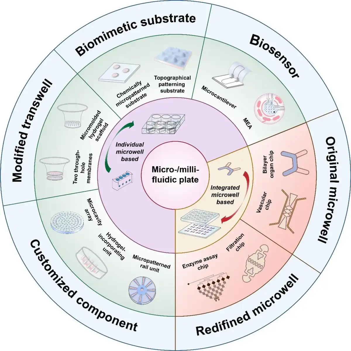
Figure 2.
Two mainstream strategies for micro-/millifluidic plate design according to microwells are regarded as individual or integrated units. Specifically, in micro-/millifluidic plates where microwells are treated as individual units, the microwells serve as containers that provide space for integrating functional inserts, including customized components, modified transwells, biomimetic substrates, and biosensors. In micro-/millifluidic plates where microwells are defined as integrated units, the microwells are regularly connected by microchannels, forming multiple parallel microfluidic chips, and the final micro-/millifluidic plates. In this type of micro-/millifluidic plate, the original microwell format can be preserved or redefined.
2.1 Micro-/millifluidic plate design based on individual microwell
In micro-/millifluidic plates where microwells are treated as individual units, the microwells serve as containers that provide space for integrating functional inserts, including customized components,[24–25–26] modified transwells,[27–28–29] biomimetic substrates,[30–31–32] and biosensors.[33,34] Each microwell is thus endowed with specific functions, allowing it to act as an independent micro-/millifluidic device for various biomedical applications. Typically, customized components divide the microwells into multiple horizontal regions for cell culture, such as left or right regions,[35,36] inner or outer regions,[37,38] and self-defined regions.[39] Different regions are either separated from each other or interconnected by microchannels,[40,41] micropillars,[42] and liquid rails.[43,44] A notable example is the microcavity array, a design that consists of hundreds of independently and regularly arranged microcavities within a single microwell.[45] It provides a promising strategy to overcome the limitations of the conventional organoid culture system based on the Matrigel dome, which lacks robustness in morphogenesis and compatibility with high-content analysis.[46] Both our group and Decembrini et al.[47] employed hydrogel-based microcavities as artificial niches, providing consistent physical and biological microenvironments to direct the generation of the liver,[48,49] retinal,[47] cerebral,[50] and colorectal cancer[51] organoids with uniform sizes and morphologies. In addition to the microcavity array, a 3D-printed inhibiting insert[52] and hanging drop dripper[53–54–55] integrated into the micro-/millifluidic plate have also been demonstrated to improve the cell spheroid and organoid homogeneity by unifying the spatial location and cell concentration. However, both inserts are limited by the throughput capacity predetermined by the microtiter plate format. In contrast to the microcavity array, the component with connected regions aims to construct histotypic and organotypic models with spatially-defined heterocellular architectures such as neurovascular units,[56] neuromuscular junctions,[57] and vascularized tumor tissues.[58,59] In this kind of customized component, heterogeneous cells are precisely positioned in different regions with connecting microstructures promoting cell–cell interactions and guiding some special physiological processes like angiogenesis,[60] axon elongation,[57] and immune cell directional migration.[61]
Transwell is another type of insert that provides 2 distinct vertical regions for cell culture[62] and it can be modified to perform a wider range of tasks in reconstituting the microenvironment in vivo, such as dynamic culture[63,64] and chemical gradient generation.[27,65,66] Compared with customized components, the porous membranes on transwells provide a more adequate exchange of nutrients and gas, minimize hypoxia injury, and improve the maturity of models.[67–68–69–70–71] However, the limited optical transparency of these membranes is a potential stumbling block for in situ observation. Moreover, transwells restrict microwell organization to up-down regions, limiting the potential for more complex cell arrangements.
Unlike customized components and modified transwells, which prefer to compartmentalize the microwells using physical boundaries to achieve the differential arrangement of heterogeneous cells, the micropatterned substrates employ invisible boundaries to construct heterocellular architectures with micron-level resolution analogous to that found in native tissues and organs. Different types of cells are precisely localized using removable micropatterned stencils[72] and further coating the substrates with extracellular matrix to provide selective binding sites for specific cells.[73–74–75] In addition to chemical modification of the substrate to accomplish the heterocellular arrangement, substrates can also be granted topographical cues that are crucial for regulating cell fate.[76–77–78] For instance, integrating biomimetic polydimethylsiloxane (PDMS) substrate that mimicked the curvature of glomerular capillaries improved podocyte differentiation efficiency.[79]
The final category, biosensor, represents a versatile class of inserts capable of monitoring multiple physiologically relevant tissue properties in a real-time, continuous, and noninvasive manner.[80,81] Currently, the main transduction principles applied to micro-/millifluidic plates are based on electrical and mechanical signal propagation.[82–83–84] The microelectrode array (MEA) is a common biosensor that is widely used for recording electrical activities in tissues, such as neural and cardiac tissues.[85–86–87] Traditional MEAs are often based on opaque printed circuit board (PCB) substrates, which pose significant challenges for microscopic observation. To address this challenge, transparent glass or thermoplastic-based substrates are used as alternatives that offer additional assay flexibility, including bright-field imaging and immunofluorescence.[88] However, these MEAs still face other limitations, such as reduced capability to monitor 3D tissues,[89] and the metal electrodes often have short operational lifespans due to saturation or degradation.[81] Except for electrical activity, mechanical forces generated by tissues such as cardiac and skeletal muscle are also essential to their function.[90,91] Mechanical biosensors, such as microcantilevers or microwires, are commonly used in this context. These sensors bend in response to tissue contraction, and the resulting contraction forces can be measured by electronically or optically detecting the deflection of the sensors.[92–93–94]
In general, the advantage of individual microwell-based micro-/millifluidic plates is that they can inherit and even increase the throughput of the original microtiter plate. The functional inserts can be designed independently from the microwells, allowing them to be easily installed in users’ standardized microtiter plates and detached for subsequent analysis. However, the fluid flow, which serves as a key hallmark in microfluidic devices, has not been widely demonstrated in this kind of micro-/millifluidic plate, probably due to lacking sufficient integration space and unified design for the fluid control system. In most cases, the fluid is driven manually, either by connecting to a syringe pump[63] or employing an orbital shaker,[66] which potentially reduces the compatibility and usability of the micro-/millifluidic plate.
2.2 Micro-/millifluidic plate design based on integrated microwell
For micro-/millifluidic plates where microwells are defined as integrated units, the microwells are regularly connected by microchannels, forming multiple parallel microfluidic chips, and the final micro-/millifluidic plates.[95,96] Microwells serve diverse purposes, including liquid handling,[97,98] cell culture,[99] biochemical reaction,[100,101] and sample detection.[102,103] In most cases, the original microwell format is preserved, with microchannels integrated among or within the microwells. For example, a typical micro-/millifluidic plate may feature each chip containing 3 microwells, with up to 128 independent chips formed within a 384-well micro-/millifluidic plate. The microwells at either end function work as inlets and outlets for culture medium perfusion, while the middle microwells serve as the core, embedded with scaffolds designed to mimic native tissues.[104–105–106–107] Specifically, a perfusable tubular scaffold termed “AngioTube” was suspended across the tissue culture microwell and connected to both inlet and outlet.[108] It provides mechanical stability for a vessel lumen and supports the self-assembly of various parenchymal tissues, such as the liver,[109] cardiac,[108] kidney,[110] lung,[111] and solid tumors.[112] The scaffold’s walls featured nanopores and microholes to enhance cell–cell interactions and improve biomolecular exchange between endothelial and parenchymal cells.[113] Moreover, microcantilevers were strategically integrated into the scaffold to noninvasively probe the contraction of the tissues. PREDICT-96 micro-/millifluidic plate is another example that adapts and extends organ-on-a-chip (OoC) systems to a 384-well plate format.[114] Unlike the AngioTube, PREDICT-96 utilizes bilayer chips with 2 overlapping microchannels positioned between 2 sets of inlets and outlets. Different cell types were cultured on opposing sides of a basement membrane, while the micropores on the membrane promoted cell–cell interactions. Furthermore, a recirculatory flow has been demonstrated by employing a programmable pneumatic pump positioned in the plate lid that was fluidically coupled to the micro-/millifluidic plate via a set of vertical tubes that extend into the microwells.[115–116–117]
In addition to retaining the original microwells, they also can be removed or redefined to accommodate specific design requirements for microfluidic chips with well-defined configurations, such as double-T junction,[23] Christmas tree-shaped network,[118] parallel microwell array,[119–120–121] and culture chamber with three assembly ridges.[122] Essentially, this kind of micro-/millifluidic plate can be regarded as an array of conventional microfluidic chips. However, optimizing the number and arrangement of these chips is crucial to ensure compatibility with standard microtiter plates. For instance, in a micro-/millifluidic plate, 96 microwells were replaced by filtration devices of the same number, while each device was positioned at a 45-degree angle to accommodate a long filtration channel. Cell suspensions were loaded into the upper right inlets and driven through the micropillar arrays using air pressure. Cells that were poorly deformable could not pass through the micropillar gaps, resulting in the occlusion of these gaps. The micropillar arrays were strategically placed in the positions of the original microwells, allowing for high-throughput cellular imaging and quantification of the occluded gap percentage.[123]
In summary, this kind of micro-/millifluidic plates possess the ability to perform complex tasks. On the one hand, the available space for chip design extends beyond the microwells to encompass the entire plate, allowing for the integration of more functional structures into the chip. On the other hand, each microwell within the chip serves a unique function, and together, they collaborate to perform complex tasks. Despite these significant advantages, these derivative micro-/millifluidic plates still face challenges in fully achieving both functional complexity and multiplexing simultaneously due to the fixed dimensions of the microwell plate prototype. Depending on the application, an arbitrary number of chips (ranging from 4 to 192) are arranged in parallel on a single micro-/millifluidic plate.[118,124] Looking forward, multilayer structure design may offer a promising solution to balance the throughput and the functional complexity of these micro-/millifluidic plates.[125]
3. Liquid-driven systems for micro-/millifluidic plate
Liquid perfusion is a hallmark of microfluidic chips. In POCT systems, for example, precise fluid control is critical for sample loading, separation, and reaction processes.[126] In OoC systems, medium perfusion mimics the circulatory system, ensuring the transport of nutrients and oxygen while facilitating waste removal. Furthermore, mechanical force, like fluid shear stress, is proved to be a fundamental regulator of cellular behaviors.[127,128] To drive liquid perfusion, a variety of pumps have been adapted for use with micro-/millifluidic plates. The pumps can be categorized into 2 types: active pumps and passive pumps, based on whether they require external power sources to move the liquid (Figure 3 and Table 3).[129]
Table 3
Comparison of active and passive pumps in micro-/millifluidic plates.
| Parameter | Active pump | Passive pump |
|---|---|---|
| Velocity range | 1 nL min−1–1 mL min−1 | 100 nL min−1–100 μL min−1 |
| Sustaining perfusion time | Hours to weeks | Minutes to hours |
| Liquid storage volume | A few milliliters to tens of milliliters | 10 μL–1 mL |
| Potential for external equipment integration | Syringe pump, peristaltic pump, pneumatic pump, vacuum pump | Perfusion rocker |
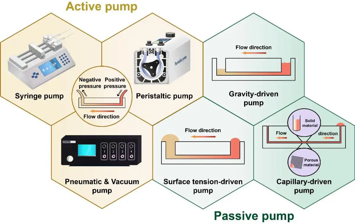
Figure 3.
Two kinds of liquid-driven systems are used for micro-/millifluidic plates including active pumps (syringe pump, peristaltic pump, pneumatic pump, and vacuum pump) and passive pumps (gravity-driven pump, surface tension-driven pump, and capillary-driven pump).
3.1 Active pump
Conventional syringe pumps,[130–131–132–133] peristaltic pumps,[134,135] pneumatic pumps,[136] and vacuum pumps[137,138] are all active pumps with prominent advantages in accurate fluid control in micro-/millifluidic plates.[129,139] These active pumps are also amenable to generating complex flow patterns, such as pulsatile and recirculatory flow.[140,141] For instance, Satoh et al.[142] developed an OoC system using a pneumatic pump to control medium circulation across multiple culture chambers in a unidirectional manner. This medium circulation mechanism relied on the synergistic effects of the pneumatic pump, the elevated inlet, and the Laplace valve. Among them, the pneumatic pump was programmed to periodically apply pressure to tandem culture chambers for medium actuation. The elevated inlet and the Laplace valve were designed to prevent the medium from traveling back to the upstream chamber and stop the introduction of the gas into the microchannel, respectively. Although active pumps are still used as mainstream liquid-driven systems in micro-/millifluidic plates, these pumps are bulky and difficult to massively scale up.[143] Moreover, active pumps typically contain multiple auxiliary components, such as power sources, fluid connections, and tubing, which require fluid handling expertise. These cumbersome components may ultimately hinder the widespread adoption of such systems by biomedical end-users.[18] Therefore, enhancing usability should be a key consideration in the design of liquid-driven systems for micro-/millifluidic plates.
3.2 Passive pump
As an alternative to active pumps, passive pumps provide a solution for the limitations mentioned. Based on their advantages, including high level of integration, simplicity of operation, and portability, passive pumps have been widely adopted and applied to a large extent in biomedical applications.[144] In current micro-/millifluidic plates, passive pumping methods such as gravity,[99,145–146–147–148] surface tension,[149,150] and capillary force[151–152–153] have been successfully demonstrated for fluid actuation.
Gravity-driven pumps typically employ reservoirs filled with liquid at different heights to achieve fluid actuation. During the working process, the fluid flows continuously from the higher inlet to the lower outlet until the liquid levels in the reservoirs equalize. A continuous flow can be maintained over long experimental periods by regularly refilling the open inlet or placing the micro-/millifluidic plate on a perfusion rocker to create a hydraulic head difference between the inlet and outlet.[154] However, as the hydraulic head difference decreases, the flow rate gradually diminishes.[129] To maintain a constant flow rate, autonomous droplet dispensers are ingeniously integrated into the inlets to balance the volumes of the replenished and drained fluid.[154,155] The prominent advantages of gravity-driven pumps include simplified system design, the elimination of complex auxiliary components, and improved usability. Additionally, air bubbles are easily removed in micro-/millifluidic plates with open microwell configurations.[156] Despite these benefits, the drawbacks of gravity-driven pumps include a limited range of flow rates and the inability to generate pulsatile flow.
The surface tension effect is another strategy utilized for fluid actuation, which is generated by the internal pressure difference between 2 liquid drops with unequal volume. According to the Young–Laplace equation, smaller liquid drops have higher internal pressure than larger ones, and this pressure difference drives the fluid and dictates the flow direction.[157] Similar to gravity-driven pumps, surface tension-driven pumps require only simple components (an inlet, an outlet, and a connection microchannel) for system operation, rendering them easy to integrate into micro-/millifluidic plates with a high degree of parallelism. Surface tension-driven pumps also function as an open system with standardized interfaces, allowing automatic liquid filling at the inlet via robotic liquid handlers. Surface tension-driven pumps have already been used to support dynamic cell cultures in micro-/millifluidic plates for applications such as drug screening and disease diagnosis.[158–159–160] While they allow for the refilling of liquid to maintain continuous flow, surface tension-driven pumps are not suitable for long-term perfusion culture due to the limited volume of the liquid drops, which is typically in the microliter range.[161,162]
A capillary-driven pump operates on the principle of capillary action that allows fluid to move through narrow channels or porous materials. Based on the substrate material, capillary-driven pumps based on solid material and porous material have been widely used in micro-/millifluidic plates.[163] The capillary-driven pump was initially proposed in the context of a Si microfluidic chip.[164] In microfluidic systems, the spontaneous liquid transport through microchannels is governed by capillary forces arising from the interaction between liquid surface tension and solid interface geometry. Flow cessation occurs upon complete channel filling.[165] To address this limitation, absorbent matrices have been strategically implemented at channel termini to sustain continuous flow cycles. For example, when liquid is introduced into microwells of 96-well ELISA-optimized micro-/millifluidic plates, it propagates through spiral microchannels via capillarity. The terminal absorbent pads, exhibiting enhanced capillary potential compared with the microchannel network, continuously extract liquid from the system. This configuration enables sequential introduction of samples and reagents through the microchannels, thereby completing the analytical workflow.[166] In addition to solid materials, capillary-driven pumps have also been closely associated with the use of porous materials. Paper is a common-used porous material on which hydrophobic boundaries can be created by using techniques such as photolithography[167] and wax printing,[168–169–170] guiding fluid through patterned hydrophilic regions.[171] Except for paper, functionalized powders have also been demonstrated as ideal materials to form porous microzones on hydrophobic polymer plates.[172] The porous capillary-driven pump eliminates the need for narrow channels and prevents bubble entrapment.[165] However, it primarily relies on stochastic capillary flow within pore spaces, indicating that the inherent properties of porous materials, such as the porosity, will determine the fluid behaviors and cannot be regulated once the pumps are fabricated.[163,173]
Simplifying fluid handling challenges in micro-/millifluidic plate design through passive pumping strategies is an inevitable trend that will accelerate adoption. Advances in passive pumps will eliminate the need for auxiliary components and improve the integration of micro-/millifluidic plates. Passive pumps effectively lower the barrier to entry for the end-users, as they only require an automated liquid handler to operate. A prime example is the paper micro-/millifluidic plate used in POCT, where the liquid-driven mechanism is hidden, allowing users to focus solely on interpreting test results.[174,175] Another advantage of passive pump-based micro-/millifluidic plate is that they are easy to integrate, which helps establish standards for plate design, manufacturing, and testing within the industrial supply chain. This ultimately reduces costs and makes micro-/millifluidic plates more accessible.[1,176] While current active pumps offer superior fluid control and the ability to generate complex flow patterns, passive pumps gradually match these capabilities through the introduction of functional microstructures. For instance, continuous unidirectional perfusion with recirculation has been achieved in a gravity-driven flow system by incorporating supporting microchannels and passive valves.[177,178]
4. Applications for micro-/millifluidic plate
The development of standardized and high-throughput microfluidic devices aimed at enhancing the capabilities of end-users in biomedical research has always been a core goal for microfluidic engineers. The emerging micro-/millifluidic plate is highly compatible with these criteria, laying a foundation for its wide adoption throughout the biomedical community. Meanwhile, finding killer applications is equally important to trigger the broad adoption of these micro-/millifluidic plates. By definition, a killer application describes a product that has higher desirable properties than its predecessor and thus becomes indispensable.[179,180] In this perspective, micro-/millifluidic plates used in OoC and POCT may become the killer applications that propel microfluidics into the biomedical field (Figure 4).[18,181,182]
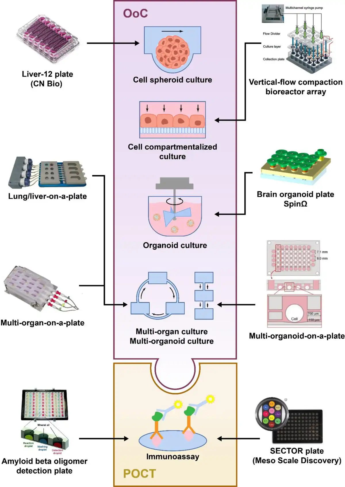
Figure 4.
Typical applications of micro-/millifluidic plates for biomedical research including OoC and POCT.
4.1 OoC
A preclinical study is a necessary stage in the drug development pipeline, where the primary goal is to evaluate the efficacy and safety of drug candidates to eliminate ineffective and highly toxic candidates as soon as possible. To do so, the drug candidates undergo a series of stringent tests in 2D static cell cultures and animal models.[183] However, more than 80% of drug candidates fail to demonstrate efficacy and safety in clinical trials,[184] indicating the current models often cannot faithfully predict human responses to these drug candidates.[185] The limitations of these models have prompted the urgent need to develop models with better predictive ability. To meet this need, OoC systems were hatched through the convergence of microtechnology and biology.[186–187–188] On the one hand, the OoC systems recapitulate key tissue- and organ-level functions by mimicking physiologically relevant microenvironments in microfluidic devices. On the other hand, the species difference can be eliminated by using cells of human origin in OoC systems. OoC is a promising in vitro model bridging the gap between traditional 2D cell cultures and animal models. Despite their promise, most OoC systems currently operate at very low throughput, allowing only a limited number of replicates at a time.[189] Consequently, it is difficult to meet the requirements of practical pharmaceutical applications that need to screen thousands of drug candidates in a short time.[190] Developing novel OoC systems based on microtiter plate format can effectively increase the number of replicates per plate, promote the faster transition from personalized to standardized design, and enhance the repeatability and reproducibility of the system.
Up to now, micro-/millifluidic plates have been utilized in the development of both single-organ and multiorgan systems, depending on the target applications.[191–192–193] Single-organ micro-/millifluidic plates are commonly used to study specific tissue and organ functions without involving the complexity of other organ systems. For example, the liver is the largest internal organ in the human body. It performs various essential biological functions, such as the synthesis of proteins and lipids, secretion of bile, and metabolism of drugs.[194] However, hepatocytes cultured as monolayers typically exhibit nonpolarized and rapidly lose their liver-specific functions.[195] The PhysioMimix micro-/millifluidic plate integrates 12 chips in parallel, enabling the dynamic culture of hepatocytes to maintain their phenotypes and functions by providing continuous oxygen and nutrients as well as biomechanical stimuli through the fluid shear stress.[196] Additionally, by replacing the laminar flow with vertical flow, the high flow resistance of porous membranes will induce pressure inside culture chambers, which exerts compaction forces on hepatocytes. This mechanical compaction simulates the pressure experienced by hepatocytes inside the abdominal cavity. As a result, the hepatocytes exhibit accelerated repolarization, in vivo-like cuboidal morphology, and better liver metabolic and synthetic functions.[197] Moreover, biophysical cues like arterial strain waveform[198] and oxygen tension[199] have also been proven to play an important role in regulating the phenotypes and functions of vascular smooth muscle cells and alveolar basal epithelial cells, respectively. Except for biochemical and biophysical cues, the spatial arrangement of the cellular structure also has a significant impact on regulating the microenvironment of the embedded cells.[200,201] By converging OoC and dielectrophoretic bioassembly, cells can be spatially arranged to form liver lobules and then transferred and cultured in 96-well micro-/millifluidic plates.[202] Similarly, advanced extrusion-based bioprinting[203,204] and acoustic bioassembly[205–206–207] techniques have also been demonstrated to construct functional tissues with predetermined cellular structures in micro-/millifluidic plates, such as cerebral cortex, liver lobule, cardiac, and tumor.
Different from single-organ micro-/millifluidic plates, multiorgan micro-/millifluidic plates are suitable for investigating the interactions between different tissues and organs.[208] To investigate the potential toxicity linked to metabolism by the liver, at least one other OoC is connected to the liver-on-a-chip. For example, a lung–liver micro-/millifluidic plate has been developed to assess both acute and chronic toxicity of pathogens, where lung tissues are cocultured with liver tissues. The micro-/millifluidic plate had 4 multiorgan chips, and each chip comprised 2 adjacent compartments to house the lung and liver tissues. Peristaltic pumps were employed to drive the culture medium in a circulatory manner. The liver’s detoxification ability was validated using aflatoxin B1 (AFB1), demonstrating that cocultured liver tissues reduced the AFB1 toxicity in lung tissues, indicating liver-mediated detoxification decreased lung tissue cytotoxicity[209] In addition to toxicity screening, liver-target organ micro-/millifluidic plate also opens possibilities for testing prodrugs that only become biologically active after liver metabolism. The bioactivation process for prodrugs named capecitabine and cyclophosphamide targeting colorectal cancer has been recapitulated in liver-colorectal cancer micro-/millifluidic plates.[41,110] In these 2 micro-/millifluidic plates, capecitabine and cyclophosphamide were transformed into metabolites 5-fluorouracil and 4-hydroxycyclophosphamide respectively, and then acted on colorectal cancer located at the compartments downstream.
Different from OoC, organoid follows intrinsic developmental programs and forms by self-organization and differentiation of stem cells.[210–211–212] Organoids represent a complementary yet distinct approach to achieving the same goal as OoCs: replicating key aspects of native organ structures and functions in vitro.[213–214–215] The convergence of OoC in organoid is leading to technological advances that allow proper biochemical and biophysical microenvironments and consequently overcome the limitations of this nascent technology such as lack of reproducibility, significant heterogeneity, and limited level of maturity and function.[216,217] As a striking example, 3D-printed miniaturized spinning bioreactors, called SpinΩ, were developed to fit standard 12-well microtiter plates and used for generating forebrain organoids with high reproducibility as well as minimized heterogeneity and variability.[218] Above the cover, a single electric motor drove 12 spinning shafts through a set of interconnected gears. The parameters of spinning shafts and leaves were systematically optimized to dynamically culture organoids in the suspension medium while preventing their aggregation at the center of each microwell. The forebrain organoids recapitulated critical features of human cortical development, including progenitor zone organization, neurogenesis, and especially a distinct human-specific outer radial glia cell layer. These organoids could also present pathologic features of microcephaly when infected with Zika virus. Together, SpinΩ provided a versatile system for modeling human brain development and disease and for drug screening. Furthermore, OoC also provides opportunities to mimic biological interactions at higher levels of organization, which remains a critical challenge in current organoids. In practice, liver, intestinal, and stomach organoids were cultured in 3 adjacent compartments connected via microchannels. Gravity-driven flow was generated by employing a perfusion rocker and further allows communication between different organoids. Bile acid homeostasis regulated by the interactions between liver organoid and intestine organoid is proved in such a multiorganoid micro-/millifluidic plate.[219]
4.2 POCT
POCT plays a vital role in the improvement of clinical outcomes in medical healthcare management.[220] In 2022, the global market of POCT has reached US$43.2 billion, with projections to grow to US$72.0 billion by 2027. Lab-on-a-chip (LOC) is considered one of the important technological drivers to transform the POCT industry. Especially the micro-/millifluidic plate with high throughput and sensitivity is now paving the way for the next generation of POCT platforms.[221] For example, an ELISA micro-/millifluidic plate based on a 96-well format has been developed for immunological detection of Staphylococcal Enterotoxin B at concentrations as low as 0.1 ng mL−1.[222–223–224] The plate contained 3 layers of functional structures for sample loading, detection, and removal, and thus could provide a sample-to-answer process for immunological assays. Similarly, a droplet-based magnetic bead immunoassay was proposed to simplify washing steps and reduce sample or reagent consumption during the ELISA testing process. The immune reaction occurred within a small droplet in the reaction microwell and the antibody-conjugated magnetic beads were driven and manipulated by an XY-motorized magnet array stage to accomplish the following washing and detection step in the other 2 connected microwells, respectively. By using such a micro-/millifluidic plate, less than 5 pg/mL synthetic amyloid beta oligomers were detected as a model analyte for the early diagnosis of Alzheimer disease.[102] To further increase the detection throughput of the device, Meso Scale Discovery launched a multiplexed electrochemiluminescent micro-/millifluidic plate that enabled to simultaneously measure up to 10 analytes in the same 96-well. The plate also enabled highly sensitive wash-free sandwich immunoassays in complex sample matrices by using SULFO-TAG labels on the antibody, which emitted light when electrochemically stimulated at the carbon electrode surface.[225]
4.3 Other applications
In addition to the mainstream applications aforementioned, some micro-/millifluidic plates are also applied in cell biology for purposes such as cell transfection,[226] cell proliferation,[134] and cell agglomeration.[227] Cell transfection involves the deliberate introduction of exogenous molecules into living eukaryotic cells.[228] Compared with classical chemical transfection strategies, electroporation offers a nontoxic, cost-effective, and efficient alternative. Despite its advantages, traditional electroporation systems are not well-suited for high-throughput experiments due to their cumbersome manual operations. To meet this challenge, a standard 96-well suspended-drop electroporation device was developed in which biomolecules were able to be introduced into difficult-to-transfect cell types, such as primary neurons and differentiated neutrophils.[229] This device contained 2 machined gold-coated copper pieces and each of them had 96 electrodes. Vertical electrode pairs created by these plates function as suspended electroporation chambers, allowing for rapid and high-throughput cell transfection. However, the electroporation parameters for each chamber were not customizable. This issue could be mitigated by integrating a PCB into the suspended-drop electroporation system.[226] The PCB incorporates 8-row electrodes and 12-column electrodes mounted on its top and bottom surfaces, which are connected to 2 electrodes within each electroporation chamber. This modification provides individually addressable capabilities, enabling the transfection of various cell types with a wide range of biomolecules.
5. Commercialization for micro-/millifluidic plate
The degree of innovation and adoptability are both crucial for the commercial success of a technology.[230] Although innovative microfluidics has offered several solutions for biomedical research, few products have achieved widespread adoption and commercial success.[231] One major aspect that hinders the growth of microfluidics is the lack of standards necessary for harmonizing microfluidic engineers and end-users. Micro-/millifluidic plates offer a promising tool to surmount the barriers to commercialization. The chip design based on the microtiter plate format not only follows the original commercial standards but also attracts massive loyal users of this classic device. Moreover, the micro-/millifluidic plates target niche markets where other existing technologies cannot meet the needs of the users and thus create a foundation for successful commercialization.
Numerous companies have now participated in market competitions (Table 4). Among them, MIMETAS, founded in 2013, is one of the representative companies, which occupies the commercial space through its unique technological advantages. MIMETAS successfully raised US$21.0 million in a Series B funding round in 2018. Currently, the company has launched 4 OrganoPlate platforms (OrganoPlate 2‑lane 96,[232–233–234–235] OrganoPlate 3‑lane 40,[236,237] OrganoPlate 3‑lane 64, and OrganoPlate Graft[238]), supporting up to 96 tissue and organ models on a single micro-/millifluidic plate. The standardized microtiter plate format makes the OrganoPlate compatible with robotic liquid handlers and broad imaging and testing equipment. In all types of micro-/millifluidic plates, the phaseguides are the unique core microstructures that enable precise and barrier-free definition of hydrogels and cells in spatial based on meniscus pinning effect,[239,240] and consequently improve the ability of the platform in studying cell–cell interactions,[241] migration,[242,243] invasion,[244] transport,[245] and angiogenesis.[236,246] Moreover, gravity-driven pumps are employed and a commercial perfusion rocker, OrganoFlow, is used together to provide continuous medium flow with minimal settings required. Based on these advantages, the OrganoPlate enables the construction of perfused tubules (eg, intestinal epithelium tube,[247–248–249] renal tubule,[250] and blood vessel[251]) and barrier tissues (eg, blood–brain barrier,[252] glomerular filtration barrier,[253] and blood–retinal barrier[254]) without artificial membranes. These advanced models will further be used in investigating drug adsorption, the loss of barrier function caused by drug toxicity, and the permeability of compounds.
Table 4
Summary of micro-/millifluidic plate start-ups and their core products.
| Company | Selected products | Applications |
|---|---|---|
| MIMETAS | OrganoPlate 2‑lane 96 OrganoPlate 3‑lane 40 OrganoPlate 3‑lane 64 OrganoPlate Graft OrganoReady products (latest trade name) | OoC |
| CN Bio | Liver-12 & -48 plates (MPS-LC12 & MPS-LC48) Barrier Plate (MPS-T12) Dual-Organ Plate (MPS-TL6) | OoC |
| Draper | PREDICT96 | OoC |
| 4Design Biosciences | Vascularized Micro-Organ Platform | OoC |
| CellAsic | CellASIC ONIX Micro-/millifluidic plates | OoC |
| AMSBIO | Reinnervate Perfusion Plate | OoC |
| Alvetex Scaffold Wellplates | ||
| InSphero | Akura 96 or 384 Plate | OoC |
| 3D Biomatrix | Perfecta3D Hanging Drop Plates | OoC |
| DAXIANG | IBAC (Integrated Biomimetic Array Chip) M IBAC (Integrated Biomimetic Array Chip) O IBAC (Integrated Biomimetic Array Chip) S1 | OoC |
| CytoNiche | 3D FloTrix microSPIN | OoC |
| Neurosetta | RosetteArray | OoC |
| MyCartis | MyCartis Evalution | POCT |
| QuantaMatrix | QMAC-dRAST Panel QMAC-DST PLUS Panel | POCT |
| Meso Scale Discovery | SECTOR Plates | POCT |
| QuickPlex Plates | ||
| Microfluidic ChipShop | Fluidic 102 Fluidic 600 Fluidic 627 | Cell-based assays, hybridization assays, chemical synthesis |
| Unchained Labs | Lunatic Stunner | Protein and nucleic acid quantification, gene therapy |
| Anatrace | High-Throughput Crystal Former Plate (CF-HT2) | Crystal growth |
| m2p-labs | Microfluidic FlowerPlate Microfluidic Round Well Plate | Synthetic biology, clone screening, media optimization, bioprocess development, anaerobic fermentations |
Except for MIMETAS, companies like 4Design Biosciences, Draper, and CN Bio have also developed their micro-/millifluidic plates with prominent advantages. The 4Design Biosciences Vascularized Micro-Organ Platform supports 16 parallel chips with 3 tissue models in series as replicates. The chip allows clear localization and visualization of cell interactions, such as the vascularization of tumors and the perfusion of the medium through the microvascular network.[255,256] Draper’s PREDICT96 employs 192 integrated pumps that allow for exquisite control of upper and lower microchannel flow conditions for each organ model. Sensors and probes in the plates enable real-time monitoring of tissue functions and evaluating the response to drug exposures.[257,258] PhysioMimix multichip plates developed by CN Bio support up to 48 tissue scaffolds as single-tissue replicates[259] or interconnected in multitissue circuits as demonstrated with tissue scaffolds and Transwell inserts.[260] These micro-/millifluidic plates are versatile platforms with immense potential to enhance the capability in disease modeling, drug safety evaluation, and pharmacokinetics study. Especially a lung-on-a-chip based on PREDICT96 has been successfully demonstrated in investigating severe acute respiratory syndrome coronavirus 2 (SARS-CoV-2) infection and replication, and further evaluating the antiviral efficacy of 3 drugs named Paxlovid, Molnupiravir, and Remdesivir in COVID-19 treatment.[261,262]
6. Challenges and perspectives
According to Emergen Research, the global microfluidics market reached US$20.14 billion in 2021 and is projected to grow at a compound annual growth rate of 16.1% through 2030. By 2030, the market size is expected to reach US$77.28 billion, with the biomedical field being a key driver of this growth. Micro-/millifluidic plates are emerging as powerful tools for biomedical research, combining the high throughput and standardized format of microtiter plates with the advanced functionalities of micro-/millifluidic devices. However, several challenges need to be addressed to fully unlock their potential and facilitate broader adoption.
6.1 Standardization of micro-/millifluidic plates
Existing standards, such as ANSI/SLAS, ISO, and ASTM standards, are loosely applied to micro-/millifluidic plates. These standards primarily address plate designs and general laboratory practices but do not fully address the unique challenges of micro-/millifluidic systems, such as fluid dynamics, surface chemistry, and integration with external systems. As a result, micro-/millifluidic plates are still in the early stages of standardization. Future efforts should focus on refining existing standards and developing new, comprehensive guidelines that cover the entire lifecycle of micro-/millifluidic plates.[263] Establishing consistent performance evaluation criteria and quality control standards will be crucial to ensure the reproducibility, reliability, and scalability of microfluidic systems. A collaborative effort between researchers, industry leaders, and standardization bodies is necessary to address these needs.
6.2 Materials and manufacturing strategies
The materials and manufacturing strategies for micro-/millifluidic plates have not been standardized. During the research phase, efficiency and ease of fabrication are prioritized to facilitate rapid prototyping and experimentation. However, transitioning to large-scale production requires considerations such as market demand, cost efficiency, and manufacturing scalability. These differing priorities often result in discrepancies in material selection and fabrication techniques, making it challenging to bridge the gap between laboratory development and commercial viability.
Silicone rubber, particularly PDMS, is widely used in the fabrication of micro- and millifluidic plates due to its optical transparency, gas permeability, and biocompatibility[264]. However, the tendency of PDMS to absorb small molecules can affect the temporal and spatial concentrations of chemicals, compromising the reliability of experimental results, particularly in clinical diagnosis and drug testing.[265–266–267] Furthermore, the existing fabrication techniques for PDMS-based plates are unsuitable for large-scale production due to challenges in scaling-up manufacturing process.[143]
Thermoplastics like polystyrene (PS) and cyclic olefin copolymer are promising alternatives for both laboratory experiments and commercial production. Fabrication techniques such as hot embossing,[268] laser engraving,[269] and micromilling[270] can optimize microstructures during the laboratory stage. Once the final design is determined, injection molding can enable rapid prototyping for industrial-scale production.[271] However, injection molding is unsuitable for plates with complex microstructures or functional features due to limitations in material flexibility and filling narrow, deep cavities.[272] Advances in 3D printing technology offer solutions to these challenges by enabling the rapid and cost-effective fabrication of intricate structures, enhancing design flexibility, and accelerating prototyping.[273,274] Future innovations in high-resolution printing and multimaterial integration will be key to bridging the gap between research prototypes and commercial products.
6.3 User accessibility and integration
The interaction between end-users and micro-/millifluidic platforms is critical for their adoption.[275] Many current plates rely on multiple external devices, such as liquid-driven pumps, microscopes, and computers, which require professional skills and experience to operate. This reliance complicates automation and scalability.[276] Future efforts should focus on integrating micro-/millifluidic plates with automated experimental platforms and intelligent analysis systems. By incorporating advanced sensors and real-time data analysis, these systems can continuously acquire, process, and interpret complex data, enabling intelligent adjustments during experiments without human intervention.[277,278] Artificial intelligence (AI) can optimize data collection, analysis, and decision-making, significantly enhancing automation, accelerating workflows, and improving accuracy.[279–280–281–282] For instance, Tebon et al. and Al Shihabi et al.[283,284] developed a high-throughput drug screening platform combining bioprinting, high-speed live cell interferometry (HSLCI) imaging, and machine learning. This platform continuously monitors bioprinted tumor organoids via HSLCI imaging, while AI-driven segmentation and tracking enable precise, label-free mass measurements and automated analysis.[283,284] Future AI-driven micro-/millifluidic plates will benefit from more intuitive tools and standardized algorithm selection guidelines. Advances in explainable AI and automated model training will further enhance AI’s reliability and accelerate its adoption in high-throughput screening and precision analysis.[285] Additionally, replacing multiple external devices with smaller, integrated components within the micro-/millifluidic platform will streamline workflows, reduce the need for specialized skills, and make the technology more scalable and accessible.
6.4 Future directions
Significant progress remains to be made before micro-/millifluidic plates can be fully integrated into the biomedical research pipeline. Microfluidic engineers have already made strides in design strategies, materials, fabrication techniques, and liquid-driven systems, which are essential for expanding the adoption of this technology. These efforts aim to build a broad user base and facilitate academic advancements. Moreover, micro-/millifluidic plates have advanced beyond the proof-of-concept stage and are now positioned to demonstrate their value in key biomedical applications, such as drug development and clinical diagnostics.
Conflicts of interests
The authors declare that they have no conflicts of interest.
Funding
This study was funded the National Natural Science Foundation of China (Grant No. 82272173).
Author contributions
Longjun Gu: Conceptualization, writing—original draft, writing—review & editing, visualization. Peidi Liu: Visualization, writing—review & editing. Wen Zhao and Yuhang Fan: Writing—review & editing. Yuwen Wang: Visualization. Pu Chen: Conceptualization, funding acquisition, supervision, writing—review & editing.
References
- [1] Reyes DR, van Heeren H, Guha S, et al. Accelerating innovation and commercialization through standardization of microfluidic-based medical devices. Lab Chip. 2021;21(1):9–21.
- [2] Parrish J, Lim K, Zhang B, et al. New frontiers for biofabrication and bioreactor design in microphysiological system development. Trends Biotechnol. 2019;37(12):1327–1343.
- [3] Wu J, Kumar-Kanojia A, Hombach-Klonisch S, et al. A radial microfluidic platform for higher throughput chemotaxis studies with individual gradient control. Lab Chip. 2018;18(24):3855–3864.
- [4] ANSI_SLAS_1-2004_FootprintDimensions. American National Standards Institute & Society for Laboratory Automation and Screening; 2004.
- [5] ANSI_SLAS_2-2004_HeightDimensions. American National Standards Institute & Society for Laboratory Automation and Screening; 2004.
- [6] ANSI_SLAS_3-2004_BottomOutsideFlangeDimensions. American National Standards Institute & Society for Laboratory Automation and Screening; 2004.
- [7] ANSI_SLAS_4-2004_WellPositions. American National Standards Institute & Society for Laboratory Automation and Screening; 2004.
- [8] ASNI_SLAS_6-2012_WellBottomElevation. American National Standards Institute & Society for Laboratory Automation and Screening; 2012.
- [9] ISO-7581. International Organization for Standardization; 2023.
- [10] ISO-10993-5-2009. International Organization for Standardization; 2009.
- [11] ASTM D6400-23. American Society for Testing and Materials; 2023.
- [12] USP 1031. United States Pharmacopeia; 1985.
- [13] Storck J, Del Razek A, Zimmermann ER. Effect of polyvinyl chloride plastic on the growth and physiology of human umbilical vein endothelial cells. Biomaterials. 1996;17(18):1791–1794.
- [14] Skehan P, Storeng R, Scudiero D, et al. New colorimetric cytotoxicity assay for anticancer-drug screening. J Natl Cancer Inst. 1990;82(13):1107–1112.
- [15] Sarkkinen HK, Halonen PE, Arstila PP, et al. Detection of respiratory syncytial, parainfluenza type 2, and adenovirus antigens by radioimmunoassay and enzyme immunoassay on nasopharyngeal specimens from children with acute respiratory disease. J Clin Microbiol. 1981;13(2):258–265.
- [16] Sapsford KE, Francis J, Sun S, et al. Miniaturized 96-well ELISA chips for Staphylococcal Enterotoxin B detection using portable colorimetric detector. Anal Bioanal Chem. 2009;394(2):499–505.
- [17] Corman VM, Landt O, Kaiser M, et al. Detection of 2019 novel coronavirus (2019-nCoV) by real-time RT-PCR. Euro Surveill. 2020;25(3):2000045.
- [18] Ching T, Toh Y-C, Hashimoto M, et al. Bridging the academia-to-industry gap: organ-on-a-chip platforms for safety and toxicology assessment. Trends Pharmacol Sci. 2021;42(9):715–728.
- [19] Becker H. One size fits all? Lab Chip. 2010;10(15):1894–1897.
- [20] van Heeren H. Standards for connecting microfluidic devices? Lab Chip. 2012;12(6):1022–1025.
- [21] Mastrangeli M, van den Eijnden-van Raaij J. Organs-on-chip: the way forward. Stem Cell Rep. 2021;16(9):2037–2043.
- [22] Gerlach A, Knebel G, Guber AE, et al. Microfabrication of single-use plastic microfluidic devices for high-throughput screening and DNA analysis. Microsyst Technol. 2002;7(5-6):265–268.
- [23] Guber AE, Heckele M, Herrmann D, et al. Microfluidic lab-on-a-chip systems based on polymers - fabrication and application. Chem Eng J. 2004;101(1-3):447–453.
- [24] Maher SP, Conway AJ, Roth A, et al. An adaptable soft-mold embossing process for fabricating optically-accessible, microfeature-based culture systems and application toward liver stage antimalarial compound testing. Lab Chip. 2020;20(6):1124–1139.
- [25] Irimia D, Toner M. Spontaneous migration of cancer cells under conditions of mechanical confinement. Integr Biol (Camb). 2009;1(8-9):506–512.
- [26] Park SE, Kang S, Paek J, et al. Geometric engineering of organoid culture for enhanced organogenesis in a dish. Nat Methods. 2022;19(11):1449–1460.
- [27] Sip CG, Bhattacharjee N, Folch A. Microfluidic transwell inserts for generation of tissue culture-friendly gradients in well plates. Lab Chip. 2014;14(2):302–314.
- [28] Bang S, Tahk D, Choi YH, et al. 3D microphysiological system‐inspired scalable vascularized tissue constructs for regenerative medicine. Adv Funct Mater. 2021;32(1):2105475.
- [29] Liu N, Zhu Y, Yu K, et al. Functional blood-brain barrier model with tight connected minitissue by liquid substrates culture. Adv Healthc Mater. 2022;12(4):e2201984.
- [30] Hortobagyi GN. Simple and rapid process for single cell micro-patterning. Lab Chip. 2009;20(2):123.
- [31] Xu X, Jiang S, Gu L, et al. High-throughput bioengineering of homogenous and functional human-induced pluripotent stem cells-derived liver organoids via micropatterning technique. Front Bioeng Biotechnol. 2022;10:937595.
- [32] Vesga-Castro C, Mosqueira-Martín L, Ubiria-Urkola P, et al. Development of an in vitro platform for the analysis of contractile and calcium dynamics in single human myotubes. Lab Chip. 2024;24(20):4741–4754.
- [33] Eaton M, Que Z, Zhang J, et al. Multi-electrode array of sensory neurons as an in vitro platform to identify the nociceptive response to pharmaceutical buffer systems of injectable biologics. Pharm Res. 2021;38(7):1179–1186.
- [34] Segeritz P, Kolesnik K, Scott DJ, et al. Quantitative mechanical stimulation of GPR68 using a novel 96 well flow plugin. Lab Chip. 2024;24(6):1616–1625.
- [35] Lee Y, Choi JW, Yu J, et al. Microfluidics within a well: an injection-molded plastic array 3D culture platform. Lab Chip. 2018;18(16):2433–2440.
- [36] Kim J, Song Y, Jolly AL, et al. High‐throughput microfluidic 3D outer blood‐retinal barrier model in a 96‐well format: analysis of cellular interactions and barrier function in retinal health and disease. Adv Mater Technol. 2024;9(22):2400634.
- [37] Ko J, Lee Y, Lee S, et al. Human ocular angiogenesis-inspired vascular models on an injection-molded microfluidic chip. Adv Healthc Mater. 2019;8(15):e1900328.
- [38] Kim MK, Park J, Tak S, et al. A long-term storable gel-laden chip composite built in a multi-well plate enabling in situ cell encapsulation for high-throughput liver model. Biofabrication. 2024;16(2):025020.
- [39] Alvarez-Garcia YR, Ramos-Cruz KP, Agostini-Infanzón RJ, et al. Open multi-culture platform for simple and flexible study of multi-cell type interactions. Lab Chip. 2018;18(20):3184–3195.
- [40] Park J, Koito H, Li J, et al. Microfluidic compartmentalized co-culture platform for CNS axon myelination research. Biomed Microdevices. 2009;11(6):1145–1153.
- [41] Ma WY, Hsiung L-C, Wang C-H, et al. A novel 96well-formatted micro-gap plate enabling drug response profiling on primary tumour samples. Sci Rep. 2015;5:9656.
- [42] Lee SR, Hyung S, Bang S, et al. Modeling neural circuit, blood-brain barrier, and myelination on a microfluidic 96 well plate. Biofabrication. 2019;11(3):035013.
- [43] Berry SB, Zhang T, Day JH, et al. Upgrading well plates using open microfluidic patterning. Lab Chip. 2017;17(24):4253–4264.
- [44] Shin N, Kim Y, Ko J, et al. Vascularization of iNSC spheroid in a 3D spheroid‐on‐a‐chip platform enhances neural maturation. Biotechnol Bioeng. 2021;119(2):566–574.
- [45] Vrij EJ, Espinoza S, Heilig M, et al. 3D high throughput screening and profiling of embryoid bodies in thermoformed microwell plates. Lab Chip. 2016;16(4):734–742.
- [46] Sato T, Vries RG, Snippert HJ, et al. Single Lgr5 stem cells build crypt-villus structures in vitro without a mesenchymal niche. Nature. 2009;459(7244):262–265.
- [47] Decembrini S, Hoehnel S, Brandenberg N, et al. Hydrogel-based milliwell arrays for standardized and scalable retinal organoid cultures. Sci Rep. 2020;10(1):10275.
- [48] Jiang S, Xu F, Jin M, et al. Development of a high-throughput micropatterned agarose scaffold for consistent and reproducible hPSC-derived liver organoids. Biofabrication. 2022;15(1):015006.
- [49] Fan H, Shang J, Li J, et al. High-throughput formation of pre-vascularized hiPSC-derived hepatobiliary organoids on a chip via nonparenchymal cell grafting. Adv Sci. 2025;12(8):e2407945.
- [50] Acharya P, Joshi P, Shrestha S, et al. Uniform cerebral organoid culture on a pillar plate by simple and reproducible spheroid transfer from an ultralow attachment well plate. Biofabrication. 2024;16(2):025005.
- [51] Brandenberg N, Hoehnel S, Kuttler F, et al. High-throughput automated organoid culture via stem-cell aggregation in microcavity arrays. Nat Biomed Eng. 2020;4(9):863–874.
- [52] Oksdath Mansilla M, Salazar-Hernandez C, Perrin SL, et al. 3D-printed microplate inserts for long term high-resolution imaging of live brain organoids. BMC Biomed Eng. 2021;3(1):6.
- [53] Zhao L, Xiu J, Liu Y, et al. A 3D printed hanging drop dripper for tumor spheroids analysis without recovery. Sci Rep. 2019;9(1):19717.
- [54] Marimuthu M, Rousset N, St-Georges-Robillard A, et al. Multi-size spheroid formation using microfluidic funnels. Lab Chip. 2018;18(2):304–314.
- [55] Frey O, Misun PM, Fluri DA, et al. Reconfigurable microfluidic hanging drop network for multi-tissue interaction and analysis. Nat Commun. 2014;5:4250.
- [56] Yu YJ, Kim YH, Na K, et al. Hydrogel-incorporating unit in a well: 3D cell culture for high-throughput analysis. Lab Chip. 2018;18(17):2604–2613.
- [57] Yamamoto K, Yamaoka N, Imaizumi Y, et al. Development of a human neuromuscular tissue-on-a-chip model on a 24-well-plate-format compartmentalized microfluidic device. Lab Chip. 2021;21(10):1897–1907.
- [58] Ko J, Ahn J, Kim S, et al. Tumor spheroid-on-a-chip: a standardized microfluidic culture platform for investigating tumor angiogenesis. Lab Chip. 2019;19(17):2822–2833.
- [59] Lee S, Lim J, Yu J, et al. Engineering tumor vasculature on an injection-molded plastic array 3D culture (IMPACT) platform. Lab Chip. 2019;19(12):2071–2080.
- [60] Yu J, Lee S, Song J, et al. Perfusable micro-vascularized 3D tissue array for high-throughput vascular phenotypic screening. Nano Converg. 2022;9(1):16.
- [61] Park D, Son K, Hwang Y, et al. High-throughput microfluidic 3D cytotoxicity assay for cancer immunotherapy (CACI-IMPACT Platform). Front Immunol. 2019;10:1133.
- [62] Lee S, Kim S, Ahn J, et al. Membrane-bottomed microwell array added to transwell insert to facilitate non-contact co-culture of spermatogonial stem cell and STO feeder cell. Biofabrication. 2020;12(4):045031.
- [63] Rauti R, Ess A, Le Roi B, et al. Transforming a well into a chip: a modular 3D-printed microfluidic chip. APL Bioeng. 2021;5(2):13.
- [64] Schimek K, Hsu H-H, Boehme M, et al. Bioengineering of a full-thickness skin equivalent in a 96-well insert format for substance permeation studies and organ-on-a-chip applications. Bioengineering. 2018;5(2):43.
- [65] Wang Y, Kim R, Gunasekara DB, et al. Formation of human colonic crypt array by application of chemical gradients across a shaped epithelial monolayer. Cell Mol Gastroenterol Hepatol. 2018;5(2):113–130.
- [66] Chong HB, Youn J, Shin W, et al. Multiplex recreation of human intestinal morphogenesis on a multi-well insert platform by basolateral convective flow. Lab Chip. 2021;21(17):3316–3327.
- [67] Ao Z, Cai H, Wu Z, et al. Human spinal organoid-on-a-chip to model nociceptive circuitry for pain therapeutics discovery. Anal Chem. 2022;94(2):1365–1372.
- [68] Ao Z, Cai H, Havert DJ, et al. One-stop microfluidic assembly of human brain organoids to model prenatal cannabis exposure. Anal Chem. 2020;92(6):4630–4638.
- [69] Kim D, Lee SJ, Youn J, et al. A deep and permeable nanofibrous oval-shaped microwell array for the stable formation of viable and functional spheroids. Biofabrication. 2021;13(3):035050.
- [70] Udomluck N, Kim S-H, Cho H, et al. Three-dimensional cartilage tissue regeneration system harnessing goblet-shaped microwells containing biocompatible hydrogel. Biofabrication. 2019;12(1):015019.
- [71] Tian C, Cai H, Ao Z, et al. Engineering human midbrain organoid microphysiological systems to model prenatal PFOS exposure. Sci Total Environ. 2024;947:174478.
- [72] Xu J, Wang X, Li X, et al. High-throughput cell migration assay under combinatorial chemical environments by a novel 24-well-plate based device. Biomed Microdevices. 2020;22(2):40.
- [73] March S, Ng S, Velmurugan S, et al. A microscale human liver platform that supports the hepatic stages of Plasmodium falciparum and vivax. Cell Host Microbe. 2013;14(1):104–115.
- [74] Khetani SR, Bhatia SN. Microscale culture of human liver cells for drug development. Nat Biotechnol. 2008;26(1):120–126.
- [75] Berger DR, Ware BR, Davidson MD, et al. Enhancing the functional maturity of induced pluripotent stem cell-derived human hepatocytes by controlled presentation of cell-cell interactions in vitro. Hepatology. 2015;61(4):1370–1381.
- [76] Kolind K, Leong KW, Besenbacher F, et al. Guidance of stem cell fate on 2D patterned surfaces. Biomaterials. 2012;33(28):6626–6633.
- [77] Bao M, Xie J, Huck WTS. Recent advances in engineering the stem cell microniche in 3D. Adv Sci (Weinh). 2018;5(8):1800448.
- [78] Huethorst E, Cutiongco MF, Campbell FA, et al. Customizable, engineered substrates for rapid screening of cellular cues. Biofabrication. 2020;12(2):025009.
- [79] Korolj A, Laschinger C, James C, et al. Curvature facilitates podocyte culture in a biomimetic platform. Lab Chip. 2018;18(20):3112–3128.
- [80] Feiner R, Dvir T. Tissue–electronics interfaces: from implantable devices to engineered tissues. Nat Rev Mater. 2017;3(1):1.
- [81] Kavand H, Nasiri R, Herland A. Advanced materials and sensors for microphysiological systems: focus on electronic and electrooptical interfaces. Adv Mater. 2022;34(17):e2107876.
- [82] Eichler M, Jahnke H-G, Krinke D, et al. A novel 96-well multielectrode array based impedimetric monitoring platform for comparative drug efficacy analysis on 2D and 3D brain tumor cultures. Biosens Bioelectron. 2015;67:582–589.
- [83] Thavandiran N, Hale C, Blit P, et al. Functional arrays of human pluripotent stem cell-derived cardiac microtissues. Sci Rep. 2020;10(1):6919.
- [84] Soucy JR, Bindas AJ, Koppes AN, et al. Instrumented microphysiological systems for real-time measurement and manipulation of cellular electrochemical processes. iScience. 2019;21:521–548.
- [85] Hayes HB, Nicolini AM, Arrowood CA, et al. Novel method for action potential measurements from intact cardiac monolayers with multiwell microelectrode array technology. Sci Rep. 2019;9(1):11893.
- [86] Meng Q, Zhang W, Wang X, et al. Human forebrain organoids reveal connections between valproic acid exposure and autism risk. Transl Psychiatry. 2022;12(1):130.
- [87] Cai H, Ao Z, Tian C, et al. Engineering human spinal microphysiological systems to model opioid-induced tolerance. Bioact Mater. 2023;22:482–490.
- [88] Schmidt S, Frank R, Krinke D, et al. Novel PMMA based 96-well microelectrode arrays for bioelectronic high throughput monitoring of cells in a live mode. Biosens Bioelectron. 2022;202:114012.
- [89] Yang X, Forró C, Li TL, et al. Kirigami electronics for long-term electrophysiological recording of human neural organoids and assembloids. Nat Biotechnol. 2024;42(12):1836–1843.
- [90] Vesga-Castro C, Aldazabal J, Vallejo-Illarramendi A, et al. Contractile force assessment methods for in vitro skeletal muscle tissues. Elife. 2022;11:e77204.
- [91] Lind JU, Yadid M, Perkins I, et al. Cardiac microphysiological devices with flexible thin-film sensors for higher-throughput drug screening. Lab Chip. 2017;17(21):3692–3703.
- [92] Zhao Y, Wang EY, Davenport LH, et al. A multimaterial microphysiological platform enabled by rapid casting of elastic microwires. Adv Healthc Mater. 2019;8(5):e1801187.
- [93] Gracioso Martins AM, Wilkins MD, Ligler FS, et al. Microphysiological system for high-throughput computer vision measurement of microtissue contraction. ACS Sens. 2021;6(3):985–994.
- [94] Wu Q, Xue R, Zhao Y, et al. Automated fabrication of a scalable heart-on-a-chip device by 3D printing of thermoplastic elastomer nanocomposite and hot embossing. Bioact Mater. 2024;33:46–60.
- [95] Kim JY, Fluri DA, Kelm JM, et al. 96-well format-based microfluidic platform for parallel interconnection of multiple multicellular spheroids. J Lab Autom. 2015;20(3):274–282.
- [96] Lockhart EJ, Horowitz LF, Rodríguez A, et al. Drug testing of monodisperse arrays of live microdissected tumors using a valved multiwell microfluidic platform. Lab Chip. 2024;24(10):2683–2699.
- [97] Chen SY, Hung PJ, Lee PJ. Microfluidic array for three-dimensional perfusion culture of human mammary epithelial cells. Biomed Microdevices. 2011;13(4):753–758.
- [98] Falconnet D, She J, Tornay R, et al. Rapid, sensitive and real-time multiplexing platform for the analysis of protein and nucleic-acid biomarkers. Anal Chem. 2015;87(3):1582–1589.
- [99] Lee PJ, Ghorashian N, Gaige TA, et al. Microfluidic system for automated cell-based assays. JALA Charlottesv Va. 2007;12(6):363–367.
- [100] Choi CJ, Cunningham BT. A 96-well microplate incorporating a replica molded microfluidic network integrated with photonic crystal biosensors for high throughput kinetic biomolecular interaction analysis. Lab Chip. 2007;7(5):550–556.
- [101] Park DS, Chen P-C, You BH, et al. Titer plate formatted continuous flow thermal reactors for high throughput applications: fabrication and testing. J Micromech Microeng. 2010;20(5):055003.
- [102] Park MC, Kim M, Lim GT, et al. Droplet-based magnetic bead immunoassay using microchannel-connected multiwell plates (muCHAMPs) for the detection of amyloid beta oligomers. Lab Chip. 2016;16(12):2245–2253.
- [103] Sanjay ST, Dou M, Sun J, et al. A paper/polymer hybrid microfluidic microplate for rapid quantitative detection of multiple disease biomarkers. Sci Rep. 2016;6:30474.
- [104] Lin DSY, Rajasekar S, Marway MK, et al. From model system to therapy: scalable production of perfusable vascularized liver spheroids in “open-top” 384-well plate. ACS Biomater Sci Eng. 2021;7(7):2964–2972.
- [105] Savoji H, Davenport Huyer L, Mohammadi MH, et al. 3D printing of vascular tubes using bioelastomer prepolymers by freeform reversible embedding. ACS Biomater Sci Eng. 2020;6(3):1333–1343.
- [106] Rajasekar S, Lin DSY, Zhang F, et al. Subtractive manufacturing with swelling induced stochastic folding of sacrificial materials for fabricating complex perfusable tissues in multi-well plates. Lab Chip. 2022;22(10):1929–1942.
- [107] Rajasekar S, Lin DSY, Abdul L, et al. IFlowPlate-a customized 384-well plate for the culture of perfusable vascularized colon organoids. Adv Mater. 2020;32(46):e2002974.
- [108] Lai BFL, Lu RXZ, Davenport Huyer L, et al. A well plate-based multiplexed platform for incorporation of organoids into an organ-on-a-chip system with a perfusable vasculature. Nat Protoc. 2021;16(4):2158–2189.
- [109] Lai BF, Huyer LD, Lu RX, et al. InVADE: integrated vasculature for assessing dynamic events. Adv Funct Mater. 2017;27(46):1703524.
- [110] Xie R, Korolj A, Liu C, et al. h-FIBER: microfluidic topographical hollow fiber for studies of glomerular filtration barrier. ACS Cent Sci. 2020;6(6):903–912.
- [111] Lu RXZ, Lai BFL, Rafatian N, et al. Vasculature-on-a-chip platform with innate immunity enables identification of angiopoietin-1 derived peptide as a therapeutic for SARS-CoV-2 induced inflammation. Lab Chip. 2022;22(6):1171–1186.
- [112] Lai Benjamin FL, Lu Rick X, Hu Y, et al. Recapitulating pancreatic tumor microenvironment through synergistic use of patient organoids and organ-on-a-chip vasculature. Adv Funct Mater. 2020;30(48):2000545.
- [113] Liu C, Campbell SB, Li J, et al. High throughput omnidirectional printing of tubular microstructures from elastomeric polymers. Adv Healthc Mater. 2022;11(23):e2201346.
- [114] Gard AL, Luu RJ, Miller CR, et al. High-throughput human primary cell-based airway model for evaluating influenza, coronavirus, or other respiratory viruses in vitro. Sci Rep. 2021;11(1):14961.
- [115] Bale SS, Manoppo A, Thompson R, et al. A thermoplastic microfluidic microphysiological system to recapitulate hepatic function and multicellular interactions. Biotechnol Bioeng. 2019;116(12):3409–3420.
- [116] Azizgolshani H, Coppeta JR, Vedula EM, et al. High-throughput organ-on-chip platform with integrated programmable fluid flow and real-time sensing for complex tissue models in drug development workflows. Lab Chip. 2021;21(8):1454–1474.
- [117] Rogers MT, Gard AL, Gaibler R, et al. A high-throughput microfluidic bilayer co-culture platform to study endothelial-pericyte interactions. Sci Rep. 2021;11(1):12225.
- [118] Kang JH, Park J-K. Development of a microplate reader compatible microfluidic device for enzyme assay. Sens Actuators B. 2005;107(2):980–985.
- [119] Chen Y, Gao D, Liu H, et al. Drug cytotoxicity and signaling pathway analysis with three-dimensional tumor spheroids in a microwell-based microfluidic chip for drug screening. Anal Chim Acta. 2015;898:85–92.
- [120] Lohasz C, Rousset N, Renggli K, et al. Scalable microfluidic platform for flexible configuration of and experiments with microtissue multiorgan models. SLAS Technol. 2019;24(1):79–95.
- [121] Reed-McBain CA, Turaga RV, Zima SRT, et al. Microfluidic device with reconfigurable spatial temporal gradients reveals plastic astrocyte response to stroke and reperfusion. Lab Chip. 2023;23(18):3945–3960.
- [122] Busche M, Tomilova O, Schütte J, et al. HepaChip-MP - a twenty-four chamber microplate for a continuously perfused liver coculture model. Lab Chip. 2020;20(16):2911–2926.
- [123] Gill NK, Ly C, Nyberg KD, et al. A scalable filtration method for high throughput screening based on cell deformability. Lab Chip. 2019;19(2):343–357.
- [124] Khnouf R, Beebe DJ, Fan ZH. Cell-free protein expression in a microchannel array with passive pumping. Lab Chip. 2009;9(1):56–61.
- [125] Parrish J, Lim KS, Baer K, et al. A 96-well microplate bioreactor platform supporting individual dual perfusion and high-throughput assessment of simple or biofabricated 3D tissue models. Lab Chip. 2018;18(18):2757–2775.
- [126] Wang P, Yuan S, Yang N, et al. A comprehensive review on non-active micro-pumps for microfluidic platforms. J Micromech Microeng. 2021;31(9):093001.
- [127] Panciera T, Azzolin L, Cordenonsi M, et al. Mechanobiology of YAP and TAZ in physiology and disease. Nat Rev Mol Cell Biol. 2017;18(12):758–770.
- [128] Vining KH, Mooney DJ. Mechanical forces direct stem cell behaviour in development and regeneration. Nat Rev Mol Cell Biol. 2017;18(12):728–742.
- [129] Byun CK, Abi-Samra K, Cho Y-K, et al. Pumps for microfluidic cell culture. Electrophoresis. 2014;35(2-3):245–257.
- [130] Park DS, Hupert ML, Witek MA, et al. A titer plate-based polymer microfluidic platform for high throughput nucleic acid purification. Biomed Microdevices. 2008;10(1):21–33.
- [131] Witek MA, Hupert ML, Park DS-W, et al. 96-well polycarbonate-based microfluidic titer plate for high-throughput purification of DNA and RNA. Anal Chem. 2008;80(9):3483–3491.
- [132] Chen PC, Park DS, You B-H, et al. Titer-plate formatted continuous flow thermal reactors: design and performance of a nanoliter reactor. Sens Actuators B Chem. 2010;149(1):291–300.
- [133] Chang TC, Mikheev AM, Huynh W, et al. Parallel microfluidic chemosensitivity testing on individual slice cultures. Lab Chip. 2014;14(23):4540–4551.
- [134] Lecault V, Vaninsberghe M, Sekulovic S, et al. High-throughput analysis of single hematopoietic stem cell proliferation in microfluidic cell culture arrays. Nat Methods. 2011;8(7):581–586.
- [135] Zhang W, Gu Y, Hao Y, et al. Well plate-based perfusion culture device for tissue and tumor microenvironment replication. Lab Chip. 2015;15(13):2854–2863.
- [136] Tan K, Keegan P, Rogers M, et al. A high-throughput microfluidic microphysiological system (PREDICT-96) to recapitulate hepatocyte function in dynamic, re-circulating flow conditions. Lab Chip. 2019;19(9):1556–1566.
- [137] Huang SB, Wang S-S, Hsieh C-H, et al. An integrated microfluidic cell culture system for high-throughput perfusion three-dimensional cell culture-based assays: effect of cell culture model on the results of chemosensitivity assays. Lab Chip. 2013;13(6):1133–1143.
- [138] Gumuscu B, Albers HJ, van den Berg A, et al. Compartmentalized 3D tissue culture arrays under controlled microfluidic delivery. Sci Rep. 2017;7(1):3381.
- [139] Vunjak-Novakovic G, Ronaldson-Bouchard K, Radisic M. Organs-on-a-chip models for biological research. Cell. 2021;184(18):4597–4611.
- [140] Domansky K, Inman W, Serdy J, et al. Perfused multiwell plate for 3D liver tissue engineering. Lab Chip. 2010;10(1):51–58.
- [141] Coppeta JR, Mescher MJ, Isenberg BC, et al. A portable and reconfigurable multi-organ platform for drug development with onboard microfluidic flow control. Lab Chip. 2016;17(1):134–144.
- [142] Satoh T, Sugiura S, Shin K, et al. A multi-throughput multi-organ-on-a-chip system on a plate formatted pneumatic pressure-driven medium circulation platform. Lab Chip. 2017;18(1):115–125.
- [143] De Stefano P, Bianchi E, Dubini G. The impact of microfluidics in high-throughput drug-screening applications. Biomicrofluidics. 2022;16(3):13.
- [144] Narayanamurthy V, Jeroish ZE, Bhuvaneshwari KS, et al. Advances in passively driven microfluidics and lab-on-chip devices: a comprehensive literature review and patent analysis. RSC Adv. 2020;10(20):11652–11680.
- [145] Kim YS, Asif A, Chethikkattuveli Salih AR, et al. Gravity-based flow efficient perfusion culture system for spheroids mimicking liver inflammation. Biomedicines. 2021;9(10):1369.
- [146] Qin W, Yang Z, Yin J, et al. Effect assessment of aurantio-obtusin on novel human renal glomerular endothelial cells model using a microfluidic chip. Nutrients. 2022;14(21):4615.
- [147] Chen Z, He S, Zilberberg J, et al. Pumpless platform for high-throughput dynamic multicellular culture and chemosensitivity evaluation. Lab Chip. 2019;19(2):254–261.
- [148] Sotra A, Jozani KA, Zhang B. A vascularized crypt-patterned colon model for high-throughput drug screening and disease modelling. Lab Chip. 2023;23(15):3370–3387.
- [149] Echeverria V, Meyvantsson I, Skoien A, et al. An automated high-content assay for tumor cell migration through 3-dimensional matrices. J Biomol Screen. 2010;15(9):1144–1151.
- [150] Montanez-Sauri SI, Sung KE, Puccinelli JP, et al. Automation of three-dimensional cell culture in arrayed microfluidic devices. J Lab Autom. 2011;16(3):171–185.
- [151] Cheng CM, Martinez AW, Gong J, et al. Paper-based ELISA. Angew Chem Int Ed Engl. 2010;49(28):4771–4774.
- [152] Murdock RC, Shen Li, Griffin DK, et al. Optimization of a paper-based ELISA for a human performance biomarker. Anal Chem. 2013;85(23):11634–11642.
- [153] Kozak KR, Wang J, Lye M, et al. Micro-volume wall-less immunoassays using patterned planar plates. Lab Chip. 2013;13(7):1342–1350.
- [154] Kim T, Cho YH. A pumpless cell culture chip with the constant medium perfusion-rate maintained by balanced droplet dispensing. Lab Chip. 2011;11(10):1825–1830.
- [155] Kim T, Doh I, Cho Y-H. A 3D tumor spheroid chip with the pharmacokinetic drug elimination model developed by balanced droplet dispensing. Sens Actuators B. 2012;174:436–440.
- [156] Goral VN, Zhou C, Lai F, et al. A continuous perfusion microplate for cell culture. Lab Chip. 2013;13(6):1039–1043.
- [157] Walker G, Beebe DJ. A passive pumping method for microfluidic devices. Lab Chip. 2002;2(3):131–134.
- [158] Berthier E, Surfus J, Verbsky J, et al. An arrayed high-content chemotaxis assay for patient diagnosis. Integr Biol. 2010;2(11-12):630–638.
- [159] Puccinelli JP, Su X, Beebe DJ. Automated high-throughput microchannel assays for cell biology: operational optimization and characterization. JALA Charlottesv Va. 2010;15(1):25–32.
- [160] Su X, Young EWK, Underkofler HAS, et al. Microfluidic cell culture and its application in high-throughput drug screening: cardiotoxicity assay for hERG channels. J Biomol Screen. 2011;16(1):101–111.
- [161] Meyvantsson I, Warrick JW, Hayes S, et al. Automated cell culture in high density tubeless microfluidic device arrays. Lab Chip. 2008;8(5):717–724.
- [162] Zhang H, Whalley RD, Ferreira AM, et al. High throughput physiological micro-models for in vitro pre-clinical drug testing: a review of engineering systems approaches. Prog Biomed Eng. 2020;2(2):022001.
- [163] Xu LF, Wang A, Li X, et al. Passive micropumping in microfluidics for point-of-care testing. Biomicrofluidics. 2020;14(3):16.
- [164] Juncker D, Schmid H, Drechsler U, et al. Autonomous microfluidic capillary system. Anal Chem. 2002;74(24):6139–6144.
- [165] Park J, Han DH, Park JK. Towards practical sample preparation in point-of-care testing: user-friendly microfluidic devices. Lab Chip. 2020;20(7):1191–1203.
- [166] Kai J, Puntambekar A, Santiago N, et al. A novel microfluidic microplate as the next generation assay platform for enzyme linked immunoassays (ELISA). Lab Chip. 2012;12(21):4257–4262.
- [167] Carrilho E, Phillips ST, Vella SJ, et al. Paper microzone plates. Anal Chem. 2009;81(15):5990–5998.
- [168] Wang S, Ge L, Song X, et al. Paper-based chemiluminescence ELISA: lab-on-paper based on chitosan modified paper device and wax-screen-printing. Biosens Bioelectron. 2012;31(1):212–218.
- [169] Wang S, Ge L, Song X, et al. Simple and covalent fabrication of a paper device and its application in sensitive chemiluminescence immunoassay. Analyst. 2012;137(16):3821–3827.
- [170] Rosenfeld T, Bercovici M. Amplification-free detection of DNA in a paper-based microfluidic device using electroosmotically balanced isotachophoresis. Lab Chip. 2018;18(6):861–868.
- [171] Martinez AW, Phillips ST, Butte MJ, et al. Patterned paper as a platform for inexpensive, low-volume, portable bioassays. Angew Chem Int Ed Engl. 2007;46(8):1318–1320.
- [172] Tian J, Li X, Shen W. Printed two-dimensional micro-zone plates for chemical analysis and ELISA. Lab Chip. 2011;11(17):2869–2875.
- [173] Olanrewaju A, Beaugrand M, Yafia M, et al. Capillary microfluidics in microchannels: from microfluidic networks to capillaric circuits. Lab Chip. 2018;18(16):2323–2347.
- [174] Ozer T, McMahon C, Henry CS. Advances in paper-based analytical devices. In: Bohn PW, Pemberton JE, eds. Annual Review Of Analytical Chemistry, Vol 13. Annual Reviews; 2020:85–109.
- [175] Hu J, Wang SQ, Wang L, et al. Advances in paper-based point-of-care diagnostics. Biosens Bioelectron. 2014;54:585–597.
- [176] Battat S, Weitz DA, Whitesides GM. An outlook on microfluidics: the promise and the challenge. Lab Chip. 2022;22(3):530–536.
- [177] Wang YI, Shuler ML. UniChip enables long-term recirculating unidirectional perfusion with gravity-driven flow for microphysiological systems. Lab Chip. 2018;18(17):2563–2574.
- [178] Sung JH, Wang YI, Narasimhan Sriram N, et al. Recent advances in body-on-a-chip systems. Anal Chem. 2019;91(1):330–351.
- [179] Becker H. Hype, hope and hubris: the quest for the killer application in microfluidics. Lab Chip. 2009;9(15):2119–2122.
- [180] Blow N. Microfluidics: in search of a killer application. Nat Methods. 2007;4(8):665–670.
- [181] Cong HJ, Zhang N. Perspectives in translating microfluidic devices from laboratory prototyping into scale-up production. Biomicrofluidics. 2022;16(2):15.
- [182] Teixeira Carvalho DJ, Moroni L, Giselbrecht S. Clamping strategies for organ-on-a-chip devices. Nat Rev Mater. 2023;8:147–164.
- [183] DiMasi JA, Grabowski HG, Hansen RW. Innovation in the pharmaceutical industry: new estimates of R&D costs. J Health Econ. 2016;47:20–33.
- [184] Alteri E, Guizzaro L. Be open about drug failures to speed up research. Nature. 2018;563(7731):317–319.
- [185] Pound P, Ritskes-Hoitinga M. Is it possible to overcome issues of external validity in preclinical animal research? Why most animal models are bound to fail. J Transl Med. 2018;16(1):304.
- [186] Huh D, Matthews BD, Mammoto A, et al. Reconstituting organ-level lung functions on a chip. Science. 2010;328(5986):1662–1668.
- [187] Wu Q, Liu J, Wang X, et al. Organ-on-a-chip: recent breakthroughs and future prospects. Biomed Eng Online. 2020;19(1):9.
- [188] Zhou Y, Qiao H, Xu F, et al. Bioengineering of a human physiologically relevant microfluidic blood-cerebrospinal fluid barrier model. Lab Chip. 2023;23(13):3002–3015.
- [189] Low LA, Mummery C, Berridge BR, et al. Organs-on-chips: into the next decade. Nat Rev Drug Discov. 2021;20(5):345–361.
- [190] Dove A. Drug screening - beyond the bottleneck. Nat Biotechnol. 1999;17(9):859–863.
- [191] Milani N, Parrott N, Ortiz Franyuti D, et al. Application of a gut-liver-on-a-chip device and mechanistic modelling to the quantitative in vitro pharmacokinetic study of mycophenolate mofetil. Lab Chip. 2022;22(15):2853–2868.
- [192] Lozito TP, Alexander PG, Lin H, et al. Three-dimensional osteochondral microtissue to model pathogenesis of osteoarthritis. Stem Cell Res Ther. 2013;4:6.
- [193] Lin H, Lozito TP, Alexander PG, et al. Stem cell-based microphysiological osteochondral system to model tissue response to interleukin-1beta. Mol Pharm. 2014;11(7):2203–2212.
- [194] Agarwal T, Banerjee D, Konwarh R, et al. Recent advances in bioprinting technologies for engineering hepatic tissue. Mater Sci Eng C Mater Biol Appl. 2021;123:112013.
- [195] Treyer A, Musch A. Hepatocyte polarity. Compr Physiol. 2013;3(1):243–287.
- [196] Docci L, Milani N, Ramp T, et al. Exploration and application of a liver-on-a-chip device in combination with modelling and simulation for quantitative drug metabolism studies. Lab Chip. 2022;22(6):1187–1205.
- [197] Zhu L, Xia H, Wang Z, et al. A vertical-flow bioreactor array compacts hepatocytes for enhanced polarity and functions. Lab Chip. 2016;16(20):3898–3908.
- [198] Lee J, Wong M, Smith Q, et al. A novel system for studying mechanical strain waveform-dependent responses in vascular smooth muscle cells. Lab Chip. 2013;13(23):4573–4582.
- [199] Peng CC, Liao W-H, Chen Y-H, et al. A microfluidic cell culture array with various oxygen tensions. Lab Chip. 2013;13(16):3239–3245.
- [200] Ovsianikov A, Khademhosseini A, Mironov V. The synergy of scaffold-based and scaffold-free tissue engineering strategies. Trends Biotechnol. 2018;36(4):348–357.
- [201] Gu L, Jiang S, Xu X, et al. Size- and density-dependent acoustic differential bioassembly of spatially-defined heterocellular architecture. Biofabrication. 2022;15(1):015019.
- [202] Macdonald NP, Menachery A, Reboud J, et al. Creating tissue on chip constructs in microtitre plates for drug discovery. RSC Adv. 2018;8(18):9603–9610.
- [203] Maloney E, Clark C, Sivakumar H, et al. Immersion bioprinting of tumor organoids in multi-well plates for increasing chemotherapy screening throughput. Micromachines (Basel). 2020;11(2):208.
- [204] Hwang HH, You S, Ma X, et al. High throughput direct 3D bioprinting in multiwell plates. Biofabrication. 2021;13(2):025007.
- [205] Ren T, Chen P, Gu L, et al. Soft ring-shaped cellu-robots with simultaneous locomotion in batches. Adv Mater. 2020;32(8):e1905713.
- [206] Wang J, Soto F, Ma P, et al. Acoustic fabrication of living cardiomyocyte-based hybrid biorobots. ACS Nano. 2022;16(7):10219–10230.
- [207] Gu L, Zhao W, Fan Y, et al. Multifrequency control of Faraday wave bioassembly for constructing multiscale hPSC-derived neuronal networks. BioRxiv. 2023:2023.12.01.569533.
- [208] Picollet-D’hahan N, Zuchowska A, Lemeunier I, et al. Multiorgan-on-a-chip: a systemic approach to model and decipher inter-organ communication. Trends Biotechnol. 2021;39(8):788–810.
- [209] Bovard D, Sandoz A, Luettich K, et al. A lung/liver-on-a-chip platform for acute and chronic toxicity studies. Lab Chip. 2018;18(24):3814–3829.
- [210] Gu L, Cai H, Chen L, et al. Functional neural networks in human brain organoids. BME Front. 2024;5:0065.
- [211] Li K, Gu L, Cai H, et al. Human brain organoids for understanding substance use disorders. Drug Metab Pharmacokinet. 2024;58:101031.
- [212] Li K, Gu L, Cai H, Guo F. Organoid computing: leveraging organoid neural networks for artificial intelligence. In: Human Brain Organoids: Scientific and Ethical Considerations. Cham: Springer Nature Switzerland; 2024:165–182.
- [213] Park SE, Georgescu A, Huh D. Organoids-on-a-chip. Science. 2019;364(6444):960–965.
- [214] Lou YR, Leung AW. Next generation organoids for biomedical research and applications. Biotechnol Adv. 2018;36(1):132–149.
- [215] Seiler ST, Mantalas GL, Selberg J, et al. Modular automated microfluidic cell culture platform reduces glycolytic stress in cerebral cortex organoids. Sci Rep. 2022;12(1):20173.
- [216] Hofer M, Lutolf MP. Engineering organoids. Nat Rev Mater. 2021;6(5):402–420.
- [217] Garreta E, Kamm RD, Chuva de Sousa Lopes SM, et al. Rethinking organoid technology through bioengineering. Nat Mater. 2021;20(2):145–155.
- [218] Qian X, Nguyen HN, Song MM, et al. Brain-region-specific organoids using mini-bioreactors for modeling ZIKV exposure. Cell. 2016;165(5):1238–1254.
- [219] Jin Y, Kim J, Lee JS, et al. Vascularized liver organoids generated using induced hepatic tissue and dynamic liver-specific microenvironment as a drug testing platform. Adv Funct Mater. 2018;28(37):1801954.
- [220] Chin CD, Linder V, Sia SK. Commercialization of microfluidic point-of-care diagnostic devices. Lab Chip. 2012;12(12):2118–2134.
- [221] Vashist SK, Luppa PB, Yeo LY, et al. Emerging technologies for next-generation point-of-care testing. Trends Biotechnol. 2015;33(11):692–705.
- [222] Yang M, Sun S, Kostov Y, et al. A simple 96 well microfluidic chip combined with visual and densitometry detection for resource-poor point of care testing. Sens Actuators B Chem. 2011;153(1):176–181.
- [223] Yang M, Sun S, Kostov Y, et al. An automated point-of-care system for immunodetection of staphylococcal enterotoxin B. Anal Biochem. 2011;416(1):74–81.
- [224] Sun S, Yang M, Kostov Y, et al. ELISA-LOC: lab-on-a-chip for enzyme-linked immunodetection. Lab Chip. 2010;10(16):2093–2100.
- [225] Gowan SM, Hardcastle A, Hallsworth AE, et al. Application of meso scale technology for the measurement of phosphoproteins in human tumor xenografts. Assay Drug Dev Technol. 2007;5(3):391–401.
- [226] Xu Y, Lu Y, Xing W. An individually addressable suspended-drop electroporation system for high-throughput cell transfection. Lab Chip. 2014;14(4):686–690.
- [227] Kurashina Y, Takemura K, Friend J. Cell agglomeration in the wells of a 24-well plate using acoustic streaming. Lab Chip. 2017;17(5):876–886.
- [228] Kim TK, Eberwine JH. Mammalian cell transfection: the present and the future. Anal Bioanal Chem. 2010;397(8):3173–3178.
- [229] Guignet EG, Meyer T. Suspended-drop electroporation for high-throughput delivery of biomolecules into cells. Nat Methods. 2008;5(5):393–395.
- [230] Zhang B, Radisic M. Organ-on-a-chip devices advance to market. Lab Chip. 2017;17(14):2395–2420.
- [231] Blow N. Microfluidics: the great divide. Nat Methods. 2009;6(9):683–686.
- [232] Jang M, Neuzil P, Volk T, et al. On-chip three-dimensional cell culture in phaseguides improves hepatocyte functions in vitro. Biomicrofluidics. 2015;9(3):034113.
- [233] Wevers NR, van Vught R, Wilschut KJ, et al. High-throughput compound evaluation on 3D networks of neurons and glia in a microfluidic platform. Sci Rep. 2016;6:38856.
- [234] Lanz HL, Saleh A, Kramer B, et al. Therapy response testing of breast cancer in a 3D high-throughput perfused microfluidic platform. BMC Cancer. 2017;17(1):709.
- [235] Liu L, Koo Y, Russell T, et al. Three-dimensional brain-on-chip model using human iPSC-derived GABAergic neurons and astrocytes: butyrylcholinesterase post-treatment for acute malathion exposure. PLoS One. 2020;15(3):e0230335.
- [236] van Duinen V, Zhu D, Ramakers C, et al. Perfused 3D angiogenic sprouting in a high-throughput in vitro platform. Angiogenesis. 2019;22(1):157–165.
- [237] Kurosawa T, Sako D, Tega Y, et al. Construction and functional evaluation of a three-dimensional blood-brain barrier model equipped with human induced pluripotent stem cell-derived brain microvascular endothelial cells. Pharm Res. 2022;39(7):1535–1547.
- [238] Jung O, Tung Y-T, Sim E, et al. Development of human-derived, three-dimensional respiratory epithelial tissue constructs with perfusable microvasculature on a high-throughput microfluidics screening platform. Biofabrication. 2022;14(2):025012.
- [239] Vulto P, Podszun S, Meyer P, et al. Phaseguides: a paradigm shift in microfluidic priming and emptying. Lab Chip. 2011;11(9):1596–1602.
- [240] Vulto P, Joore J. Adoption of organ-on-chip platforms by the pharmaceutical industry. Nat Rev Drug Discov. 2021;20(12):961–962.
- [241] Bircsak KM, DeBiasio R, Miedel M, et al. A 3D microfluidic liver model for high throughput compound toxicity screening in the OrganoPlate(R). Toxicology. 2021;450:152667.
- [242] de Haan L, Suijker J, van Roey R, et al. A microfluidic 3D endothelium-on-a-chip model to study transendothelial migration of T cells in health and disease. Int J Mol Sci. 2021;22(15):8234.
- [243] Spijkers XM, Pasteuning-Vuhman S, Dorleijn JC, et al. A directional 3D neurite outgrowth model for studying motor axon biology and disease. Sci Rep. 2021;11(1):2080.
- [244] Trietsch SJ, Israëls GD, Joore J, et al. Microfluidic titer plate for stratified 3D cell culture. Lab Chip. 2013;13(18):3548–3554.
- [245] Wevers NR, Kasi DG, Gray T, et al. A perfused human blood-brain barrier on-a-chip for high-throughput assessment of barrier function and antibody transport. Fluids Barriers CNS. 2018;15(1):23.
- [246] van Duinen V, Stam W, Mulder E, et al. Robust and scalable angiogenesis assay of perfused 3D human iPSC-derived endothelium for anti-angiogenic drug screening. Int J Mol Sci. 2020;21(13):4804.
- [247] Trietsch SJ, Naumovska E, Kurek D, et al. Membrane-free culture and real-time barrier integrity assessment of perfused intestinal epithelium tubes. Nat Commun. 2017;8(1):262.
- [248] Beaurivage C, Naumovska E, Chang YX, et al. Development of a gut-on-a-chip model for high throughput disease modeling and drug discovery. Int J Mol Sci. 2019;20(22):5661.
- [249] Hagiwara Y, Kumagai H, Ouwerkerk N, et al. A novel in vitro membrane permeability methodology using three-dimensional caco-2 tubules in a microphysiological system which better mimics in vivo physiological conditions. J Pharm Sci. 2022;111(1):214–224.
- [250] Schutgens F, Rookmaaker MB, Margaritis T, et al. Tubuloids derived from human adult kidney and urine for personalized disease modeling. Nat Biotechnol. 2019;37(3):303–313.
- [251] van Duinen V, van den Heuvel A, Trietsch SJ, et al. 96 perfusable blood vessels to study vascular permeability in vitro. Sci Rep. 2017;7(1):18071.
- [252] Koo Y, Hawkins BT, Yun Y. Three-dimensional (3D) tetra-culture brain on chip platform for organophosphate toxicity screening. Sci Rep. 2018;8(1):2841.
- [253] Petrosyan A, Cravedi P, Villani V, et al. A glomerulus-on-a-chip to recapitulate the human glomerular filtration barrier. Nat Commun. 2019;10(1):3656.
- [254] Ragelle H, Dernick K, Khemais S, et al. Human retinal microvasculature-on-a-chip for drug discovery. Adv Healthc Mater. 2020;9(21):e2001531.
- [255] Liu Y, Sakolish C, Chen Z, et al. Human in vitro vascularized micro-organ and micro-tumor models are reproducible organ-on-a-chip platforms for studies of anticancer drugs. Toxicology. 2020;445:152601.
- [256] Phan DTT, Wang X, Craver BM, et al. A vascularized and perfused organ-on-a-chip platform for large-scale drug screening applications. Lab Chip. 2017;17(3):511–520.
- [257] Kann SH, Shaughnessey EM, Coppeta JR, et al. Measurement of oxygen consumption rates of human renal proximal tubule cells in an array of organ-on-chip devices to monitor drug-induced metabolic shifts. Microsyst Nanoeng. 2022;8:109.
- [258] Shaughnessey EM, Kann SH, Azizgolshani H, et al. Evaluation of rapid transepithelial electrical resistance (TEER) measurement as a metric of kidney toxicity in a high-throughput microfluidic culture system. Sci Rep. 2022;12(1):13182.
- [259] Ortega-Prieto AM, Skelton JK, Wai SN, et al. 3D microfluidic liver cultures as a physiological preclinical tool for hepatitis B virus infection. Nat Commun. 2018;9(1):682.
- [260] Chen WLK, Edington C, Suter E, et al. Integrated gut/liver microphysiological systems elucidates inflammatory inter-tissue crosstalk. Biotechnol Bioeng. 2017;114(11):2648–2659.
- [261] Fisher CR, Medie FM, Luu RJ, et al. Predicting clinical outcomes of SARS-CoV-2 drug treatments with a high throughput human airway on chip platform. bioRxiv. 2022.
- [262] Fisher CR, Medie FM, Luu RJ, et al. SARS-CoV-2 viral replication in a high throughput human primary epithelial airway organ model. bioRxiv. 2021:2021–2006.
- [263] Gurkan UA, Wood DK, Carranza D, et al. Next generation microfluidics: fulfilling the promise of lab-on-a-chip technologies. Lab Chip. 2024;24(7):1867–1874.
- [264] Halldorsson S, Lucumi E, Gómez-Sjöberg R, et al. Advantages and challenges of microfluidic cell culture in polydimethylsiloxane devices. Biosens Bioelectron. 2015;63:218–231.
- [265] Leung CM, de Haan P, Ronaldson-Bouchard K, et al. A guide to the organ-on-a-chip. Nat Rev Methods Primers. 2022;2(1):33.
- [266] Probst C, Schneider S, Loskill P. High-throughput organ-on-a-chip systems: current status and remaining challenges. Curr Opin Biomed Eng. 2018;6:33–41.
- [267] Li C-W, Zhu Y, Zhan J, et al. Separation of polystyrene nanoparticles in polydimethylsiloxane microfluidic devices with a combined titania and sodium dodecyl sulfate inner coating. Microchim Acta. 2017;184(7):2227–2239.
- [268] Jiang J, Zhan J, Yue W, et al. A single low-cost microfabrication approach for polymethylmethacrylate, polystyrene, polycarbonate and polysulfone based microdevices. RSC Adv. 2015;5(45):36036–36043.
- [269] Gu L, Yu G, Li CW. A fast and low-cost microfabrication approach for six types of thermoplastic substrates with reduced feature size and minimized bulges using sacrificial layer assisted laser engraving. Anal Chim Acta. 2018;997:24–34.
- [270] Guckenberger DJ, de Groot TE, Wan AMD, et al. Micromilling: a method for ultra-rapid prototyping of plastic microfluidic devices. Lab Chip. 2015;15(11):2364–2378.
- [271] Berthier E, Young EW, Beebe D. Engineers are from PDMS-land, biologists are from polystyrenia. Lab Chip. 2012;12(7):1224–1237.
- [272] Ma Y, Sun X, Cai Z, et al. Transformation gap from research findings to large-scale commercialized products in microfluidic field. Mater Today Bio. 2024;29:101373.
- [273] Gonzalez G, Roppolo I, Pirri CF, et al. Current and emerging trends in polymeric 3D printed microfluidic devices. Addit Manuf. 2022;55:102867.
- [274] Wu X, Shi W, Liu X, et al. Recent advances in 3D-printing-based organ-on-a-chip. EngMedicine. 2024;1(1):100003.
- [275] Sackmann EK, Fulton AL, Beebe DJ. The present and future role of microfluidics in biomedical research. Nature. 2014;507(7491):181–189.
- [276] Volpatti LR, Yetisen AK. Commercialization of microfluidic devices. Trends Biotechnol. 2014;32(7):347–350.
- [277] Ma S, Zhao H, Galan EA. Integrating engineering, automation, and intelligence to catalyze the biomedical translation of organoids. Adv Biol (Weinh). 2021;5(8):e2100535.
- [278] Deng S, Li C, Cao J, et al. Organ-on-a-chip meets artificial intelligence in drug evaluation. Theranostics. 2023;13(13):4526–4558.
- [279] Zare Harofte S, Soltani M, Siavashy S, et al. Recent advances of utilizing artificial intelligence in lab on a chip for diagnosis and treatment. Small. 2022;18(42):e2203169.
- [280] Liu J, Du H, Huang L, et al. AI-powered microfluidics: shaping the future of phenotypic drug discovery. ACS Appl Mater Interfaces. 2024;16(30):38832–38851.
- [281] Galan EA, Zhao H, Wang X, et al. Intelligent microfluidics: the convergence of machine learning and microfluidics in materials science and biomedicine. Matter. 2020;3(6):1893–1922.
- [282] Liu H, Xu H, Zhu Y, et al. A large model-derived algorithm for complex organoids with internal morphogenesis and digital marker derivation. Anal Chem. 2024;96(49):19258–19266.
- [283] Tebon PJ, Wang B, Markowitz AL, et al. Drug screening at single-organoid resolution via bioprinting and interferometry. Nat Commun. 2023;14(1):3168.
- [284] Al Shihabi A, Tebon PJ, Nguyen HTL, et al. The landscape of drug sensitivity and resistance in sarcoma. Cell Stem Cell. 2024;31(10):1524–1542.e4.
- [285] Zhou J, Dong J, Hou H, et al. High-throughput microfluidic systems accelerated by artificial intelligence for biomedical applications. Lab Chip. 2024;24(5):1307–1326.
This article is available under Open Access.
© 2025 The Authors