Precision synthesis strategies and emerging applications of bioorthogonal click chemistry in tissue engineering and regenerative medicine
Authors
Yulong Hu, Yi Chen, Hai Tang, Guanxin Zhang, Minjie Ma, Ziyin Pan, Qingfeng Bai, Weikang Lin, Runfeng Cao, Lei Wang, Boyu Xu, Long Wang, Lei Zhang, Ye Gu, Minglei Yang, YunLang She, Weiyan Sun*, Chang Chen*
- aDepartment of Thoracic Surgery, Shanghai Pulmonary Hospital, Tongji University School of Medicine, Shanghai, China
- bShanghai Engineering Research Center of Lung Transplantation, Shanghai Pulmonary Hospital, Tongji University, Shanghai, China
- cThe Institute of Cardiothoracic Surgery, Changhai Hospital, Naval Medical University, Shanghai, China
- dDepartment of Thoracic Surgery, The First Hospital of Lanzhou University, Lanzhou, China
- eThe International Science and Technology Cooperation Base for Development and Application of Key Technologies in Thoracic Surgery, Lanzhou, China
- fMedical Quality Control Center in Thoracic Surgery, Lanzhou, China
- gDepartment of Endoscopy Center, Shanghai Pulmonary Hospital, Tongji University School of Medicine, Shanghai, China
- hDepartment of Cardiothoracic Surgery, Ningbo No.2 Hospital, Ningbo, Zhejiang, China.
* Correspondence: Address: Chang Chen, Department of Thoracic Surgery, Shanghai Pulmonary Hospital, Tongji University School of Medicine, Shanghai 200433, China. Email: chenthoracic@163.com (C. Chen); Weiyan Sun, Department of Thoracic Surgery, Shanghai Pulmonary Hospital, Tongji University School of Medicine, Shanghai 200433, China. Email: sweiyan1@163.com (W.Y. Sun).
MedMat · 2025 · Vol. 2 · No. 1 · pp. 33-54
Abstract
Bioorthogonal click chemistry is a reaction that covalently connects 2 components via clickable groups at room or body temperature, without side reactions or by-products. It solves the common problems of traditional organic chemistry, such as harsh reaction conditions, slow reaction rate, and extremely low yield. Importantly, the specificity between clickable groups does not affect other biochemical reactions in the life system during the reaction. Therefore, it not only makes the organic synthesis reaction more simple, accurate, and efficient but also successfully introduces it into organisms, enabling people to intervene and study a series of biological processes in the life system. Due to its unique advantages in the construction of biomaterials and the transformation of cells, bioorthogonal click chemistry plays an increasingly important role in the field of tissue engineering and regenerative medicine and has made considerable progress. Herein, we present the latest progress of bioorthogonal click chemistry in the synthesis and functionalization of biomaterials and the interaction between biomaterials and cells. In addition, we also introduced examples of the application of these strategies in the repair of bone, skin, nerve, and other tissue or organ damage and discussed the future development direction of these strategies as cross-linking tools in the field of tissue engineering and regenerative medicine.
Translations
Long abstracts in additional languages. The English article is the version of record.
中文zh-Hans
生物正交点击化学作为一种能够在室温或体温下通过可点击基团共价连接两个组分的反应,其核心优势在于无副产物且不影响生命系统中的其他生化过程。传统有机合成常面临反应条件苛刻、速率缓慢及产率极低的挑战,而该技术有效解决了这些痛点。本文旨在系统综述生物正交点击化学在组织工程与再生医学领域的最新进展,重点探讨其在构建生物材料及细胞转化中的独特作用,并分析其如何实现对生命过程的精准干预与研究,为相关领域的发展提供理论框架与实践指导。
本综述深入剖析了该技术在生物材料合成与功能化方面的策略,详细阐述了可点击基团在温和条件下的特异性反应机制。文章重点介绍了如何利用这一技术将有机合成的精确性与高效性引入活体系统,从而实现对细胞行为的调控及生物材料的精准修饰。通过梳理现有文献,我们总结了多种基于点击化学的交联工具及其设计原则,特别是针对骨、皮肤和神经等组织修复的应用案例,展示了该技术在材料表面改性及细胞微环境构建中的关键应用路径与具体实施方案。
研究发现,生物正交点击化学在促进生物材料与细胞相互作用方面展现出显著优势,其高特异性确保了反应过程中不干扰正常的生理生化活动。该技术不仅简化了有机合成步骤,更实现了在复杂生命体系内的精准操作,为组织修复提供了强有力的工具。综述中列举的骨、皮肤及神经损伤修复案例表明,基于点击化学的策略能够有效提升材料的功能性,促进细胞粘附与分化,从而加速受损组织的再生过程,验证了其作为新型交联工具的卓越潜力和科学价值。
尽管生物正交点击化学在组织工程领域已取得显著进展,但其未来仍面临诸多挑战与发展方向。本文讨论了该技术在临床转化中的潜在局限性,并展望了其作为交叉连接工具的未来应用前景。随着研究的深入,预计该技术将在更多类型的器官修复中发挥关键作用,推动再生医学向更精准、更高效的方向发展。我们强调,持续优化反应条件与生物相容性将是未来工作的重点,以进一步拓展其在复杂组织工程中的应用边界,实现从实验室研究到临床治疗的跨越。
Françaisfr
La chimie click bioorthogonale est une réaction qui connecte covalentement deux composants via des groupes cliquables à température ambiante ou corporelle, sans réactions secondaires ni sous-produits. Elle résout les problèmes courants de la chimie organique traditionnelle, tels que les conditions de réaction sévères, le taux de réaction lent et un rendement extrêmement faible. L'importance fondamentale réside dans le fait que la spécificité entre les groupes cliquables n'affecte pas d'autres réactions biochimiques au sein du système vivant pendant la réaction. Par conséquent, elle rend non seulement la synthèse organique plus simple, précise et efficace, mais permet également son introduction réussie dans les organismes vivants pour intervenir et étudier une série de processus biologiques.
Cette revue présente le progrès récent de la chimie click bioorthogonale dans la synthèse et la fonctionnalisation des biomatériaux ainsi que l'interaction entre ces derniers et les cellules. Nous avons examiné en détail les stratégies utilisées pour construire des matériaux biocompatibles, mettant l'accent sur la transformation cellulaire facilitée par cette technologie. L'article explore comment les groupes cliquables permettent une modification précise de surfaces biologiques sans perturber le métabolisme cellulaire, illustrant ainsi les mécanismes moléculaires qui sous-tendent ces avancées et offrant un cadre pour comprendre l'intégration de la chimie synthétique dans des systèmes vivants complexes.
Les résultats principaux indiquent que la chimie click bioorthogonale joue un rôle croissant dans le domaine de l'ingénierie tissulaire et de la médecine régénérative grâce à ses avantages uniques. Les exemples d'applications présentés concernent spécifiquement la réparation des dommages aux os, à la peau et aux nerfs, démontrant comment ces stratégies permettent une intervention ciblée sur les processus biologiques vitaux. L'analyse montre que l'utilisation de ces outils de réticulation améliore considérablement la fonctionnalité des biomatériaux et leur interaction avec les cellules, validant ainsi leur potentiel pour accélérer le processus de régénération tissulaire tout en maintenant une haute spécificité biologique.
En conclusion, bien que cette technologie ait réalisé des progrès considérables, son développement futur nécessite encore d'être exploré. Nous discutons des directions futures du développement de ces stratégies comme outils de réticulation dans le domaine de l'ingénierie tissulaire et de la médecine régénérative. Les limitations actuelles sont identifiées pour orienter les recherches ultérieures vers une meilleure biocompatibilité et efficacité clinique. L'objectif est d'étendre l'utilisation de ces méthodes à des réparations d'autres tissus ou organes, en s'appuyant sur leur capacité unique à intervenir dans la vie biologique sans effets secondaires indésirables, ouvrant ainsi de nouvelles perspectives pour les thérapies régénératives avancées.
Españoles
La química click bioortogonal es una reacción que conecta covalentemente dos componentes mediante grupos clicables a temperatura ambiente o corporal, sin reacciones secundarias ni subproductos. Resuelve los problemas comunes de la química orgánica tradicional, como condiciones de reacción severas, tasas de reacción lentas y rendimientos extremadamente bajos. Es importante destacar que la especificidad entre los grupos clicables no afecta otras reacciones bioquímicas en el sistema vivo durante la reacción. Por lo tanto, no solo hace que la síntesis orgánica sea más simple, precisa y eficiente, sino que también permite su introducción exitosa en organismos vivos, habilitando a las personas para intervenir y estudiar una serie de procesos biológicos en el sistema vital.
En este artículo presentamos los últimos avances de la química click bioortogonal en la síntesis y funcionalización de biomateriales, así como en la interacción entre estos materiales y células. Se examinan detalladamente las estrategias empleadas para construir biomateriales con grupos clicables que permiten una modificación precisa sin perturbar el metabolismo celular. El enfoque se centra en cómo esta tecnología facilita la transformación celular y mejora la biocompatibilidad de los implantes, ofreciendo un marco teórico sólido para comprender su integración en sistemas vivos complejos y demostrando su potencial como herramienta versátil en ingeniería tisular.
Los hallazgos principales indican que la química click bioortogonal juega un papel cada vez más importante en el campo de la ingeniería tisular y la medicina regenerativa debido a sus ventajas únicas. Los ejemplos presentados incluyen aplicaciones específicas para reparar daños óseos, cutáneos y nerviosos, demostrando cómo estas estrategias permiten una intervención precisa en procesos biológicos vitales sin efectos adversos. La revisión confirma que el uso de estos métodos mejora significativamente la funcionalidad de los biomateriales y su interacción con las células, validando así su potencial para acelerar el proceso de regeneración tisular manteniendo alta especificidad bioquímica.
A pesar del progreso considerable logrado, se discuten las direcciones futuras de desarrollo de estas estrategias como herramientas de reticulación en ingeniería tisular. Se identifican limitaciones actuales y se proponen áreas prioritarias para investigación futura, enfocándose en optimizar la biocompatibilidad y eficacia clínica. El objetivo es expandir el uso de estos métodos a reparaciones de otros tejidos u órganos, aprovechando su capacidad única para intervenir en procesos vitales sin subproductos indeseables. La química click bioortogonal se posiciona como una tecnología clave que abrirá nuevas perspectivas para terapias regenerativas avanzadas y tratamientos médicos innovadores.
日本語ja
生体直交クリック化学は、室温または体温下でクリッカブル基を介して2つの成分を共有結合させる反応であり、副反応や副生成物を伴わないという特徴があります。これは従来の有機合成が抱える過酷な反応条件、低速の反応速度、および極めて低い収率といった一般的な問題を解決するものです。特に重要なのは、クリッカブル基間の特異性が反応中に生命系内の他の生化学的プロセスに影響を与えない点です。したがって、この技術は有機合成をより単純で正確かつ効率的にするだけでなく、生物体内への導入に成功し、生命系における一連の生物学的過程に対する介入と研究を可能にしました。
本稿では、組織工学および再生医学分野における生体直交クリック化学の最新動向について概説します。特に、バイオマテリアルの合成・機能化ならびにバイオマテリアルと細胞間の相互作用に関する進歩に焦点を当てています。クリッカブル基を用いた温和な条件下での特異的反応メカニズムを通じて、有機合成の精度と効率性を生体システムへ導入する方法が詳述されています。また、骨、皮膚、神経などの組織損傷修復における具体的な適用事例を紹介し、これらの戦略がどのように細胞変換や材料設計に応用されているかを体系的に整理しました。
主要な知見として、生体直交クリック化学はバイオマテリアルの構築と細胞の変形において独自の利点を有しており、組織工学および再生医学分野でますます重要な役割を果たしていることが示されました。この技術の応用例である骨、皮膚、神経損傷の修復では、クリッカブル基間の高い特異性が生命系内の他の反応を妨げないため、精密な介入が可能となります。これにより、有機合成が単純化され、生体システム内での正確かつ効率的な操作が実現し、組織再生プロセスにおける材料と細胞の相互作用が大幅に改善されることが確認されました。
本レビューはこれらの戦略の将来の開発方向についても議論しており、特に交聯ツールとしての応用可能性について言及しています。現在の進歩にもかかわらず、臨床転化に向けた課題や限界も存在します。今後は、より複雑な組織損傷への適用拡大と反応条件の最適化が求められます。生体直交クリック化学は、生命系における生物学的プロセスを妨げずに介入できるという特性を活かし、再生医学分野において新たな可能性を開く重要なツールとして期待されています。今後の研究では、これらの戦略の実用性をさらに高め、組織工学および再生医療への貢献を拡大していくことが重要です。
العربيةar
الكيمياء النقرية غير المتفاعلة حيوياً هي تفاعل يربط مكونين عبر مجموعات قابلة للنقر بشكل تساهمي في درجة حرارة الغرفة أو الجسم، دون حدوث تفاعلات جانبية أو نواتج ثانوية. إنها تحل المشاكل الشائعة للكيمياء العضوية التقليدية، مثل ظروف التفاعل القاسية ومعدل التفاعل البطيء والإنتاج المنخفض للغاية. من المهم أن خصوصية المجموعات القابلة للنقر لا تؤثر على العمليات الكيميائية الحيوية الأخرى في نظام الحياة أثناء التفاعل. لذلك، فهي لا تجعل الاصطناع العضوي أكثر بساطة ودقة وكفاءة فحسب، بل تمكن أيضاً من إدخالها بنجاح إلى الكائنات الحية، مما يسمح بالتدخل ودراسة سلسلة من العمليات البيولوجية داخل النظام الحي.
نقدم هنا أحدث التقدم في مجال الكيمياء النقرية غير المتفاعلة حيوياً فيما يتعلق بتخليق وتوصيف المواد الحيوية والتفاعل بين هذه المواد والخلايا. يركز هذا الاستعراض على استراتيجيات بناء المواد الحيوية التي تستخدم مجموعات قابلة للنخر، مع التركيز على كيفية تمكين التحويل الخلوي بدقة عالية دون الإضرار بالعمليات الفسيولوجية الطبيعية. يتم تحليل الآليات الجزيئية التي تسمح بتطبيق الاصطناع العضوي في الأنظمة الحية المعقدة، مما يوفر إطاراً نظرياً لفهم دور هذه التقنية كجسر بين الكيمياء والبيولوجيا التطبيقية في هندسة الأنسجة.
تُظهر النتائج الرئيسية أن للكيمياء النقرية غير المتفاعلة حيوياً دورًا متزايد الأهمية في مجال هندسة الأنسجة والطب التجديدي بفضل مزاياه الفريدة. تم تقديم أمثلة تطبيقية لاستراتيجيات هذه التقنية في إصلاح تلف العظام والجلد والأعصاب، مما يوضح قدرتها على التدخل الدقيق في العمليات البيولوجية الحيوية دون عواقب سلبية. تؤكد المراجعة أن استخدام أدوات الربط المتقاطع القائمة على الكيمياء النقرية يحسن بشكل كبير من وظيفة المواد الحيوية وتفاعلها مع الخلايا، مما يعزز عملية تجديد الأنسجة ويثبت قيمته العلمية العالية.
على الرغم من التقدم الكبير المحرز، يناقش هذا الاستعراض الاتجاهات المستقبلية لتطوير هذه الاستراتيجيات كأدوات ربط متقاطع في مجال هندسة الأنسجة والطب التجديدي. يتم تحديد القيود الحالية واقتراح مجالات بحثية أولوية لتحسين التوافق الحيوي والفعالية السريرية. الهدف هو توسيع نطاق استخدام هذه الأساليب لإصلاح أنواع أخرى من تلف الأعضاء، مستفيدين من قدرتها الفريدة على التدخل في العمليات الحيوية دون نواتج ثانوية غير مرغوب فيها، مما يفتح آفاقاً جديدة للعلاجات التجديدية المتقدمة والطب الدقيق.
Keywords
Full Text
1. Introduction
The concept of click chemistry was proposed by Kolb et al in 2001.[1] It refers to a type of conjugation reaction that has a fast reaction rate and mild reaction conditions and generates a single reaction product. In 2002, Rostovtsev et al[2] and Tornøe et al[3] independently reported copper (I)-catalyzed azide–alkyne cycloaddition (CuAAC). This reaction conforms fully to the click chemistry standard and has since become a classic exemplar of the concept. Currently, this method is widely applied in developing therapeutic compounds, synthesis of innovative materials, and other applications.
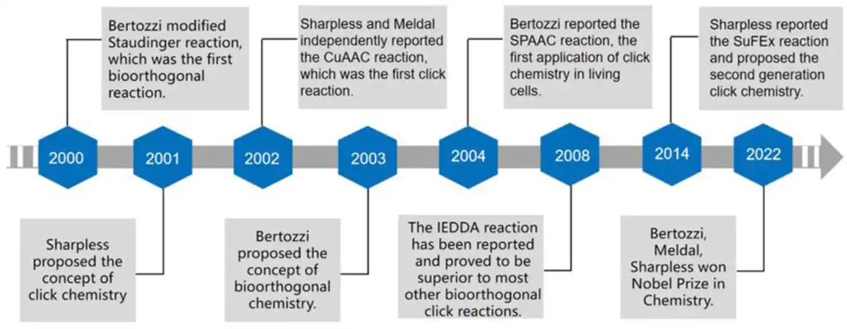
The concept of bioorthogonal chemistry was formally proposed by Dube and Bertozzi and Lemieux et al in 2003,[4,5] which refers to a set of labeling reactions that are both rapid and selective, and applied to living systems without interfering with their biochemical processes, thereby rendering them “orthogonal.” In 2000, the Saxon and Bertozzi[6] introduced the Staudinger ligation reaction between azide and triphenylphosphine, the first bioorthogonal reaction. Although this reaction has been widely used in biomarkers, it has disadvantages such as a slow reaction rate. In contrast, CuAAC exhibits superior reaction kinetics, whereby azide and alkyne are bioorthogonal. However, the cytotoxicity of monovalent copper ions limits its application in organisms. In 2004, Agard et al[7] reported the strain-promoted azide–alkyne cycloaddition (SPAAC) reaction between azide and cyclooctyne, also known as copper-free click chemistry. The strain-promoted cycloaddition reaction improves biocompatibility and is widely used in living cells or in vivo biomarkers. Figure 1 shows the milestones in the development of bioorthogonal click chemistry.
Figure 1.
Milestones in the development of bioorthogonal click chemistry.
Since then, click chemistry and bioorthogonal chemistry have been unified and become one of the most important chemical reaction tools in the fields of chemical synthesis, biomarkers, material preparation, and so on. In 2014, Dong et al[8] introduced the sulfur (VI)–fluoride exchange (SuFEx) reaction, regarded as the “second-generation click chemistry.” In addition, a series of new bioorthogonal click chemistry have been developed, such as inverse electron-demand Diels–Alder (IEDDA)[9] and photoinitiated thiol-ene reaction,[10] which continue to promote the development of this field. Compared with traditional methods for cell or material modification, bioorthogonal click chemistry has unique advantages: (1) The reaction rate is fast, for example, IEDDA has the fastest reaction rate reported so far, which is between 102 and 106 M−1s−1.[11,12] (2) The reaction selectivity is strong, side reactions are less, and the yield is significantly higher than that of traditional organic reactions. (3) The reaction conditions are mild, easy to perform in aqueous solvents, and do not require the participation of toxic catalysts.[1] (4) The reaction product is stable because of the covalent linkage between clickable groups.
In addition to the above commonalities, there are subtle differences between the characteristics of different biological orthogonal click reactions, which makes them have different application ranges. Due to its good chemical kinetic characteristics and low cost, CuAAC still plays an important role in biomaterial synthesis. However, copper ions require a high concentration when used as a catalyst (20–50 Μm),[13] which is highly cytotoxic. Research showed that the median lethal concentrations of copper in PC3 and HeLa cell lines were 97.1 and 85.5 μg/mL, respectively, which limits the application of CuAAC to the in vitro synthesis of biomaterials and cannot be directly applied in organisms. Therefore, CuAAC cannot be directly applied to organisms.[14] SPAAC is often used to modify cells, which requires higher biocompatibility. Importantly, the sialic acid pathway, a method for the expression of azide groups on the cell membrane surface, has been developed and widely used, which makes the application of SPAAC at the cellular level more convenient. For example, the reaction of azide-labeled cell surface glycans with fluorescently labeled cyclooctyne in vivo realized the first bioimaging application of bioorthogonal click chemistry in living systems.[15] However, the reaction rate of azide with dibenzocyclooctyne (DBCO) (k2 = 0.2–0.5 M−1 s−1) is relatively slow.[16] Therefore, attention turned to another stable aliphatic cyclooctyne called bicyclo[6.1.0]nonyne (BCN), which offers accelerated reaction rate constants up to 2.9 M−1s−1.[17] IEDDA, which has an ultrafast reaction rate (k2 = 104 M−1 s−1) and excellent biocompatibility, is widely used in the fields of drug modification and cell targeting. In response to this reaction, scientists have also carried out a lot of research. In response to this reaction, scientists have also carried out a lot of research. Holt et al[18] found that there is a secondary interaction between IEDDA cycloaddition products, which enhances the performance of covalently cross-linked hydrogels. The researchers also modified the reaction to achieve reversible control of tetrazine bioorthogonal reactivity.[19] So far, IEDDA is less used for cell modification. On the one hand, it may be because of its high application cost and on the other hand, the clickable group size of IEDDA is larger than that of azide group. However, its excellent reaction kinetics characteristics are still concerned by a large number of researchers. Recently, researchers used appended tetrazine-caged boron dipyrromethene–based fluorescent probe and a maleimide-substituted BCN to modify the membrane of macrophage and cancer cells, respectively. The 2 kinds of cells can be connected by IEDDA reaction, which could promote and facilitate the detection of intercellular interactions.[20] Finally, the thiol-ene reaction is easily initiated by light, which can be used as a unique click reaction to achieve the time and space regulation of the reaction and for the construction or functionalization of complex biological materials. However, ultraviolet (UV) light can trigger undesirable responses, including cellular apoptosis, and therefore, efforts have been concentrated on the design of visible and near-infrared (NIR) light-induced photoclick reactions. Groundbreaking work by Song et al[21,22] introduced the photoinduced cycloaddition of tetrazoles and alkenes, which also initiated remarkable progress in photoclick chemistry, such as light-initiated thiol-yne reactions,[23] azide–alkyne cycloadditions,[24,25] and azirine ligation.[26] The representative bioorthogonal click reactions and their features have been summarized in Figure 2.
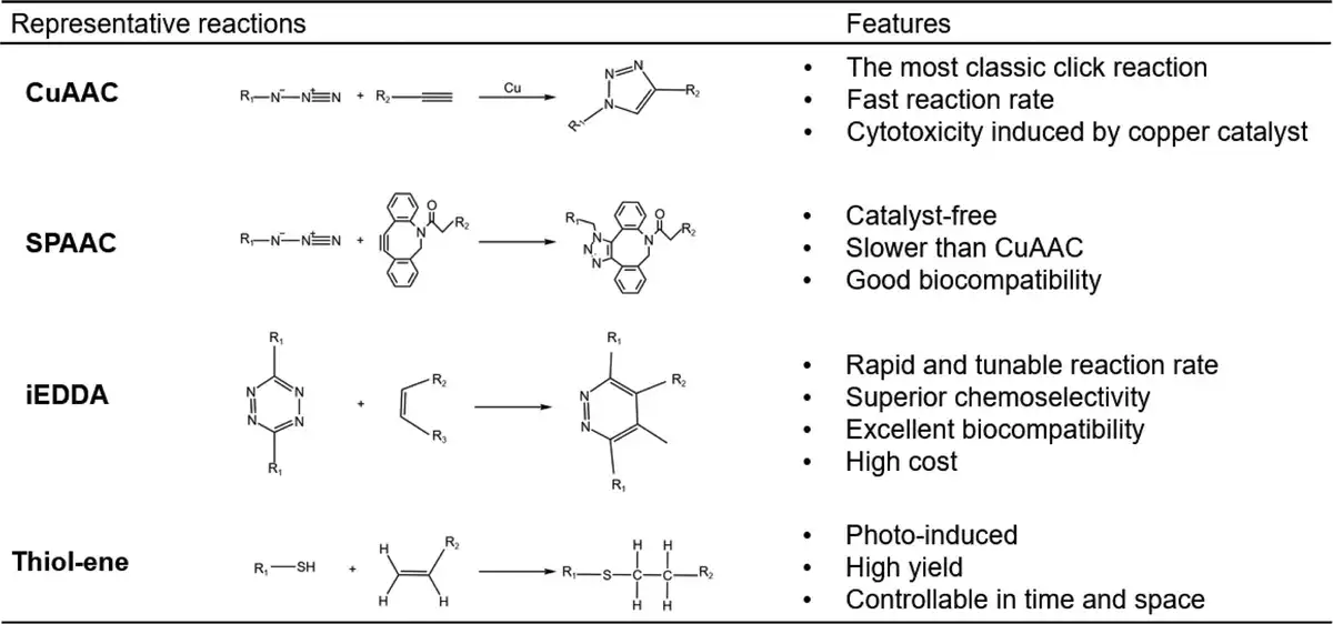
Figure 2.
Summary of the characteristics of representative click reactions.
Based on these advantages, bioorthogonal click chemistry is becoming more commonly used within the field of tissue engineering and regenerative medicine. First, compared with traditional organic synthesis reactions, the high efficiency of bioorthogonal click reaction improves the rate and yield of the biomaterial synthesis process, enabling researchers to obtain biomaterials with ideal properties in a short time. Additionally, the bioorthogonal click reaction can be performed without harsh reaction conditions or the use of toxic catalysts. This makes it possible to introduce bioactive substances during the synthesis of biomaterials and effectively improve the functionality of implants in vivo.[27] Importantly, bioorthogonal click chemistry, as a safe and efficient cross-linking tool, can on the one hand enable cells to interact with biomaterials in vitro,[28] so that the 2 functions are synergistic and complementary, and on the other hand, the orthogonality of the click reaction allows it to be transferred to the organism. Even if the organism comprises various molecules such as proteins and lipids, the reaction is not affected by these molecules and their functional groups. It can proceed rapidly in the body and the reaction is still precise and controllable.
Tissue engineering requires the participation of biological materials, seed cells, and extracellular signal molecules. In simple terms, biomaterials carry cells or signal molecules to simulate the structure and function of damaged tissues or organs to the greatest extent on the basis of structural support for tissue regeneration. However, there is still a certain gap between the structure and function of tissue-engineered grafts and natural tissues and organs due to the simple combination of the 3. Bioorthogonal click chemistry has attracted the attention of scientists as a safe and efficient “connection tool.” In addition to excellent reaction kinetics and good biocompatibility, its orthogonality can achieve complex and coordinated interactions among biomaterials, cells, and extracellular signal molecules in tissue-engineered grafts, thereby building a suitable environment for tissue or organ regeneration for transplanted cells or host cells. Initially, bioorthogonal click reactions were mainly used to construct or functionalize biomaterials suitable for cell culture. Elbert et al[29] constructed peptide-functionalized gels by Michael addition reaction. They mixed aqueous solutions containing polyethylene glycol (PEG)-polyacrylate, and albumin solid particles with aqueous solutions containing PEG-dithiol, and PEG-polyacrylate, and PEG-dithiol were rapidly cross-linked to form hydrogels. Both did not interact with the loaded protein in this process. This early study provided a new strategy for the construction of extracellular matrix (ECM) analogs. Later, Ossipov and Hilborn introduced alkynyl and azide groups into polyvinyl alcohol, respectively, and realized the bioorthogonal click chemical cross-linking hydrogel through CuAAC.[30] The hydrogel prepared by this method performs better in terms of gel time, modulus, and swelling degree. Since then, the DeForest et al[31] used SPAAC to realize the encapsulation of living cells in the process of constructing hydrogels by bioorthogonal click reaction for the first time and combined biomolecules in specific parts of the gel through thiol-ene reaction to realize the spatial and temporal regulation of cell microenvironment, thereby controlling cell motility or intracellular signal transduction and other cell functions. Bioorthogonal click chemistry better simulates extracellular characteristics at different times and spaces by introducing different functional cues such as integrin-binding peptides, matrix metalloproteinases (MMP)-degradable peptides, or cytokines into hydrogels.[32] In addition, DeForest et al[33] and DeForest and Anseth[34] carried out a lot of research on the construction of ECM analog gel by thiol-ene click reaction and found that the properties and functions of the gel network can be finely controlled by adjusting various factors such as exposure time, initiator concentration, and stoichiometric ratio of reactive functional groups, which provides inspiration and guidance for the follow-up research of bioorthogonal click chemistry in the field of tissue engineering.
With the continuous development of tissue engineering and regenerative medicine, the role of bioorthogonal click chemistry has evolved from simple cell microenvironment construction to promoting the regeneration of complex tissue structures. Its excellent chemical properties such as good biocompatibility, high reaction efficiency, and specificity enable scientists to meet the regeneration needs of different tissues and organs through a variety of strategies. Therefore, this review discusses the application of bioorthogonal click chemistry in tissue engineering and regenerative medicine in recent years (Figure 3). In the first part, the application strategies of bioorthogonal click chemistry in tissue engineering and regenerative medicine were reviewed. The second part reviews the application of bioorthogonal click chemistry in promoting tissue or organ regeneration. Finally, we summarized and prospected the application of bioorthogonal click chemistry in the field of tissue engineering and regenerative medicine.
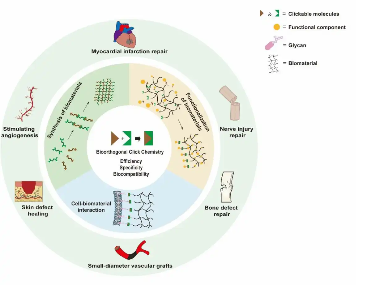
Figure 3.
Illustration showing strategies and applications of bioorthogonal click chemistry.
2. Strategies of bioorthogonal click chemistry in tissue engineering and regenerative medicine
First, clickable groups, such as azide or cyclooctyne groups, were introduced into biomaterials, biologically active substances, or cells. Subsequently, bioorthogonal click reactions between them under physiological conditions can achieve (1) synthesis of biomaterials (Table 1); (2) functionalization of biomaterials based on various bioactive substances (Table 2); and (3) interaction between cells and biomaterials (Table 3). Therefore, in this section, we will discuss how bioorthogonal click chemistry can be used in tissue engineering and regenerative medicine.
Table 1
Summary of strategies and applications in the synthesis of biomaterials.
| Reaction | Precursor | Characteristics | Disease/injury | Regeneration effect | References |
|---|---|---|---|---|---|
| SPAAC | Collagen | Cell-loaded | Corneal defect | Re-epithelialization of corneal tissue | Chen et al[45] |
| HA, collagen | In situ cross-linking | Corneal defect | Completely restored corneal curvature | Lee et al[46] | |
| PCL, PPF | Adjustability | Bone defect | New bone formation; vascularization | Liu et al[47] | |
| POSS, PCL, PPF | Adaptability of defect situation | Bone defect | Osteogenesis; vascularization | Liu et al[48] | |
| PEG | Controllable release of Chol-miR-26a | Bone defect | Higher new bone volume | Gan et al[49] | |
| PEG | Continuous release of the bone protection factor | Bone defect | Better osseointegration; osteogenic differentiation | Deng et al[50] | |
| HA, PEG | Slow release of peptides | Skin defect | Re-epithelialization, collagen deposition, and microvessel formation | Wang et al[51] | |
| GelMA | Self-adhesive | Nerve injury | Reconnected nerves with faster conduction velocity and shorter latency | Zhang et al[52] | |
| ELR | HE5 and HRGD loaded | Myocardial infarction | Better cardiac function | Contessotto et al[53] | |
| IEDDA | CS | Tissue adhesion | Skin defect | Faster healing speed; better healing quality | Li et al[54] |
| Thiol-ene | SA, PEG | Sequential release | Skin defect | Smaller volume of hypertrophic scar tissue | Shen et al[55] |
| PHA, PEGDA | Amphipathicity, fatigue-resistance | Vascular graft | Long-term intravascular patency | Hou et al[56] | |
| PEG, magnetic particle | Magnetic response | Skin defect | Fast healing of diabetic wounds | Shou et al[57] | |
| Thiol-maleimide | SA, CS | Rapid gelling rate | Esophageal injury | Epithelialization; ECM remodeling | Lei et al58] |
| Amino-alkyne | HA-ADH, DA-PEG | pH-sensitive responsive | Skin defect | Less local inflammation; faster healing speed | Jin et al[59] |
DA-PEG, dipropiolate ester of polyethylene glycol; GelMA, gelatin methacryloyl; HA, hyaluronic acid; HA-ADH, hyaluronic acid-adipic acid dihydrazide; HE5, a protease cleavage site; HRGD, functional domains for cell adhesion; SA, sodium alginate.
Table 2
Summary of strategies and applications in functionalization of biomaterials.
| Type | Biomaterials | Functional component | Reaction | Disease/injury | Regeneration effect | References |
|---|---|---|---|---|---|---|
| Polymeric materials | PLLA, PLSA | Heparin | Thiol-ene | Vascular graft | ECs and SMCs infiltration | Navarro et al[67] |
| POSS | PEG, KGN, HSPC, fluorescence | Thiol-ene | Bone defect | Delayed joint space narrowing and increased content of cartilage matrix | Yao et al[71] | |
| PCL | N-thiocarboxyanhydride | CuAAC | Angiogenesis | Larger neovascularization area | Yao et al[72] | |
| Silk fibroin | Heparin disaccharide | CuAAC | Cartilage defect | Cartilage regeneration and integration | He et al[73] | |
| Polypeptide | PLLA | BVLD | Thiol-ene | Myocardial infarction | Better endothelialization and endothelial function | Zhang et al[74] |
| Alginate | α1 peptide | CuAAC | Angiogenesis | Enhanced blood flow recovery properties | Jia et al[75] | |
| Collagen | Enzyme recognition peptide | CuAAC | Angiogenesis | Formation of blood and lymphatic vessels in the absence of exogenous VEGF | Rütsche et al[76] | |
| PCL | VEGF mimetic peptide | Thiol-ene | Angiogenesis | More blood vessels around the scaffold | Yao et al[77] | |
| HA, AT-EHBPE | CP50 peptide | Thiol-epoxy | Myocardial infarction | Better cardiac function; Neovascularization | Zou et al[78] | |
| Protein | PCL | TGF-β binding peptide, BMP-2 | CuAAC | Bone defect | Differentiation of hMSCs | Beeren et al[79] |
| PLLA | VEGF | Thiol-ene | Vascular graft | Better endothelialization and endothelial function | Zhang et al[68] | |
| Ti | BMP-2 | SPAAC | Bone defect | Osteogenesis; osseointegration | Wang et al[65] | |
| ECM | EGF | IEDDA | Controllable cell migration | Bailey et al[80] | ||
| EV | Collagen | EV | SPAAC | Angiogenesis | Host vascular ingrowth | Xing et al[69] |
| Collagen | EV | SPAAC | Hao et al[81] | |||
| Bacteria | FeSA | BL | Boronic acid, vicinal-diol | Inflammatory bowel disease | Faster healing of damaged colon | Cao et al[70] |
EGF, epidermal growth factors; hMSCs, human adipose-derived mesenchymal stem cells; SMC, smooth muscle cells.
Table 3
Summary of strategies and applications in cell–biomaterial interaction.
| Biomaterial cellularization | ||||||
|---|---|---|---|---|---|---|
| Cells | Reaction | Biomaterials | Acquired characteristics | Regeneration effect | References | |
| Macrophage | SPAAC | PCL, PEG | Improved capture efficiency and survival rate | Regeneration microenvironment regulation | Mao et al[85] | |
| ASCs | SPAAC | Alginate | Controllable differentiation of ASCs in vivo | Neatly arranged, long and thick muscle fibers | Ueda et al[86] | |
| NPCs | SPAAC | PEG | Enhancement of multiple-factor release | Less cell demand and better therapeutic effect | Oh et al[87] | |
| NPCs, astrocyte | SPAAC | Collagen | Covalent conjugation to collagen fibers | More neurons and endogenous axon regeneration | Liu et al[84] | |
| Cell tracking | ||||||
|---|---|---|---|---|---|---|
| Cells | Reaction | Imaging probe | Imaging technique | Observation | References | |
| hASCs | SPAAC | DBCO-Cy5 | FMT | Tracking cell migration to the lesion for up to 14 d | Lee et al[88] | |
| hEPCs | SPAAC | DBCO-Cy5 | FMT | Tracking the cells for 28 d and predicting the therapeutic effect | Lim et al[89] | |
| hASCs | SPAAC | BCN-dual-NPs | NIRF, MR | Tracking cell migration to the lesion for up to 14 d | Lim et al[90] | |
| Chondrocytes | SPAAC | DBCO-650 | NIRF | Tracking chondrocytes for up to 28 d with stronger NIRF intensity | Yoon et al[91] | |
| Tissue or organ targeting | ||||||
|---|---|---|---|---|---|---|
| Cells | Reaction | Direct click molecules | Indirect linker molecules | Target tissues | Results | References |
| Chondrocytes | IEDDA | TCO-Tz | TCO-(ApoPep-1-PEG), Tz-modified chondrocytes | Injured cartilage tissue | More chondrocytes and richer ECM in damage area | Co et al[92] |
| Endogenous stem cells | SPAAC | Az-DBCO | Az-(PEG-CD34), DBCO-(PEG-CD41) | Myocardial infarction area | More cardiomyocyte regeneration and angiogenesis | Li et al[93] |
| Mesenchymal stem cell | HA | Hydrazide-aldehyde | Hydrazide-(hyaluronic acid), aldehyde-(dextran sponge) | Myocardial infarction area | Better cardiac function and angiogenesis | Wu et al[94] |
hEPCs, human embryonic stem cells-derived endothelial progenitor cells.
2.1 Synthesis of biomaterials
In recent years, researchers have made great efforts in the development of biomaterials. To better meet the needs of tissue repair and regeneration, it is urgent to achieve accurate control of the structure and function of polymers, which poses great difficulties in the design and manufacture of polymers. The traditional polymerization method not only has a slow reaction rate but also is difficult to accurately control the polymer structure and separate by-products. Bioorthogonal click chemistry relies on its modular linking mode, which means that the reaction between click groups is not affected by the material precursor connected to it. This simplifies the polymer synthesis process. In addition, the reaction between clickable groups is highly selective, which minimizes the incidence of side reactions, and the structure and properties of the synthesized materials can be better predicted.[35,36] Finally, its good biosafety allows functional substances or cells[37] to be introduced into biomaterials and the reaction to be successfully transferred from the laboratory to the body. The introduction of functional substances can either be pregrafted onto the polymer precursor or mixed into the polymer during the cross-linking process. For example, hydrogels are widely used as cell scaffolds[38,39] or drug carriers[40,41] in tissue engineering and regenerative medicine due to their physical and chemical properties, including high water content,[42] adjustable mechanical properties,[43,44] and ease of chemical modification.[60] However, the traditional cross-linking process of hydrogels requires UV irradiation and the application of toxic catalysts. In this process, the activity of living cells or drugs encapsulated in them is severely compromised. Therefore, bioorthogonal click reaction groups such as azide groups and DBCO can be introduced into polymer precursors in advance, and then biomaterials can be synthesized rapidly and efficiently by click reaction under physiological conditions. For example, the NCColHA hydrogel for corneal defect can be cross-linked in situ at the defect site by mixing the 2 precursors, without external light or catalyst. In vitro experiments, there was no difference in biocompatibility between the hydrogel-coated area and the uncoated area. At the same time, the hydrogel maintains more than 90% weight at 24 hours and more than 50% weight at 48 hours, which has better stability than the pure physical hydrogel (a mixture of unmodified components).[61] In addition, the specificity and stability of the bioorthogonal click reaction can help modify the structure and mechanical properties required for the hydrogel scaffold and regulate the degradation of the hydrogel. Xu et al used SPAAC to reliably control the degradation rate without affecting mechanical and physical properties. The synthesized hydrogels showed highly predictable degradation time scales from at least 2 or 21 days to more than 250 days, respectively.[62] It is extremely difficult for traditional organic synthesis reactions. In conclusion, bioorthogonal click chemistry has unique advantages in the field of biomaterials synthesis. On the one hand, the rapid reaction rate makes the synthesis of biomaterials more efficient and on the other hand, the high selectivity of modular synthesis mode can precisely adjust the structure and properties of biomaterials.
2.1.1 Improvement of synthesis efficiency
Contessotto et al constructed a hydrogel based on elastin-like recombinants (ELRs). ELRs modified by azide groups or alkyne groups were cross-linked via SPAAC.[53] The click reaction cross-linked ELRs efficiently within 10 min at 4°C. Besides, uniform pores were formed inside the hydrogel, which was conducive to cell colonization and supply of oxygen and nutrients. Zhang et al[52] designed a drug-loaded bandage for the repair of peripheral nerve injury. The clickable groups (DBCO and azide groups) existed on the upper and lower surfaces of the bandages, respectively. When wrapped, the bioorthogonal click reaction caused the bandage to adhere quickly. On the one hand, this makes the nerve repair operation easier and significantly shortens the operation time. On the other hand, it avoids the secondary damage to the nerve during the operation to the greatest extent (Figure 4B). In addition, the extremely fast reaction rate of bioorthogonal click chemistry allows polymer precursors, such as hydrogels, to undergo rapid in situ cross-linking after being injected into the target site through a syringe.[63] Chen et al[45] injected azide-modified HA and DBCO-modified collagen into the corneal defect site, and they were able to cross-link in situ within a few minutes to fill the corneal defect. After 1 week, the corneal injury area transitioned smoothly to the adjacent normal cornea. During the entire healing process, the hydrogel did not swell or shrink and showed excellent cell compatibility and support for epithelial cells (Figure 4A). Li et al[54] introduced trans-cyclooctene (TCO)/tetrazine (Tz) into chitosan (CS) and prepared a new bioadhesive to meet the needs of surgical wound closure through IEDDA. When injected and mixed into the wound, the 2 precursors can form a solid hydrogel within 2 min. Moreover, it should be noted that the bonding strength of this adhesive is 2.3 times higher than that of traditional fibrin glue. Liu et al[48] has additionally created a system that can be injected and consists of a poly(propylene fumarate) (PPF) chain terminated by a cyclooctyne ring (PPF-BCN) and an 8-armed polyhedral oligomeric silsesquioxane (POSS) core branched by an azide-functionalized polycaprolactone (PCL) chain. When introduced to the bone defect model, the system can undergo in situ cross-linking through SPAAC following the injection of a double-tube syringe at the site of the defect. The cross-linked hydrogel exhibits superior mechanical properties to pure polymer systems, making it a viable option for hard tissue applications. Additionally, it avoids secondary damage to the injured site when the in vitro constructed block hydrogel is implanted into the body and can be shaped during the injection process to adapt to different tissue defects, allowing the hydrogel and new tissue to better integrate with the host tissue. In summary, bioorthogonal click chemistry serves as an effective cross-linking tool, enhancing the synthesis efficiency of biomaterials during in vitro synthesis and enabling direct in situ synthesis in vivo. This offers researchers greater flexibility to cater to diverse tissue regeneration requirements.
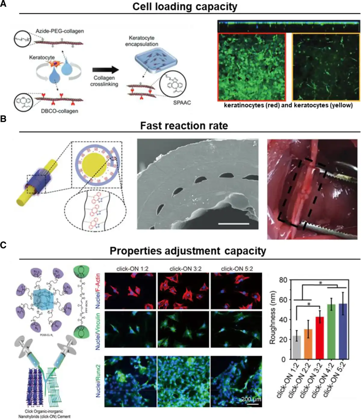
Figure 4.
Illustration showing characteristics of bioorthogonal click chemistry in the synthesis of biomaterials. (A) Coculture of human keratinocytes and keratocytes within SPAAC-cross-linked collagen gels. Adapted with permission from Lee et al.[46] (B) In upper and lower surfaces of the bandages, rapid self-adhesion via SPAAC after rolling occurs. Adapted with permission from Zhang et al.[52] (C) Hydrogels with corresponding properties can be stably obtained by adjusting different proportions of precursors during injection. Adapted with permission from Liu et al.[48]
2.1.2 Precisely controllable properties
Bioorthogonal click chemistry overcomes the shortcomings of traditional organic chemistry, such as a complex synthesis process, low yield, and excessive by-products. Compared with conventional organic synthesis methods, high selectivity of bioorthogonal click reaction reduced the generation of by-products, which allows researchers to obtain biomaterials with expected properties. Furthermore, the simple modular synthesis strategy allows researchers to precisely regulate the properties of biomaterials by adjusting the proportion of biomaterial precursors. Therefore, the synthesis method based on bioorthogonal click chemistry makes the properties of biomaterials controllable. Hou et al[56] utilized the thiol-ene click reaction between polyhydroxyalkanoates (PHA) and polyethylene glycol diacrylate (PEGDA) in producing amphiphilic organic hydrogels for small-diameter vascular grafts. The PHA organic gel exhibits contraction upon water balance and expansion upon oil balance, while the PEGDA organic gel exhibits the opposite phenomenon. The volume of PHA/PEGDA organic gels prepared by bioorthogonal click chemistry in an appropriate ratio can remain stable in both aqueous and organic phases. Liu et al[48] have developed an injectable hydrogel system. It can increase the protein adsorption capacity of the material by increasing the proportion of PCL chains. Similarly, different polymer ratios can make the compressive modulus of the material change between tens to hundreds of MPa, and the gelation time changes between 20 and 60 min (Figure 4C). Shen et al[55] also used a thiol-ene click reaction to prepare bilayered thiolated alginate/PEG diacrylate (BSSPD) hydrogels and mixed them with small extracellular vesicles (sEVs). By adjusting the ratio of alginate and PEG to give different mechanical strength and degradation rates to the upper and lower layers of the hydrogel. This realized the sequential release of sEVs to meet the repair needs of different stages of skin wound healing.
In summary, bioorthogonal click chemistry achieves controllable regulation of the properties of biomaterials, and this operation is very simple. Only by controlling the proportion of polymer precursors precisely, biomaterials with predictable properties can be stably obtained.
2.2 Functionalization of biomaterials based on various functional component
In addition to the synthesis of biomaterials, researchers have also introduced clickable groups into functional substances and biomaterials, respectively.[64] The covalent linkage between the 2 efficaciously functionalizes the biomaterials. Traditional functional modification methods mostly involve tedious chemical reactions. The complex processing technology not only increases the application cost of functional biomaterials but also has a significant negative effect on the biological activity of functional materials. Therefore, the modular synthesis mode of bioorthogonal click chemistry enables biomaterials to effectively combine multiple functional substances and ensure their biological activity, which meets the synthesis requirements of functional biomaterials. For example, Wang et al[65] grafted BMP-2 onto the surface of titanium to achieve surface modification of bone grafts. The bioorthogonal click reaction started within a few minutes, and the maximum grafting density of BMP-2 reached 140 ng cm−2, which was higher than the previous study of 50 ng cm−2.[66] In addition to the advantages in efficiency, it allows various types of substances such as peptides, proteins, extracellular vesicles, and even bacteria to be clicked to biomaterials via bioorthogonal click chemistry.
2.2.1 Click polymeric materials to biomaterials
Navarro et al[67] used thiol-ene click chemistry between the norbornene ring on poly(lactic acid-co-lactic acid) (PLSA) and the thiol group on heparin to graft the anticoagulant molecular heparin onto the tubular scaffold, achieving hydrophilicity and anticoagulation of the vascular graft. Through the bioorthogonal click reaction, the scaffold was completely modified and the original physical properties were retained (Figure 5A). Importantly, the modified tubular scaffold showed similar biocompatibility to the control group and had excellent anticoagulant properties. This suggests that the bioorthogonal click reaction does not significantly affect the biocompatibility of the scaffold and the biological activity of heparin. Yao et al[72] connected alkynyl-modified N-thiocarboxyanhydride (NTA) to an azide-modified PCL scaffold via CuAAC. The NTA attached to the PCL scaffold can release H2S, which stimulates angiogenesis. Scanning electron microscope (SEM) showed that the scaffold modification method based on bioorthogonal click chemistry had almost no effect on the morphology of PCL scaffold fibers, and the fibers remained uniform and smooth. In addition, the release of H2S can be easily regulated by changing the number of azide groups on the PCL framework. Under the accelerated conditions of the methylene blue method, H2S was released stably within 45 min. He et al[73] attached heparin disaccharides, which can specifically adsorb IL-4, to silk microcapsules (SFM) via CuAAC. Subsequently, IL-4/SFM effectively promoted the polarization of M2 macrophages in cartilage defects caused by osteoarthritis. Yao et al[71] used thiol-ene click chemistry to chemically graft polyhedral oligomeric silsesquioxane (POSS) with PEG, kartogenin (KGN), hydrogenated soybean phosphatidylcholine (HSPC), and fluorescein to form a stable and multifunctional POSS hybrid molecule (PPKHF) to prepare hydrogel microspheres (MHS@PPKHF) for in situ injection into the articular cavity for the treatment of osteoarthritis. MHS@PPKHF has excellent lubrication performance in the joint cavity, which is at least 5 times higher than that of the control group, and can effectively reduce the damage caused by mechanical friction to cartilage tissue. In addition, its excellent drug release performance retention effect in the joint cavity and penetration into the cartilage tissue can be observed by an in vivo imaging system.
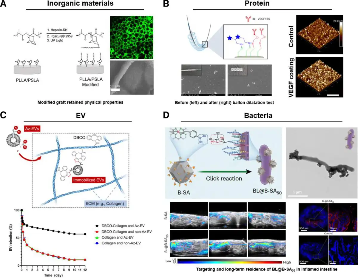
Figure 5.
Various functional components could be connected to biomaterials via bioorthogonal click chemistry. (A) Heparin was connected to the PLLA/PSLA scaffold via thiol-ene reaction. Adapted with permission from Navarro et al.[67] (B) Vascular stent was modified by VEGF via thiol-ene reaction. Adapted with permission from Zhang et al.[68] (C) EVs were connected to collagen hydrogel. Adapted with permission from Xing et al.[69] (D) Artificial enzymes–armed Bifidobacterium longum probiotics. Adapted with permission from Cao et al.[70] EVs, extracellular vesicles; PLLA, poly (L-lactic acid); PSLA, poly(spirolactic-co-lactic acid); VEGF, vascular endothelial growth factor.
2.2.2 Click polypeptides or proteins
Jia et al[75] connected α1 peptide with angiogenic capacity to alginate hydrogel via CuAAC. Compared with the control group, the bioorthogonal click chemistry showed obviously high efficiency and cell compatibility. Human umbilical vein endothelial cells (ECs) showed enhanced adhesion, spreading, and migration on α1 peptide-modified hydrogels. Yao et al[77] used a UV-induced thiol-ene click reaction to graft vascular endothelial growth factor (VEGF) mimetic peptide onto the PCL scaffold to promote angiogenesis. Coupling VEGF mimetic peptide to the PCL scaffold significantly increased the tensile strength and modulus of the scaffold without affecting its fiber morphology. Compared with the control group, the scaffold functionalized with VEGF mimetic peptide significantly improved the cell survival rate. In addition, the author investigated the covalent bonding between 7-mercapto-4-methylcoumarin and PCL scaffold under UV irradiation by light mask test. After UV irradiation, the unmasked area was functionalized and emitted fluorescence, while the masked area was not functionalized. The results prove that the efficient thiol-ene click reaction can realize the functionalization of any region of biomaterials under the control of UV light. Zou et al[78] connected the aniline tetramer (AT) grafted hyperbranched epoxy macromonomer (EHBPE) to the thiolated CP05 peptide via the thiol-epoxy click reaction. The CP50 peptide can anchor exosomes to treat ischemia/reperfusion injury after myocardial infarction. Fluorescent labeling of the exosomes showed that they were evenly distributed throughout the hydrogel. On the seventh day after the hydrogel was injected into the damaged rat heart, the fluorescence intensity of the exosomes in the hydrogel was significantly higher than that in the control group, and fluorescence could still be observed after 14 days. These results indicate that the CP05 peptide-functionalized hydrogel based on bioorthogonal click chemistry can significantly prolong the retention time of exosomes.
Zhang et al[68] utilized the thiol-ene click reaction to immobilize VEGF onto vascular scaffolds. The bioorthogonal click reaction achieved the modification of the vascular scaffold in 10 min and the VEGF coating remained intact and homogeneous after PBS solution erosion and balloon dilation experiments (Figure 5B). Crucially, the modified scaffolds also enhanced the proliferation, adhesion, and migration of human umbilical vein ECs. Wang et al[65] linked BMP-2 modified with azide and DBCO to the titanium screw’s surface. The reaction occurred within a few minutes, and the secondary modification of titanium screws by BMP-2 resulted in a significant increase in surface roughness. Additionally, the fluorescence intensity on the surface of the biomaterial remained unaffected after the incubation in DMEM/F12 for 2 weeks, even after labeling BMP-2 with a fluorescent probe. Moreover, the modified titanium screw demonstrated exceptional osteogenesis ability in vitro. Consequently, these findings suggest that the BMP-2 was firmly attached to the biomaterial through covalent bonds and preserved its authentic biological function.
2.2.3 Click EVs for long-term immobilization
Extracellular vesicles as an important ingredient of intercellular communication have been widely applied in the field of tissue engineering. Its editability makes it a carrier for a variety of bioactive substances to play a synergistic role in tissue regeneration.[82,83] Similarly, EV can also be used as a functional component to connect with biomaterials through bioorthogonal click chemistry.
Xing et al[69] achieved stable fixation of azide EV in DBCO-modified collagen hydrogel, resulting in long-term retention of EV in vivo. The high efficiency of the reaction under physiological conditions enabled the retention of more than 60% EV after 12 days, which was significantly higher than the control group’s retention of less than 15%. The method of immobilizing EV indicates the potential for enhanced drug delivery efficacy. Hao et al[81] used SPAAC to create a SILY-EV complex by covalently linking DBCO-modified EV and azide-modified collagen-binding peptide (SILY). The coupling process had a high efficiency of 70% and took place in a neutral solution without compromising the structural integrity or biological activity of the EV. The resulting complex exhibited a significant increase in the number and residence time of EV on collagen-containing tissue surfaces compared with the control group. The author pointed out in the article that this method can also be used for the modification of collagen-based biomaterials by EV. In conclusion, these results demonstrate that the binding strategy based on bioorthogonal click chemistry can achieve stable binding to biomaterials and long-term retention in vivo without affecting the structure and function of EV (Figure 5C). The azide groups can be introduced into the EV surface after treatment with tetra-acetylated N-azidoacetyl-d-mannosamine (Ac4ManNAz), which is similar to metabolic glycoengineering on the surface of the cell membrane. In addition, the surface proteins of EV can form a covalent connection with the NHS groups of DBCO-sulfo-NHS, which can also introduce azide groups into the EV surface.
2.2.4 Click bacteria for targeting and colonization
Cao et al[70] designed and created a probiotic system based on Bifidobacterium longum (BL) that features an artificial enzyme to regulate inflammation and promote gut lining restoration. To achieve this, they first prepared an iron single-atom catalyst (FeSA) through pyrolysis of an iron precursor encapsulated within a metal-organic framework (Fe@MOF). Using a boric-polyethylene glycol click reaction, they then merged BL and FeSA to generate the probiotic-artificial enzyme system (BL@B-SAx). In this system, single-atom catalyzed artificial enzymes (SAzymes) eliminate reactive oxygen species (ROS) by mimicking antioxidant enzymes and biomolecules. This not only mitigates the symptoms of intestinal inflammation but also protects BL from oxidative harm within the inflammatory zone, improving BL’s living environment. In addition, due to the superior intestinal colonization ability of BL probiotics in the colon, they play a role in reshaping the intestinal barrier function and microbiota, while ensuring that SAzymes perform long-term antioxidant treatment at the site of the disease (Figure 5D). The 2 can be covalently connected via the bioorthogonal click reaction without affecting the artificial enzyme activity and bacterial function. After oral administration, it can still reach the inflammatory colon site smoothly after digestion with gastric acid, bile, intestinal fluid, and other digestive fluids.
The earlier studies demonstrate the broad applicability of bioorthogonal click chemistry. It can efficiently and stably conjugate diverse functional substances, such as inorganic compounds, peptides, proteins, extracellular vesicles, and bacteria, to biomaterials, enhancing their functionality to meet the needs of tissue regeneration. In conclusion, the approach of biomaterial functionalization via bioorthogonal click chemistry offers an exceptional cross-linking tool for tissue engineering or regenerative medicine to establish a “functional material repository.”
2.3 Interaction between cells and biomaterials
Construction of biohybrid materials through the interaction between cells and biomaterials is a promising development direction in the field of tissue engineering and regenerative medicine. Due to good biocompatibility and stability of bioorthogonal click chemistry, bioorthogonal click chemistry can be applied to modify cells to directly participate in the reaction. In the study by Liu et al, the seeding efficiency of neural progenitor cells (NPCs) modified with azide groups on collagen scaffolds modified with DBCO groups is 2 to 2.6 times higher than that of traditional methods. Additionally, the delivery efficiency of drug-loaded nanoparticles modified with DBCO to NPCs is 2.4 to 2.6 times higher than that of the unmodified group. This highlights the significant role that bioorthogonal click chemistry plays in the interaction between cells and materials.[84] Therefore, the process of interaction between cells and biomaterials can accomplish complex functions that cannot be achieved by a single cell or biomaterials, such as (1) biomaterial cellularization, (2) cell tracking, and (3) tissue or organ targeting (Figure 6).
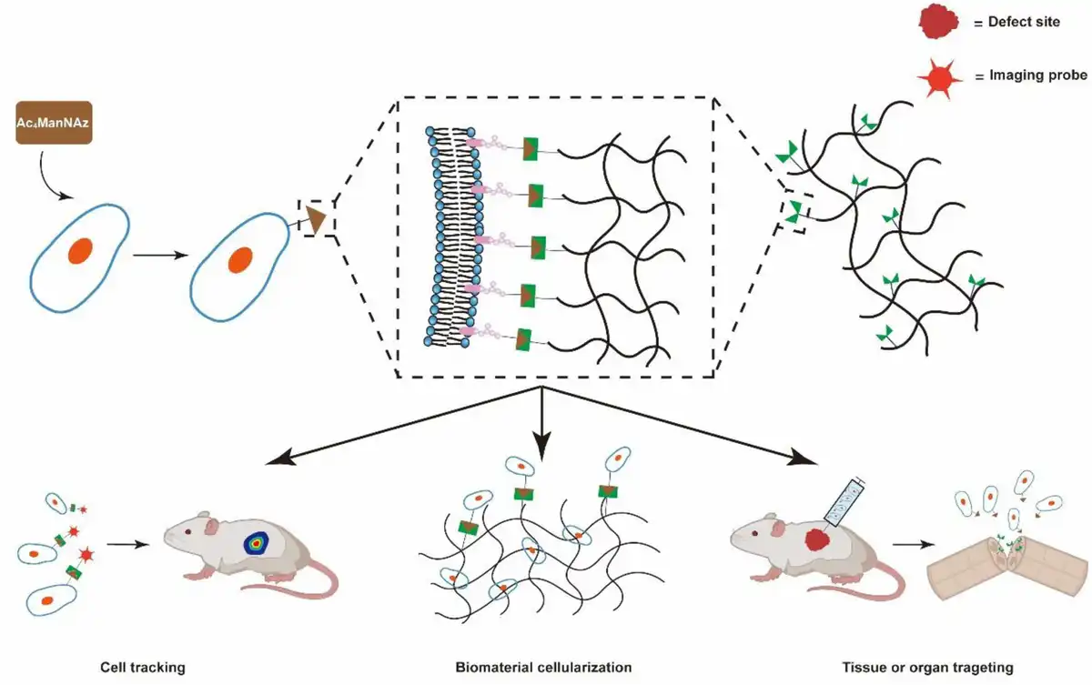
Figure 6.
Illustration showing strategies of cell–biomaterial interaction.
2.3.1 Biomaterial cellularization
Covalent binding of cells and biomaterials makes the graft closer to natural tissue in composition and function. Cell adhesion is the first phase of the interaction between cells and biomaterials, influencing the subsequent behavior of cells, including survival, proliferation, and differentiation. Using bioorthogonal click chemistry to directly modify cells to adhere to biomaterials to develop stable, covalently bound cell polymers is a suitable solution for the development of biohybrid materials. Therefore, bioorthogonal click chemistry has been increasingly applied to life systems, especially living cells, due to its good biocompatibility, that is, directly modifying the cells involved to enable them to participate in bioorthogonal click reactions. After treatment with Ac4ManNAz, cells express clickable azide groups at the glycan end of the cell membrane surface via the sialic acid pathway.[4] Subsequently, through the bioorthogonal click reaction between cells and biomaterials, on the one hand, the cellularization of biomaterials is realized, and on the other hand, more or stronger biological functions are given to cells and biomaterials.
Mao et al[85] synthesized a polymer film modified with DBCO-PCL-PEG and introduced azide groups to macrophages using metabolic glycoengineering. The resultant clickable modified film significantly enhanced cell selective capture efficiency by over 2 times. Furthermore, the cells linked chemically to the film by bioorthogonal click reaction showed no substantial change in cell viability within 7 days and demonstrated comparable cell growth rates to the control group. The findings demonstrate that bioorthogonal click chemistry-induced cell–biomaterial interaction promotes cell adhesion to biomaterials and that covalent attachment has little effect on cell viability and proliferation (Figure 7D). Ueda et al[86] employed bioorthogonal click chemistry to cross-link azide-modified adipose-derived stem cells with alkynyl-modified alginates. By altering the quantity of adipose stem cells or the concentration of alginate, the mechanical properties of the biohybrid material can be modulated to accommodate the surrounding environment. Importantly, the biohybrid materials constructed based on bioorthogonal click chemistry retained the differentiation potential of mesenchymal stem cells and enabled stem cell differentiation to be controlled by adjusting the hardness of the hydrogel (Figure 7A). Liu et al[84] labeled human NPCs and astrocytes with azide and modified longitudinally arranged collagen fibers (LACFs) and lipid nanoparticles containing Eda with DBCO groups. Subsequently, spinal cord-like grafts engineered with NPCs and human astrocytes were assembled to promote the repair of spinal cord injuries through SPAAC. The bioorthogonal click chemistry-based covalent connection not only achieved the cellularization of spinal cord-like grafts but also encouraged the adhesion and differentiation of NPCs and astrocytes on the scaffold (Figure 7C). Oh et al[87] utilized DBCO-modified PEG for enclosing NPCs modified with azide, achieving single-cell encapsulation of NPCs through SPAAC. This cell–biomaterial interaction based on bioorthogonal click chemistry enhances the release of cytokines, which in turn reduces the number of cells required for treatment (Figure 7B). Compared with the control group without any treatment, there was no significant difference in the cell survival rate of PEG-coated cells with different molecular weights. Most cells were effectively coated with polymers, as evidenced by the results from both fluorescence and projection electron microscopy.
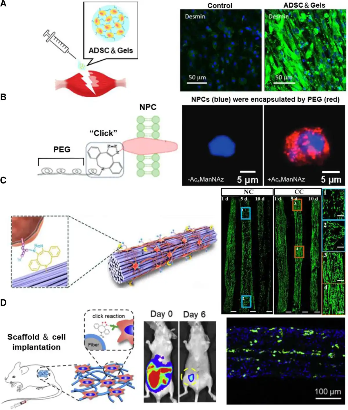
Figure 7.
Illustration showing the cellularization of biomaterial. (A) ADSC and (B) NPCs were encapsulated by biomaterials. Adapted with permission from Ueda et al[86] and Oh et al.[87] (C) NPCs were covalently bound to LACF. Adapted with permission from Liu et al.[84] (D) Higher macrophages capture the efficiency of scaffolds. Adapted with permission from Mao et al.[85]
These studies have shown that the high specificity, efficiency, and good biocompatibility of bioorthogonal click chemistry make it a suitable covalent cross-linking tool to realize the interaction between cells and biomaterials. The biohybrid materials constructed by this method can make biomaterials capture more cells in a short time and produce stronger adhesion. Furthermore, the interaction between the 2 improves cell function to some degree and facilitates tissue repair and regeneration from various perspectives.
2.3.2 Cell tracking
Transplanting stem cells into the body is a method used to regenerate tissues or organs.[95–96–97] However, this process is complex and dynamic. Therefore, monitoring the survival, proliferation, localization, migration, and differentiation of transplanted stem cells in vivo is of great significance for optimizing the delivery route and therapeutic effect. Currently, diverse direct or indirect imaging techniques have been devised to visualize and monitor transplanted stem cells in vivo. Stem cells could be labeled with imaging probes such as green fluorescent protein,[98] luciferase, fluorescent dye, quantum dots,[99–100–101] magnetic iron oxide particles,[102,103] and radioisotopes,[104,105] and then tracked using different imaging modes.[106–107–108–109] Although various marking methods have been continuously developed and improved, there are still limitations. For instance, (1) abnormal cell proliferation or cytotoxicity, (2) abnormal differentiation of stem cells, (3) dilution or loss of imaging probes, and (4) nonspecific labeling of host cells.[110,111] Therefore, a safer and more efficient labeling strategy is required for monitoring the biological processes of stem cells in vivo. The labeling strategy based on bioorthogonal click chemistry has good biocompatibility and labeling efficiency in vivo and in vitro and shows great potential for noninvasive imaging of stem cells in vivo. First, clickable chemical groups such as azide, alkynyl, and sulfhydryl groups were introduced onto the surface of stem cells through metabolic glycoengineering. These groups are selectively associated with imaging probes that contain corresponding chemical groups (DBCO or BCN) through bioorthogonal click chemistry. Due to the inherent characteristics of bioorthogonal click chemistry, such as good biocompatibility, high efficiency, and specificity of the reaction, this labeling method can achieve rapid, lasting, and uniform labeling of transplanted stem cells in vivo without causing serious side effects. Moreover, altering the quantity of metabolites can manipulate the number of nonnatural chemical groups present on the surface of the stem cells.
Lee et al and Lim et al reported a noninvasive tracking of mesenchymal stem cells in a mouse hind limb ischemia model.[88,89] First, human adipose-derived mesenchymal stem cells (hASCs) were treated with 50 μM Ac4ManNAz for 48 h to generate azide groups on the cell surface. Subsequently, the azide groups on the cell surface were coupled with near-infrared fluorescence (NIRF) dye-labeled dibenzocyclooctyne (DBCO-Cy5) via SPAAC to achieve fluorescent labeling of hASCs. In vitro, the viability of hASCs remained unchanged when treated with Ac4ManNAz (250 μM) or DBCO-Cy5 (20μM). However, in the control group, the cell viability of hASCs labeled with 10 μM or 20 μM of lipophilic indocyanine dye (DiD) significantly reduced. The authors then performed in vitro cell tracking of DBCO-Cy5 and DiD-labeled hASCs for 2 weeks. The samples were treated with DiD for 2 weeks, during which the fluorescence signal intensity of both substances decreased over time. The fluorescence signal was present in the DBCO-Cy5-hASCs group on day 14, but almost completely disappeared in the DiD-hASCs group by day 7 (Figure 8B). These results demonstrate that bioorthogonal click chemistry-based modification of mesenchymal stem cells can achieve long-term and precise in vivo tracking. Finally, the DBCO-Cy5-hASCs constructed by bioorthogonal click chemistry were intramuscularly injected into the mouse hindlimb ischemia model, and the in vivo NIRF 3-dimensional reconstruction images were obtained by fluorescence tomography system (FMT). The results showed that the NIRF signal gradually converged in the inner thigh of the ischemic hindlimb. In addition, human nuclear immunofluorescence staining was performed on ischemic lesions 2 weeks after transplantation, and the results were positive, and strong NIRF signals appeared in this area, indicating that DBCO-Cy5-labeled hASCs successfully migrated to ischemic lesions. The earlier results showed that the labeling method based on bioorthogonal click chemistry can track mesenchymal stem cells in vivo for a long time and accurately.
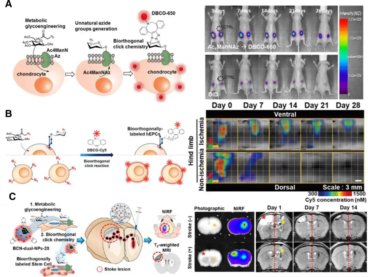
Figure 8.
Illustration showing the tracking for various types of living cells in vivo. (A) Chondrocytes were labeled through metabolic glycoengineering and SPAAC. Adapted with permission from Yoon et al.[91] (B) SPAAC-based labeling of endothelial progenitor cells for in vivo cell tracking. Adapted with permission from Lim et al.[89] (C) NIRF and MRI dual-imaging for tracking of hASCs in brain stroke lesion. Adapted with permission from Lim et al.[90] MRI, magnetic resonance imaging.
To overcome the limitations of some imaging methods and take advantage of other methods, dual-modal or multimodal imaging methods have been widely used in the imaging of implanted stem cells.[112] Recently, Lim et al[90] modified ethylene glycol chitosan nanoparticles (CNPs) with BCN groups, and then chemically conjugated BCN-modified CNPs (BCN-NPs) with NIRF dye Cy5.5 through amide bonds. Subsequently, oleic acid–coated superparamagnetic iron oxide nanoparticles (OA-Fe3O4 NPs) were physically encapsulated into Cy5.5-labeled BCN-NPs to obtain Cy5.5-labeled OA-Fe3O4 NPs-encapsulated BCN-NPs (BCN-dual-NPs) as imaging probes for NIRF/magnetic resonance (MR) dual-modality imaging systems based on bioorthogonal click chemistry. Ac4ManNAZ-pretreated hASCs were labeled with BCN-dual-NPs by SPAAC. Finally, BCN-dual-NP-labeled hASCs were implanted into a mouse model of photothrombotic stroke, which could be effectively monitored by NIRF and T2-weighted MR imaging within 14 days (Figure 8C).
In addition to labeling stem cells, Yoon et al[91] also used bioorthogonal click chemistry to label chondrocytes and implanted them subcutaneously in mice. Compared with DiD-labeled chondrocytes, chondrocytes labeled by this method continuously emitted stronger NIRF signals within 4 weeks and formed more extensive bioengineered cartilage on the chondrocyte scaffold (Figure 8A).
2.3.3 Tissue or organ targeting
Due to the high specificity of the covalent connection between clickable groups, the reaction is still precise and controllable in the presence of many biomolecules in the organism. In recent years, bioorthogonal click chemistry has gained considerable success in the design of nanoparticles for drug delivery. For example, after constructing engineered spinal cord-like grafts, Liu et al[84] intravenously injected lipid nanoparticles loaded with the neuroprotective agent edaravone. The DBCO group on the nanoparticles can precisely recognize and bind to the azide groups expressed by NPCs and astrocytes (Figure 9A). Due to the bioorthogonality of the clickable groups, this highly efficient and specific targeting strategy can also be applied to cells. First, clickable groups can be introduced into therapeutic cells through metabolic glycoengineering, and they can also be introduced into carriers that can carry cells. Subsequently, a specific bioorthogonal click reaction allows cells expressing clickable groups or molecules connected to the cell to be delivered accurately and efficiently to the target site.
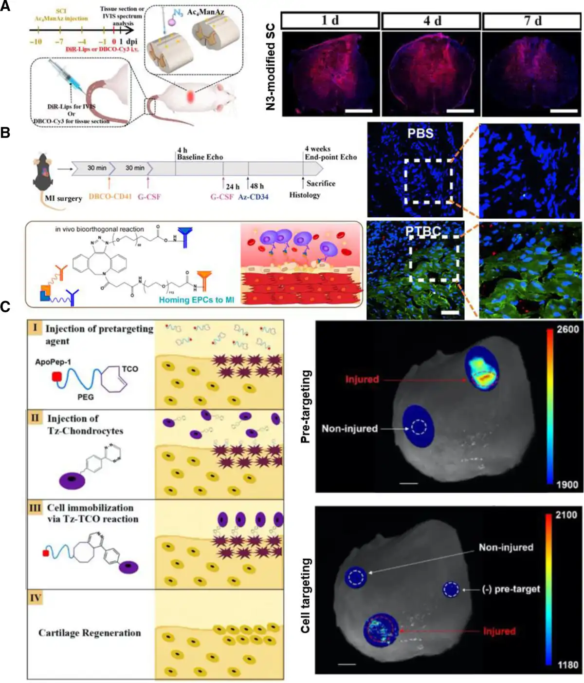
Figure 9.
Illustration showing targeting therapy of drugs and cells in vivo. (A) Illustration and immunofluorescence staining indicating successful targeting of the DBCO-Cy3 to the N3-modified SCI area after treatment. Adapted with permission from Liu et al.[84] (B) Illustration and immunofluorescence staining indicating successful homing of endogenous stem cells to the MI area after treatment (blue: nuclei, green: cardiomyocytes, red: endogenous stem cells). Adapted with permission from Li et al.[93] (C) Pretargeting apoptotic chondrocytes and Tz-chondrocyte targeting. Adapted with permission from Co et al.[92]
Li et al[93] injected DBCO-PEG-labeled CD41 and azide-PEG-labeled CD34 into a mouse myocardial infarction model at an interval of 48 h. The former is connected to platelets and aggregated in the MI region by the homing ability of platelets. After the latter recognizes and binds to endogenous stem cells, SPAAC between azide group and the DBCO group allows the stem cells to reach the infarcted area for cardiac repair (Figure 9B). Co et al[92] prepared a pretargeting agent by linking ApoPep-1 and TCO to PEG polymers. The pretargeting agent can combine with apoptotic chondrocytes and then enter into the articular cartilage injury area. Subsequently, chondrocytes containing methyltetrazine (Tz) were immobilized on the surface of TCO-coated damaged cartilage via IEDDA. The authors evaluated the targeting ability of chondrocytes to the damaged cartilage surface. The results showed that Tz-chondrocytes had a high affinity for the TCO coating on the damaged cartilage surface, and the number of Tz-chondrocytes adhered to the TCO surface increased with time, which was more than 4 times higher than that of the control group (Figure 9C). On the one hand, the application of bioorthogonal click reaction makes the cells promoting regeneration be accurately delivered to the target area. On the other hand, the covalent connection between clickable groups can make them be fixed in the target area without affecting cell activity and thus play an efficient and lasting role.
3. Applications of bioorthogonal click chemistry in promoting tissue or organ regeneration
3.1 Bone defect repair
It is an effective method of using metal, ceramic, and other polymer materials as bone substitutes for implantation into the body. It is widely used to promote the repair of bone or cartilage defects for bone fracture fixation, joint replacement, etc.[113–114–115] However, exogenous biomaterials often lack the ability to adapt to the complex physiological bone regeneration process, and surface bioengineering techniques are needed to introduce biological activities, such as osteoinductive and immunomodulatory activities. To date, various physical or chemical methods have been applied to surface bioengineering technology. For example, layer-by-layer assembly, Langmuir–Blodgett deposition silanization, acid etching, anodic oxidation, and ion doping.[116–117–118] Traditional methods have seriously affected the repair effect of implants due to a lack of long-term activity and the destruction of bioactive molecules. Due to the good biocompatibility of the bioorthogonal click reaction, this can achieve surface modification of bone grafts while ensuring the biological activity of macromolecules.
Recently, Wang et al[65] used bioorthogonal click chemistry to stably bind the bone-inducing growth factor BMP-2 as a functional coating on the titanium screws. In a series of in vivo and in vitro experiments, the coating can effectively promote the transformation of macrophages to the M2 phenotype without cytotoxicity and secrete cytokines to provide an immune microenvironment suitable for bone regeneration. Importantly, after 8 weeks of in vivo implantation, the titanium screw was stably attached to the surrounding bone tissue. In addition, the surrounding new bone density, volume, and trabecular structure characteristics were significantly better than the control group. These results confirmed that the bone graft coating using bioorthogonal click chemistry can significantly promote interface osteogenesis and enhance osseointegration in vivo (Figure 10D).
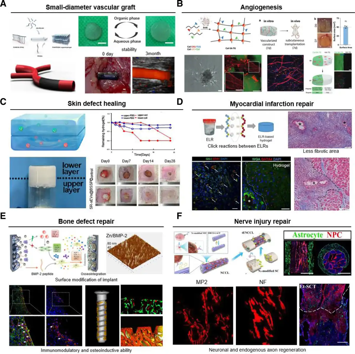
Figure 10.
Illustration showing applications and therapeutic effects of bioorthogonal click chemistry. (A) PHA/PEGDA hydrogel for small-diameter vascular graft. Adapted with permission from Hou et al.[56] (B) Hydrogels for angiogenesis. Adapted with permission from Rütsche et al.[76] (C) Sequential release of small extracellular vesicle hydrogels for scarless wound healing. Adapted with permission from Shen et al.[55] (D) ELR-based hydrogel for myocardial infarction repair. Adapted with permission from Contessotto et al.[53] (E) BMP-2 modified titanium screws for bone defect repair. Adapted with permission from ref.[65] (F) Eda-targeted engineered spinal cord-like transplant for nerve injury repair. Adapted with permission from Liu et al.[84]
In addition to surface modification, bioorthogonal click chemistry can enable the efficient cross-linking of polymer without toxic catalysts or UV irradiation. Therefore, bioorthogonal click chemistry-based hydrogels can provide in situ gelation with minimally invasive injections. Importantly, due to the orthogonality of the reaction, the in situ gelation process is not affected by other biochemistry at the defect site. Liu et al[48] developed an injectable bone cement system and observed obvious osteogenesis and vascularization at the defect site after 4 weeks of implantation in the rat cranial defect model, while the defect site of the control group was basically filled with fibrous tissue.
In summary, bioorthogonal click chemistry enables bone grafts such as hydrogels or titanium screws to achieve rapid in situ cross-linking or efficient surface modification, respectively, simplifying the construction or surface modification process of conventional bone grafts. Importantly, the covalent bond formed under physiological conditions enables bone grafts to have long-term and stable biological activity.
3.2 Skin defect healing
As a substitute for sutures in special cases, traditional adhesives still have many shortcomings, such as (1) poor tissue adhesion and tensile properties, (2) insufficient biocompatibility, and (3) uncontrollable degradation rate.[119,120] These shortcomings greatly affect the healing effect of skin wounds and limit their development and application. The ideal skin adhesive should have the advantages of safety, rapid solidification under physiological conditions, strong tissue adhesion, and promoting wound healing. The high selectivity of the bioorthogonal click results in fewer by-products, so researchers can utilize bioorthogonal click chemistry to synthesize skin adhesives suitable for different wound healing needs and introduce a variety of biologically active substances in the process.
Recently, Wang et al[51] applied a hydrogel grafted with REG peptide, a functional derivative of erythroid differentiation regulator 1, to a mouse skin wound model. The hydrogel promoted the migration of fibroblasts, keratinocytes, and ECs by activating the promigration signal of FAK. Compared with the control group, the hydrogel accelerated wound healing and improved healing quality (higher epithelial cell density, higher microvessel density, more collagen deposition). The BSSPD hydrogel prepared by Shen et al[55] via thiol-ene click chemistry can release small extracellular vesicles secreted by bone marrow mesenchymal stem cells and miR-29b-3p-rich bone marrow mesenchymal stem cells from the lower and upper layers at the early and late stages of wound healing, respectively. In the rat full-thickness skin defect model, the sequentially released small extracellular vesicle hydrogel group healed faster than the control group, and the collagen arrangement and vascular structure distribution were more uniform. In addition, in the rabbit ear skin defect model, which is more similar to human wound scar healing, the wound healing efficiency and quality are consistent with those in the rat model. Importantly, after the application of BSSPD hydrogel with sEVs released in this order, the scar thickness and scar elevation index were significantly smaller than those of the control group (Figure 10A).
The earlier results indicate that the hydrogel based on bioorthogonal click chemistry can be rapidly cross-linked in situ to achieve the injectability of tissue adhesives. It can also offer more functions to the biomaterials by adjusting the physical and chemical properties to meet the needs of skin wound healing under different conditions. In addition, the bioorthogonal click reaction under physiological conditions ensures the activity of the functional material carried out and improves the quality of wound healing while maintaining the wound healing rate.
3.3 Nerve injury repair
In the process of nerve injury, the damage of vascular structure causes pathophysiological changes such as inflammatory response, free radical production, and ion imbalance at the site of injury.[121,122] In addition, poor regeneration of endogenous nerve cells and glial scarring[123] caused by reactive proliferation of glial cells inhibit the recovery of structure and function after nerve injury. Therefore, it is a common method to repair damaged and degenerated tissues by transplanting stem cells to differentiate into mature cells or secreting molecules to induce endogenous regeneration.[124,125] Because of the orthogonality of click reaction, it does not affect cell viability and function in the process of covalently attaching cells to biomaterials.
Liu et al[84] constructed engineered spinal cord-like grafts with drug delivery functions. Compared with noncovalent attachment, astrocytes and NPCs covalently attached to collagen fibers by bioorthogonal click chemistry can better adhere, spread, and differentiate. In addition, DBCO group-modified Eda lipid nanoparticles can be delivered to the engineered graft via SPAAC to improve the microenvironment of spinal cord injury. After delivery of Eda-containing nanoparticles by bioorthogonal click chemistry, the expression levels of proapoptotic proteins and lipid peroxidation markers were significantly downregulated at the spinal cord injury site. Finally, the engineered spinal cord-like graft with drug-guided function increased the number of newborn neurons at the spinal cord injury site and promoted the regeneration of endogenous axons and blood vessels (Figure 10E). Zhang et al[52] prepared a biodegradable self-adhesive bandage (SADB) loaded with XMU-MP-1 nanoparticles, which can promote nerve repair through SPAAC. In the rat sciatic nerve transection model, the bandage can directly bind the nerve and reduce the operation time significantly. Electrophysiological evaluation and histological examination confirmed that SADB promoted myelin regeneration and nerve repair and had better neurological function recovery (faster nerve conduction velocity and shorter latency) than the control group.
The covalent binding formed by bioorthogonal click chemistry tightly connects transplanted cells and biomaterials to promote nerve regeneration without interfering with cell proliferation and differentiation. In addition, the fast reaction rate and good biocompatibility of bioorthogonal click chemistry can not only achieve targeted drug delivery but also encapsulate drugs that promote nerve regeneration during material synthesis. In view of the above advantages, injectable hydrogels based on bioorthogonal click chemistry can be constructed during spinal cord injury repair to further reduce secondary damage to the injured site and enhance graft and tissue integration,[126] thereby promoting nerve regeneration.
3.4 Angiogenesis and vascular graft
The viability and functionality of engineered tissue grafts in vivo are dependent on a rich blood vessel supply of abundant oxygen and nutrients. Thus, the vascularization of tissue-engineered grafts constitutes a critical intervention to enhance the repair and regeneration of tissues. Slow angiogenesis or insufficient vascular perfusion is a common cause of graft failure. Therefore, vascularization in tissue-engineered grafts is a key step in promoting tissue repair and regeneration.[127]
Jia et al[75] utilized peptide α1, which supports ECs to functionalize alginate hydrogels through CuAAC. The results demonstrated that the hydrogel can encourage EC adhesion, spreading, migration, and the formation of vascular networks. Additionally, the hydrogel can reduce muscle degeneration and fibrosis area by increasing arterial and capillary development in a mouse hindlimb ischemia model. Marsico et al[128] linked 2 ELRs that featured both MMP binding sites and integrin-binding sites using SPAAC to create elastin-like hydrogels. In a severe limb ischemia mouse model, there was a substantial rise in capillary density and length, small artery density, and cross-sectional area. In addition, crucial biological processes such as EC proliferation and angiogenesis were highly upregulated. It also contributed to ECM remodeling and tissue repair by controlling N-glycosylation (Figure 10F). Rütsche et al.[76] also used CuAAC to couple substrate peptides with novel collagen derivatives, which were then mediated by transpeptidase A to form hydrogels. EC mixed with the hydrogel facilitates the growth of blood vessels and lymphatic vessels without the use of VEGF. It is worth noting that extracellular vesicles have demonstrated beneficial therapeutic effects in numerous injury repair models. Xing et al[69] fixed them in collagen hydrogel by SPAAC to achieve long-term retention in vivo and observed effective angiogenesis and host vessel ingrowth after subcutaneous implantation in mice.
At present, allografts, xenografts, and synthetic grafts cannot meet the needs of vascular grafts, especially small-diameter vascular grafts. Therefore, scientists have used tissue engineering as a means to produce suitable small-diameter vascular grafts. Due to the safety and high efficiency of the reaction process, bioorthogonal click chemistry can be used as a cross-linking tool to realize the synthesis and functionalization of materials.
Navarro et al[67] used thiol-ene click chemistry to attach heparin molecules to biomimetic tubular scaffolds. After the stent was implanted in the rat model, ultrasound results showed that blood flow was smooth, and the inner diameter and total diameter of the vessel wall were almost the same as before implantation. The scaffold began to degrade and be infiltrated by host cells 1 week after implantation and was replaced by natural cells 1 month later. After 3 months, the blood vessels remained unobstructed. Histological examination showed that the cells of the remodeling scaffold were smooth muscle cells and ECs, and the new tissue was rich in natural collagen and elastin. These results confirmed the reconstruction of neovascularization. Hou et al[56] also prepared gel blood vessels by thiol-ene click reaction between unsaturated functional microorganisms PHA and PEGDA combined with 2-dimensional material graphdiyne (GDY). The application of the bioorthogonal click reaction gives the vascular graft not only better biocompatibility than traditional organic materials but also better mechanical properties. It can also remain stable in the body for a long time. After the graft was implanted into the rabbit carotid artery defect for 1 month and 3 months, the overall patency rate of the PHA/PEGDA-GDY group remained at about 90%. Histological analysis showed that inflammatory cell infiltration gradually decreased within 3 months, providing a good immune microenvironment for tissue regeneration. Importantly, the vascular graft promoted vascular cell proliferation and extracellular matrix deposition, forming a stable vascular-like tissue structure (Figure 10C).
The results of these studies showed that the construction strategy based on bioorthogonal click chemistry not only overcomes the problem of poor biocompatibility of traditional organic materials but also retains their good physical and chemical properties and supports the proliferation and differentiation of vascular regeneration-related cells. In addition, vascular grafts can be endowed with more functions through efficient bioorthogonal click reactions, making them more like natural blood vessels. In summary, bioorthogonal click chemistry allows bioactive substances that facilitate angiogenesis to be tightly bound to biomaterials to prolong their retention in the body and has little interference with their functional activity during the binding process, providing a suitable solution for tissue engineering and regenerative medicine to promote vascularization.
3.5 Myocardial infarction repair
Stent implantation is the main clinical treatment for coronary artery disease.[129] Bare stent or drug-eluting stent implantation has problems such as in-stent restenosis, late thrombosis, and incomplete endothelial healing.[130,131] Therefore, one of the major challenges in this field is how to effectively design a coating that can rapidly promote the healing of the vascular endothelium.
Zhang et al[132] employed catechol-modified natural chitosan as a carrier to construct an organic/metal ion composite polypeptide coating. The stent, coated with the polypeptide, triggered the zinc ions to release endogenous nitric oxide (NO) gas. Subsequently, the REDV peptide, which has a cell-promoting capability, was efficiently and quickly grafted onto the coating surface via thiol-ene click reaction. The coating inhibited the formation of intravascular thrombosis by immobilizing REDV peptides and catalyzing the release of endogenous NO. After 1 week of implantation into the rabbit abdominal aorta, the control group had a stent occlusion rate of over 50%, whereas the bioorthogonal click chemistry-based coated stent had no visible occlusion. Furthermore, the composite coating effectively inhibits excessive intimal proliferation by suppressing the inflammatory response and maintaining the contractile smooth muscle cell phenotype. This, in turn, promotes rapid vascular endothelial healing. The group additionally utilized bioorthogonal click reaction to integrate both bivalirudin (BVLD)[74] and VEGF[68] onto the surface of vascular scaffolds. They could maintain excellent stability and biological functionality, thus effectively promoting vascular repair.
While stent implantation is able to postpone the progression of myocardial infarction, it cannot repair the damaged myocardium. Therefore, Contessotto et al[53] designed an ELR hydrogel that mimics the ECM via SPAAC. The hydrogel contains a functional domain for cell adhesion and a protease cleavage site. In the group treated with ELRs, the ejection fraction of surgically induced nontransmural myocardial infarction significantly improved by 16.2%, and fibrosis in the ischemic core decreased by 44.3%. Furthermore, the hydrogel’s degradable properties facilitate EC sprouting and stromal cell migration, thus enhancing angiogenesis in the infarct core area (Figure 10B). Since SPAAC is performed under physiological conditions and does not use any potentially harmful catalysts or by-products, the hydrogel can be safely applied in vivo and the activity of the functional ingredients is not affected during the reaction, while the covalent bond formed between the ELRs ensures its stability. Stem cell therapy is also a promising strategy for treating myocardial infarction. Li et al[93] utilized the high specificity of bioorthogonal click chemistry to deliver endogenous stem cells to the infarcted area for cardiac repair. After 4 weeks of application in the mouse model of myocardial infarction, the experimental results showed that a large number of cardiomyocytes proliferated in the infarcted area (about 2.5 times that of the control group), cardiac function (left ventricular ejection fraction and short-axis shortening rate) was significantly improved, and interstitial and perivascular fibrosis was reduced. The findings indicated that SPAAC successfully recruited stem cells to the MI area and improved cardiac function. The bioorthogonal click reaction is highly specific and ensures efficient interaction between the DBCO group linked to platelets and the azide group linked to stem cells. Treatment with this strategy did not impact susceptible organs, including the liver, spleen, lung, and kidney, thus reflecting good biocompatibility.
In summary, the high efficiency and specificity of bioorthogonal click chemistry have enabled the surface modification of vascular scaffolds, the construction of hydrogels encapsulating bioactive substances, and the targeting of therapeutic cells to infarcted areas. Crucially, all of these can be performed under physiological conditions without any harmful catalysts or cytotoxic agents. Researchers can utilize the earlier advantages of bioorthogonal click chemistry to repair the heart after myocardial infarction from multiple perspectives, such as delaying the progression of myocardial infarction and promoting the recovery of cardiac function (Table 4).
Table 4
Summary of applications in promoting tissue or organ regeneration.
| Disease/injury | Strategy | Reaction | Regeneration effect | References |
|---|---|---|---|---|
| Bone and cartilage defect | Synthesis of biomaterials | SPAAC | New bone formation; vascularization | Liu et al[47] |
| SPAAC | Osteogenesis; vascularization | Liu et al[48] | ||
| SPAAC | Higher new bone volume | Gan et al[49] | ||
| SPAAC | Better osseointegration; osteogenic differentiation | Deng et al[50] | ||
| Functionalization of biomaterials | Thiol-ene | Delayed joint space narrowing and increased content of cartilage matrix | Yao et al[71] | |
| CuAAC | Cartilage regeneration and integration | He et al[73] | ||
| CuAAC | Differentiation of HMSC | Beeren et al[79] | ||
| SPAAC | Osteogenesis; osseointegration | Wang et al[65] | ||
| Cell–biomaterial interaction | IEDDA | More chondrocytes and richer ECM in damage area | Co et al[92] | |
| Skin defect | Synthesis of biomaterials | SPAAC | Re-epithelialization, collagen deposition, and microvessel formation | Wang et al[51] |
| IEDDA | Faster healing speed; better healing quality | Li et al[54] | ||
| Thiol-ene | Smaller volume of hypertrophic scar tissue | Shen et al[55] | ||
| Thiol-ene | Fast healing of diabetic wounds | Shou et al[57] | ||
| Amino-alkyne | Less local inflammation; faster healing speed | Jin et al[59] | ||
| Nerve injury | Synthesis of biomaterials | SPAAC | Faster conduction velocity and shorter latency | Zhang et al[52] |
| Cell–biomaterial interaction | SPAAC | Less cell demand and better therapeutic effect | Oh et al[87] | |
| SPAAC | More neurons and endogenous axons regeneration | Liu et al[84] | ||
| Vascular graft | Synthesis of biomaterials | Thiol-ene | Long-term intravascular patency | Hou et al[56] |
| Functionalization of biomaterials | Thiol-ene | ECs and SMCs infiltration | Navarro et al[67] | |
| Thiol-ene | Better endothelialization and endothelial function | Zhang et al[68] | ||
| Angiogenesis | Functionalization of biomaterials | CuAAC | Larger neovascularization area | Yao et al[72] |
| CuAAC | Enhanced blood flow recovery properties | Jia et al[75] | ||
| CuAAC | Formation of blood and lymphatic vessels in the absence of exogenous VEGF | Rütsche et al[76] | ||
| Thiol-ene | More blood vessels around the scaffold | Yao et al[77] | ||
| SPAAC | Host vascular ingrowth | Xing et al[69] | ||
| Myocardial infarction | Synthesis of biomaterials | SPAAC | Better cardiac function; | Contessotto et al[53] |
| Functionalization of biomaterials | Thiol-ene | Better endothelialization and endothelial function | Zhang et al[74] | |
| Thiol-epoxy | Better cardiac function; neovascularization | Zou et al[78] | ||
| Cell–biomaterial interaction | SPAAC | More cardiomyocytes regeneration | Li et al[93] | |
| Hydrazide-aldehyde | Better cardiac function and angiogenesis | Wu et al[94] |
4. Conclusion and prospect
In recent years, bioorthogonal click chemistry has been widely used in tissue engineering and regenerative medicine due to its advantages of good biocompatibility, fast reaction rate, high reaction specificity, and stable reaction products. This review focuses on the application of bioorthogonal click chemistry in the synthesis of biomaterials, functionalization of biomaterials, and cell–biomaterial interaction to achieve tissue repair and regeneration. Bioorthogonal click chemistry provides researchers with a modular connection strategy. It not only overcomes many shortcomings of traditional organic synthesis reactions but also successfully transforms the covalent cross-linking process into a living system. Depending on the different application scenarios, researchers tend to select specific bioorthogonal click reactions based on their unique advantages and limitations. For example, CuAAC has excellent reaction kinetics but requires copper, which is cytotoxic, as a catalyst. This limits its application mainly to the synthesis of biomaterials in vitro. As the representative of copper-free bioorthogonal click chemistry, SPAAC can be used for in situ synthesis of biomaterials or cell modification in vivo. However, its drawback is the relatively slow reaction rate. Thiol-ene click reaction has a faster reaction rate and is photoresponsive, but their use is limited in biological systems rich in thiols. Therefore, the excellent research introduced in this article is conducive to providing inspiration for researchers to choose the appropriate tools according to their needs.
In addition to the classical bioorthogonal click reaction, more reactions will be used in regenerative medicine in the future. For example, as the second generation click chemistry, SuFEx click reaction has the advantages of high selectivity, stability, and rapidity. In materials science, SuFEx reaction can be used to synthesize polymer materials with specific functions, and in the field of medicine, it can be used to design covalent drugs to improve the stability and bioavailability of drugs in vivo by forming stable sulfur–carbon bonds. With the deepening of research, SuFEx reaction is expected to play a greater role in tissue engineering and regenerative medicine.
To further promote the clinical transformation of bioorthogonal click chemistry, some challenges need to be solved. (1) Suitable equipment: when applying in situ synthesized hydrogels, the two precursors need to be fully mixed before cross-linking occurs. Many studies have used special devices, such as 2-way syringes. In the future, more devices may need to be developed to give full play to the advantages of bioorthogonal click chemistry in more scenarios. (2) High cost: the high price of clickable groups, especially in the IEDDA reaction, is a significant problem because the amount of material needed for hydrogels is generally larger than that required for nanoparticles. (3) Specific dose: for cell modification, safety issues are common and should be carefully considered. The process of introducing clickable groups, especially in the case of high-concentration drugs, is likely to affect the activity of biomolecules or cells.[133] Studies have confirmed that high concentrations (>20 μM) of Ac4ManNAz have a negative impact on the proliferation, adhesion, and signal transduction of EPCs.[134] The ability of different types of cells to express azide groups through the sialic acid pathway is different, which makes the required Ac4ManNAz concentration and the negative effects on cells different. Therefore, exploring the maximum tolerated dose of Ac4ManNAz in cells and humans and finding ways to prolong the half-life of glycans is an urgent problem to be solved in the process of bioorthogonal click chemistry to clinical transformation. (4) Long-term efficacy and biosafety: the ultimate goal of all biomaterial development is to be used in the human body to treat diseases. Before this, researchers should also determine the long-term stability and safety of bioorthogonal click reactions in the human body. For example, whether the formed covalent bond can be stably connected in the body, and whether the degradation of biomaterials with clickable groups will affect the normal physiological function of tissues and organs or smooth discharge from the body. In addition, the clickable group in the glycan expressed on the cell membrane by metabolic glycoengineering has a fast metabolic rate and a short half-life. For long-term cell tracking and cell or organ targeting, this strategy is not available.
Looking forward to the future, we believe that bioorthogonal click chemistry can be further developed from the following aspects. First, in view of the excellent biocompatibility of bioorthogonal click chemistry, cells can be used as module units and connected to each other by bioorthogonal click chemistry to construct macroscopic tissues. Recently, Lavrador et al connected cells together through azide groups and DBCO groups to form living human tissues (Cellgels). Cellgels have the ability of living cells to perceive and respond to the environment, showing the ability of autonomous tissue integration behavior, mechanical maturation, biological self-healing, biological specific adhesion, and promoting wound healing.[135] In addition, the authors have verified it in a variety of cells, including dermal fibroblasts (hDFs), umbilical cord - derived endothelial cells (hUVECs), and pancreatic cancer cells (hPANC - 1), which have the potential to extend it to any type of human cells. In the future, scientists can use bioorthogonal click chemistry to construct a variety of human cells into tissues and even use different tissues as modular units to construct organs with biological functions. Second, bioorthogonal click chemistry offers a means of intervening in tissue regeneration at the genetic level, such as exploring the differences in nucleic acid metabolism during tissue regeneration by labeling oligonucleotides[136–137–138–139] or protein,[140,141] generating engineered nucleic acid sequences by cross-linking oligonucleotides or single-stranded DNA and RNA to provide a more convenient tool for tissue engineering. Currently, scientists have utilized bioorthogonal click chemistry to effectively modify CRISPR-Cas in the field of gene editing, achieving large-scale and low-cost sgRNA synthesis.[142–143–144] Finally, physical factors should be introduced into bioorthogonal click chemistry to achieve precise regulation of tissue regeneration processes, including but not limited to ultrasound, light, temperature, and pressure. Bioorthogonal click reaction causes irreversible covalent bonds between molecules. Once the substrates contact each other, the reaction occurs immediately and irreversibly. This means that the hydrogel cannot return to the solution state, and small molecules can only be permanently connected to the scaffold. This reduces the controllability of the bioorthogonal click reaction. Therefore, precise space-time control brought by physical factors will improve the intelligence of bioorthogonal click chemistry and also enable bioorthogonal click chemistry to be applied to more complex tissue regeneration processes. For example, drug delivery and cell targeting will occur at specific tissue repair stages. At present, there are already light-triggered click reactions that can synthesize different organic structures in a fast and accurate manner under mild conditions, such as dipolar cycloadditions,[145] Diels−Alder reactions,[146] alternating radical propagation chain transfer reactions,[147] which gives the reaction space and time control. Therefore, bioorthogonal click reactions triggered by light or other physical factors may bring new opportunities for tissue engineering and regenerative medicine. In summary, with the advancement of novel theories and technologies, we believe that the future application of bioorthogonal click chemistry in tissue engineering and regenerative medicine will continue to expand and exhibit its distinct advantages.
Acknowledgments
Thanks for the Young Elite Scientists Sponsorship Program (1st Doctoral Student Special Program) by CAST.
Funding
This study was supported by the National Natural Science Foundation of China (NSFC) (No. 824B2061, 82402494), the Science and Technology Commission of Shanghai Municipality (No. 24YF2735500), the Clinical Research Plan of Shanghai Hospital Development Center (multicenter clinical research project for major diseases, No. SHDC2020CR1021B), National Key Research and Development Program of China (No. 2022YFC2407401), National Key Research and Development Program of China (No. 2024YFC3044600), the Foundation from Shanghai Municipal Health Commission (No. 202340245), Ningbo Top Medical and Health Research Program (No.2022030208).
Conflicts of interests
The authors declare that they have no conflicts of interest.
Author contributions
C.C., and W.S. are corresponding authors. Writing of original draft: Y.H., Y.C., H.T. and M.M. Review and editing: Z.P., W.L., R.C., Q.B., L.W., B.X., L.Z., Y.S., L.W., W.S., G.Z., and C.C. All authors have read and agreed to the published version of the manuscript.
References
- [1] Kolb HC, Finn MG, Sharpless KB. Click chemistry: diverse chemical function from a few good reactions. Angew Chem Int Ed Engl. 2001;40(11):2004–2021.
- [2] Rostovtsev VV, Green LG, Fokin VV, et al. A stepwise huisgen cycloaddition process: copper(I)-catalyzed regioselective “ligation” of azides and terminal alkynes. Angew Chem Int Ed Engl. 2002;41(14):2596–2599.
- [3] Tornøe CW, Christensen C, Meldal M. Peptidotriazoles on solid phase: [1,2,3]-triazoles by regiospecific copper(I)-catalyzed 1,3-dipolar cycloadditions of terminal alkynes to azides. J Org Chem. 2002;67(9):3057–3064.
- [4] Dube DH, Bertozzi CR. Metabolic oligosaccharide engineering as a tool for glycobiology. Curr Opin Chem Biol. 2003;7(5):616–625.
- [5] Lemieux GA, de Graffenried CL, Bertozzi CR. A fluorogenic dye activated by the Staudinger ligation. J Am Chem Soc. 2003;125(16):4708–4709.
- [6] Saxon E, Bertozzi CR. Cell surface engineering by a modified Staudinger reaction. Science. 2000;287(5460):2007–2010.
- [7] Agard NJ, Prescher JA, Bertozzi CR. A strain-promoted [3 + 2] azide− alkyne cycloaddition for covalent modification of biomolecules in living systems. J Am Chem Soc. 2004;126(46):15046–15047.
- [8] Dong J, Krasnova L, Finn MG, et al. Sulfur(VI) fluoride exchange (SuFEx): another good reaction for click chemistry. Angew Chem Int Ed Engl. 2014;53(36):9430–9448.
- [9] Blackman ML, Royzen M, Fox JM. Tetrazine ligation: fast bioconjugation based on inverse-electron-demand Diels−Alder reactivity. J Am Chem Soc. 2008;130(41):13518–13519.
- [10] Gress A, Völkel A, Schlaad H. Thio-click modification of poly[2-(3-butenyl)-2-oxazoline]. Macromolecules. 2007;40(22):7928–7933.
- [11] Darko A, Wallace S, Dmitrenko O, et al. Conformationally strained trans-cyclooctene with improved stability and excellent reactivity in tetrazine ligation. Chem Sci. 2014;5(10):3770–3776.
- [12] Oliveira BL, Guo Z, Bernardes GJL. Inverse electron demand Diels–Alder reactions in chemical biology. Chem Soc Rev. 2017;46(16):4895–4950.
- [13] Peschke F, Taladriz-Sender A, Andrews MJ, et al. Glutathione mediates control of dual differential bio-orthogonal labelling of biomolecules. Angew Chem Int Ed Engl. 2023;62(50):e202313063.
- [14] Ferrari M. Cancer nanotechnology: opportunities and challenges. Nat Rev Cancer. 2005;5(3):161–171.
- [15] Laughlin ST, Baskin JM, Amacher SL, et al. In vivo imaging of membrane-associated glycans in developing zebrafish. Science. 2008;320(5876):664–667.
- [16] Dommerholt J, Schmidt S, Temming R, et al. Readily accessible bicyclononynes for bioorthogonal labeling and three-dimensional imaging of living cells. Angew Chem Int Ed Engl. 2010;49(49):9422–9425.
- [17] Dommerholt J, van Rooijen O, Borrmann A, et al. Highly accelerated inverse electron-demand cycloaddition of electron-deficient azides with aliphatic cyclooctynes. Nat Commun. 2014;5(1):5378.
- [18] Holt SE, Rakoski A, Jivan F, et al. Hydrogel synthesis and stabilization via tetrazine click-induced secondary interactions. Macromol Rapid Commun. 2020;41(14):2000287.
- [19] Cao W, Wang H, Quan M, et al. Reversible control of tetrazine bioorthogonal reactivity by naphthotube-mediated host-guest recognition. Chem. 2023;9(10):2881–2901.
- [20] Xue EY, Lee ACK, Chow KT, et al. Promotion and detection of cell–cell interactions through a bioorthogonal approach. J Am Chem Soc. 2024;146(25):17334–17347.
- [21] Song W, Wang Y, Qu J, et al. A photoinducible 1,3-dipolar cycloaddition reaction for rapid, selective modification of tetrazole-containing proteins. Angew Chem Int Ed Engl. 2008;47(15):2832–2835.
- [22] Song W, Wang Y, Qu J, et al. Selective functionalization of a genetically encoded alkene-containing protein via “photoclick chemistry” in bacterial cells. J Am Chem Soc. 2008;130(30):9654–9655.
- [23] Gandavarapu NR, Azagarsamy MA, Anseth KS. Photo-click living strategy for controlled, reversible exchange of biochemical ligands. Adv Mater. 2014;26(16):2521–2526.
- [24] Adzima BJ, Tao Y, Kloxin CJ, et al. Spatial and temporal control of the alkyne–azide cycloaddition by photoinitiated Cu(II) reduction. Nat Chem. 2011;3(3):256–259.
- [25] Ritter SC, König B. Signal amplification and transduction by photo-activated catalysis. Chem Commun. 2006;7(45):4694–4696.
- [26] Lim RKV, Lin Q. Azirine ligation: fast and selective protein conjugation via photoinduced azirine–alkene cycloaddition. Chem Commun (Camb). 2010;46(42):7993–7995.
- [27] Yang L, Lin X, Zhou J, et al. Cell membrane-biomimetic coating via click-mediated liposome fusion for mitigating the foreign-body reaction. Biomaterials. 2021;271:120768.
- [28] Hafa L, Breideband L, Posada LR, et al. Light sheet-based laser patterning bioprinting produces long-term viable full-thickness skin constructs. Adv Mater. 2024;36:e2306258.
- [29] Elbert DL, Pratt AB, Lutolf MP, et al. Protein delivery from materials formed by self-selective conjugate addition reactions. J Control Release. 2001;76(1):11–25.
- [30] Ossipov DA, Hilborn J. Poly(vinyl alcohol)-based hydrogels formed by “click chemistry”. Macromolecules. 2006;39(5):1709–1718.
- [31] DeForest CA, Polizzotti BD, Anseth KS. Sequential click reactions for synthesizing and patterning three-dimensional cell microenvironments. Nat Mater. 2009;8(8):659–664.
- [32] Lutolf MP, Lauer-Fields JL, Schmoekel HG, et al. Synthetic matrix metalloproteinase-sensitive hydrogels for the conduction of tissue regeneration: engineering cell-invasion characteristics. Proc Natl Acad Sci U S A. 2003;100(9):5413–5418.
- [33] DeForest CA, Sims EA, Anseth KS. Peptide-functionalized click hydrogels with independently tunable mechanics and chemical functionality for 3D cell culture. Chem Mater. 2010;22(16):4783–4790.
- [34] DeForest CA, Anseth KS. Cytocompatible click-based hydrogels with dynamically tunable properties through orthogonal photoconjugation and photocleavage reactions. Nat Chem. 2011;3(12):925–931.
- [35] Wang Q, Cui H, Wang X, et al. Exceptional light sensitivity by thiol–ene click lithography. J Am Chem Soc. 2023;145(5):3064–3074.
- [36] Fu Y, Wu K, Alachouzos G, et al. Efficient, near-infrared light-induced photoclick reaction enabled by upconversion nanoparticles. Adv Funct Mater. 2023;33:2306531.
- [37] Mora-Boza A, Ghebrezadik SG, Leisen JE, et al. Rapid and facile light-based approach to fabricate protease-degradable poly(ethylene glycol)−norbornene microgels for cell encapsulation. Adv Healthcare Mater. 2023;12(26):2300942.
- [38] Zhou C, Wang C, Xu K, et al. Hydrogel platform with tunable stiffness based on magnetic nanoparticles cross-linked GelMA for cartilage regeneration and its intrinsic biomechanism. Bioact Mater. 2023;25:615–628.
- [39] Caldwell AS, Campbell GT, Shekiro KMT, et al. Clickable microgel scaffolds as platforms for 3D cell encapsulation. Adv Healthcare Mater. 2017;6(15):1700254.
- [40] Lin SH, Huang AP, Hsu SH, et al. Injectable, micellar chitosan self-healing hydrogel for asynchronous dual-drug delivery to treat stroke rats. Adv Funct Mater. 2023;33(45):2303853.
- [41] Kim K, Park JH, Park SH, et al. An injectable, click-cross-linked small intestinal submucosa drug depot for the treatment of rheumatoid arthritis. Adv Healthcare Mater. 2016;5(24):3105–3117.
- [42] Liu J, Mckeon L, Garcia J, et al. Additive manufacturing of Ti3C2-MXene-functionalized conductive polymer hydrogels for electromagnetic-interference shielding. Adv Mater. 2022;34(5):2106253.
- [43] Wu S, Hua M, Alsaid Y, et al. Poly(vinyl alcohol) hydrogels with broad-range tunable mechanical properties via the Hofmeister effect. Adv Mater. 2021;33(11):2007829.
- [44] Yang J-A, Yeom J, Hwang BW, et al. In situ-forming injectable hydrogels for regenerative medicine. Prog Polym Sci. 2014;39(12):1973–1986.
- [45] Chen F, Le P, Fernandes-Cunha GM, et al. Bio-orthogonally crosslinked hyaluronate-collagen hydrogel for suture-free corneal defect repair. Biomaterials. 2020;255:120176.
- [46] Lee HJ, Fernandes-Cunha GM, Na K-S, et al. Bio-orthogonally crosslinked, in situ forming corneal stromal tissue substitute. Adv Healthcare Mater. 2018;7(19):1800560.
- [47] Liu X, Gaihre B, Li L, et al. Bioorthogonal “click chemistry” bone cement with bioinspired natural mimicking microstructures for bone repair. ACS Biomater Sci Eng. 2023;9(3):1585–1597.
- [48] Liu X, Camilleri ET, Li L, et al. Injectable catalyst-free “click” organic-inorganic nanohybrid (click-ON) cement for minimally invasive in vivo bone repair. Biomaterials. 2021;276:121014.
- [49] Gan M, Zhou Q, Ge J, et al. Precise in-situ release of microRNA from an injectable hydrogel induces bone regeneration. Acta Biomater. 2021;135:289–303.
- [50] Deng J, Cohen DJ, Sabalewski EL, et al. Semaphorin 3A delivered by a rapidly polymerizing click hydrogel overcomes impaired implant osseointegration in a rat type 2 diabetes model. Acta Biomater. 2023;157:236–251.
- [51] Wang SY, Kim H, Kwak G, et al. Development of biocompatible HA hydrogels embedded with a new synthetic peptide promoting cellular migration for advanced wound care management. Adv Sci (Weinh). 2018;5(11):1800852.
- [52] Zhang J, Chen Y, Huang Y, et al. A 3D-printed self-adhesive bandage with drug release for peripheral nerve repair. Adv Sci (Weinh). 2020;7(23):2002601.
- [53] Contessotto P, Orbanić D, Da Costa M, et al. Elastin-like recombinamers-based hydrogel modulates post-ischemic remodeling in a non-transmural myocardial infarction in sheep. Sci Transl Med. 2021;13(581):eaaz5380.
- [54] Li S, Zhou J, Huang Y, et al. Injectable click chemistry-based bioadhesives for accelerated wound closure. Acta Biomater. 2020;110:95–104.
- [55] Shen Y, Xu G, Huang H, et al. Sequential release of small extracellular vesicles from bilayered thiolated alginate/polyethylene glycol diacrylate hydrogels for scarless wound healing. ACS Nano. 2021;15(4):6352–6368.
- [56] Hou J, Zhang X, Wu Y, et al. Amphiphilic and fatigue-resistant organohydrogels for small-diameter vascular grafts. Sci Adv. 2022;8(30):eabn5360.
- [57] Shou Y, Le Z, Cheng HS, et al. Mechano-activated cell therapy for accelerated diabetic wound healing. Adv Mater. 2023;35:e2304638.
- [58] Lei X-X, Hu J-J, Zou C-Y, et al. Multifunctional two-component in-situ hydrogel for esophageal submucosal dissection for mucosa uplift, postoperative wound closure and rapid healing. Bioact Mater. 2023;27:461–473.
- [59] Jin X, Shang Y, Zou Y, et al. Injectable hypoxia-induced conductive hydrogel to promote diabetic wound healing. ACS Appl Mater Interfaces. 2020;12(51):56681–56691.
- [60] Xu X, Xia X, Zhang K, et al. Bioadhesive hydrogels demonstrating pH-independent and ultrafast gelation promote gastric ulcer healing in pigs. Sci Transl Med. 2020;12(558):eaba8014.
- [61] Kang N-W, Jang K, Song E, et al. In situ-forming, bioorthogonally cross-linked, nanocluster-reinforced hydrogel for the regeneration of corneal defects. ACS Nano. 2024;18(33):21925–21938.
- [62] Xu J, Feng E, Song J. Bioorthogonally cross-linked hydrogel network with precisely controlled disintegration time over a broad range. J Am Chem Soc. 2014;136(11):4105–4108.
- [63] Koshy ST, Desai RM, Joly P, et al. Click-crosslinked injectable gelatin hydrogels. Adv Healthcare Mater. 2016;5(5):541–547.
- [64] Tournier P, Saint-Pé G, Lagneau N, et al. Clickable dynamic bioinks enable post-printing modifications of construct composition and mechanical properties controlled over time and space. Adv Sci. 2023;10(30):e2300055.
- [65] Wang T, Bai J, Lu M, et al. Engineering immunomodulatory and osteoinductive implant surfaces via mussel adhesion-mediated ion coordination and molecular clicking. Nat Commun. 2022;13(1):160.
- [66] Shi Z, Neoh KG, Kang ET, et al. Titanium with surface-grafted dextran and immobilized bone morphogenetic protein-2 for inhibition of bacterial adhesion and enhancement of osteoblast functions. Tissue Eng Part A. 2009;15(2):417–426.
- [67] Navarro RS, Jiang L, Ouyang Y, et al. Biomimetic tubular scaffold with heparin conjugation for rapid degradation in in situ regeneration of a small diameter neoartery. Biomaterials. 2021;274:120874.
- [68] Zhang B, Qin Y, Yang L, et al. An organic selenium and VEGF-conjugated bioinspired coating promotes vascular healing. Biomaterials. 2022;287:121654.
- [69] Xing Y, Yerneni SS, Wang W, et al. Engineering pro-angiogenic biomaterials via chemoselective extracellular vesicle immobilization. Biomaterials. 2022;281:121357.
- [70] Cao F, Jin L, Gao Y, et al. Artificial-enzymes-armed Bifidobacterium longum probiotics for alleviating intestinal inflammation and microbiota dysbiosis. Nat Nanotechnol. 2023;18(6):617–627.
- [71] Yao Y, Wei G, Deng L, et al. Visualizable and lubricating hydrogel microspheres via NanoPOSS for cartilage regeneration. Adv Sci. 2023;10(15):e2207438.
- [72] Yao T, Nunen T. van, Rivero R, et al. Electrospun scaffolds functionalized with a hydrogen sulfide donor stimulate angiogenesis. ACS Appl Mater Interfaces. 2022;14(25):28628–28638.
- [73] He S, Fu X, Wang L, et al. Self-assemble silk fibroin microcapsules for cartilage regeneration through gene delivery and immune regulation. Small. 2023;19(40):2302799.
- [74] Zhang B, Qin Y, Yang L, et al. A polyphenol-network-mediated coating modulates inflammation and vascular healing on vascular stents. ACS Nano. 2022;16(4):6585–6597.
- [75] Jia J, Jeon EJ, Li M, et al. Evolutionarily conserved sequence motif analysis guides development of chemically defined hydrogels for therapeutic vascularization. Sci Adv. 2020;6(28):eaaz5894.
- [76] Rütsche D, Nanni M, Rüdisser S, et al. Enzymatically crosslinked collagen as a versatile matrix for in vitro and in vivo co-engineering of blood and lymphatic vasculature. Adv Mater. 2023;35(16):2209476.
- [77] Yao T, Chen H, Wang R, et al. Thiol-ene conjugation of a VEGF peptide to electrospun scaffolds for potential applications in angiogenesis. Bioact Mater. 2023;20:306–317.
- [78] Zou Y, Li L, Li Y, et al. Restoring cardiac functions after myocardial infarction–ischemia/reperfusion via an exosome anchoring conductive hydrogel. ACS Appl Mater Interfaces. 2021;13(48):56892–56908.
- [79] Beeren IAO, Dijkstra PJ, Lourenço AFH, et al. Installation of click-type functional groups enable the creation of an additive manufactured construct for the osteochondral interface. Biofabrication. 2023;15(1):014106.
- [80] Bailey SJ, Hopkins E, Baxter NJ, et al. Diels–Alder photoclick patterning of extracellular matrix for spatially controlled cell behaviors. Adv Mater. 2023;35:e2303453.
- [81] Hao D, Lu L, Song H, et al. Engineered extracellular vesicles with high collagen-binding affinity present superior in situ retention and therapeutic efficacy in tissue repair. Theranostics. 2022;12(13):6021–6037.
- [82] Zeng J, Gu C, Sun Y, et al. Engineering of M2 macrophages-derived exosomes via click chemistry for spinal cord injury repair. Adv Healthcare Mater. 2023;12(11):2203391.
- [83] Ruan H, Li Y, Wang C, et al. Click chemistry extracellular vesicle/peptide/chemokine nanocarriers for treating central nervous system injuries. Acta Pharm Sin B. 2023;13(5):2202–2218.
- [84] Liu W, Xu B, Zhao S, et al. Spinal cord tissue engineering via covalent interaction between biomaterials and cells. Sci Adv. 2023;9(6):eade8829.
- [85] Mao D, Zhang C, Kenry, . Bio-orthogonal click reaction-enabled highly specific in situ cellularization of tissue engineering scaffolds. Biomaterials. 2020;230:119615.
- [86] Ueda N, Sawada S, Yuasa F, et al. Covalent stem cell-combining injectable materials with enhanced stemness and controlled differentiation in vivo. ACS Appl Mater Interfaces. 2022;14(47):52618–52633.
- [87] Oh B, Swaminathan V, Malkovskiy A, et al. Single-cell encapsulation via click-chemistry alters production of paracrine factors from neural progenitor cells. Adv Sci (Weinh). 2020;7(8):1902573.
- [88] Lee SY, Lee S, Lee J, et al. Non-invasive stem cell tracking in hindlimb ischemia animal model using bio-orthogonal copper-free click chemistry. Biochem Biophys Res Commun. 2016;479(4):779–786.
- [89] Lim S, Yoon HY, Park S-J, et al. Predicting in vivo therapeutic efficacy of bioorthogonally labeled endothelial progenitor cells in hind limb ischemia models via non-invasive fluorescence molecular tomography. Biomaterials. 2021;266:120472.
- [90] Lim S, Yoon HY, Jang HJ, et al. Dual-modal imaging-guided precise tracking of bioorthogonally labeled mesenchymal stem cells in mouse brain stroke. ACS Nano. 2019;13(10):10991–11007.
- [91] Yoon HI, Yhee JY, Na JH, et al. Bioorthogonal copper free click chemistry for labeling and tracking of chondrocytes in vivo. Bioconjug Chem. 2016;27(4):927–936.
- [92] Co CM, Izuagbe S, Zhou J, et al. Click chemistry-based pre-targeting cell delivery for cartilage regeneration. Regener Biomater. 2021;8(3):rbab018.
- [93] Li Z, Shen D, Hu S, et al. Pretargeting and bioorthogonal click chemistry-mediated endogenous stem cell homing for heart repair. ACS Nano. 2018;12(12):12193–12200.
- [94] Wu T, Zhang X, Liu Y, et al. Wet adhesive hydrogel cardiac patch loaded with anti-oxidative, autophagy-regulating molecule capsules and MSCs for restoring infarcted myocardium. Bioact Mater. 2023;21:20–31.
- [95] Deng C, Li Z, Lu L, et al. Sophisticated magneto-mechanical actuation promotes in situ stem cell assembly and chondrogenesis for treating osteoarthritis. ACS Nano. 2023;17:21690–21707.
- [96] Ma L, Thapa BR, Le Suer JA, et al. Airway stem cell reconstitution by the transplantation of primary or pluripotent stem cell-derived basal cells. Cell Stem Cell. 2023;30(9):1199–1216.e7.
- [97] Kurmann AA, Serra M, Hawkins F, et al. Regeneration of thyroid function by transplantation of differentiated pluripotent stem cells. Cell Stem Cell. 2015;17(5):527–542.
- [98] Wang Y, Zoneff ER, Thomas JW, et al. Hydrogel oxygen reservoirs increase functional integration of neural stem cell grafts by meeting metabolic demands. Nat Commun. 2023;14(1):457.
- [99] Kawabori M, Kuroda S, Ito M, et al. Timing and cell dose determine therapeutic effects of bone marrow stromal cell transplantation in rat model of cerebral infarct. Neuropathology. 2013;33(2):140–148.
- [100] Ranjbarvaziri S, Kiani S, Akhlaghi A, et al. Quantum dot labeling using positive charged peptides in human hematopoetic and mesenchymal stem cells. Biomaterials. 2011;32(22):5195–5205.
- [101] Yukawa H, Kagami Y, Watanabe M, et al. Quantum dots labeling using octa-arginine peptides for imaging of adipose tissue-derived stem cells. Biomaterials. 2010;31(14):4094–4103.
- [102] Yi P, Chen G, Zhang H, et al. Magnetic resonance imaging of Fe3O4@SiO2-labeled human mesenchymal stem cells in mice at 11.7 T. Biomaterials. 2013;34(12):3010–3019.
- [103] Tian F, Chen G, Yi P, et al. Fates of Fe3O4 and Fe3O4@SiO2 nanoparticles in human mesenchymal stem cells assessed by synchrotron radiation-based techniques. Biomaterials. 2014;35(24):6412–6421.
- [104] Jin Y, Kong H, Stodilka RZ, et al. Determining the minimum number of detectable cardiac-transplanted 111In-tropolone-labelled bone-marrow-derived mesenchymal stem cells by SPECT. Phys Med Biol. 2005;50(19):4445–4455.
- [105] Yoon J-K, Park B-N, Shim W-Y, et al. In vivo tracking of 111In-labeled bone marrow mesenchymal stem cells in acute brain trauma model. Nucl Med Biol. 2010;37(3):381–388.
- [106] Iskander A, Knight RA, Zhang ZG, et al. Intravenous administration of human umbilical cord blood-derived AC133+ endothelial progenitor cells in rat stroke model reduces infarct volume: magnetic resonance imaging and histological findings. Stem Cells Transl Med. 2013;2(9):703–714.
- [107] Kuliszewski MA, Fujii H, Liao C, et al. Molecular imaging of endothelial progenitor cell engraftment using contrast-enhanced ultrasound and targeted microbubbles. Cardiovasc Res. 2009;83(4):653–662.
- [108] Higuchi T, Anton M, Saraste A, et al. Reporter gene PET for monitoring survival of transplanted endothelial progenitor cells in the rat heart after pretreatment with VEGF and atorvastatin. J Nucl Med. 2009;50(11):1881–1886.
- [109] Wang X-Y, Ju S, Li C, et al. Non-invasive imaging of endothelial progenitor cells in tumor neovascularization using a novel dual-modality paramagnetic/near-infrared fluorescence probe. PLoS One. 2012;7(11):e50575.
- [110] Wen X, Wang Y, Zhang F, et al. In vivo monitoring of neural stem cells after transplantation in acute cerebral infarction with dual-modal MR imaging and optical imaging. Biomaterials. 2014;35(16):4627–4635.
- [111] Kircher MF, Gambhir SS, Grimm J. Noninvasive cell-tracking methods. Nat Rev Clin Oncol. 2011;8(11):677–688.
- [112] Xu J, Zhang Y, Liu Y, et al. Vitality-enhanced dual-modal tracking system reveals the dynamic fate of mesenchymal stem cells for stroke therapy. Small. 2022;18(47):2203431.
- [113] Tejero R, Anitua E, Orive G. Toward the biomimetic implant surface: biopolymers on titanium-based implants for bone regeneration. Prog Polym Sci. 2014;39(7):1406–1447.
- [114] Wang T, Qian S, Zha G-C, et al. Synergistic effects of titania nanotubes and silicon to enhance the osteogenic activity. Colloids Surf B. 2018;171:419–426.
- [115] Pan G, Sun S, Zhang W, et al. Biomimetic design of mussel-derived bioactive peptides for dual-functionalization of titanium-based biomaterials. J Am Chem Soc. 2016;138(45):15078–15086.
- [116] Zhao X, Wang T, Qian S, et al. Silicon-doped titanium dioxide nanotubes promoted bone formation on titanium implants. Int J Mol Sci. 2016;17(3):292.
- [117] Qin W, Li Y, Ma J, et al. Osseointegration and biosafety of graphene oxide wrapped porous CF/PEEK composites as implantable materials: the role of surface structure and chemistry. Dent Mater. 2020;36(10):1289–1302.
- [118] Chen X, Gao Y, Wang Y, et al. Mussel-inspired peptide mimicking: an emerging strategy for surface bioengineering of medical implants. Smart Mater Med. 2021;2:26–37.
- [119] Vakalopoulos KA, Wu Z, Kroese L, et al. Mechanical strength and rheological properties of tissue adhesives with regard to colorectal anastomosis: an ex vivo study. Ann Surg. 2015;261(2):323–331.
- [120] Spotnitz WD. Fibrin sealant in the United States: clinical use at the University of Virginia. Thromb Haemost. 1995;74:482–485.
- [121] Ahuja CS, Nori S, Tetreault L, et al. Traumatic spinal cord injury—repair and regeneration. Neurosurgery. 2017;80(3S):S9–S22.
- [122] Zhang Y, Al Mamun A, Yuan Y, et al. Acute spinal cord injury: pathophysiology and pharmacological intervention (review). Mol Med Rep. 2021;23(6):417.
- [123] Hara M, Kobayakawa K, Ohkawa Y, et al. Interaction of reactive astrocytes with type I collagen induces astrocytic scar formation through the integrin–N-cadherin pathway after spinal cord injury. Nat Med. 2017;23(7):818–828.
- [124] Koffler J, Zhu W, Qu X, et al. Biomimetic 3D-printed scaffolds for spinal cord injury repair. Nat Med. 2019;25(2):263–269.
- [125] Rosenzweig ES, Brock JH, Lu P, et al. Restorative effects of human neural stem cell grafts on the primate spinal cord. Nat Med. 2018;24(4):484–490.
- [126] Xiao L, Xie P, Ma J, et al. A bioinspired injectable, adhesive, and self-healing hydrogel with dual hybrid network for neural regeneration after spinal cord injury. Adv Mater. 2023;35(41):2304896.
- [127] Laschke MW, Harder Y, Amon M, et al. Angiogenesis in tissue engineering: breathing life into constructed tissue substitutes. Tissue Eng. 2006;12(8):2093–2104.
- [128] Marsico G, Jin C, Abbah SA, et al. Elastin-like hydrogel stimulates angiogenesis in a severe model of critical limb ischemia (CLI): an insight into the glyco-host response. Biomaterials. 2021;269:120641.
- [129] Fu J, Su Y, Qin Y-X, et al. Evolution of metallic cardiovascular stent materials: a comparative study among stainless steel, magnesium and zinc. Biomaterials. 2020;230:119641.
- [130] Gao P, Qiu H, Xiong K, et al. Metal-catechol-(amine) networks for surface synergistic catalytic modification: therapeutic gas generation and biomolecule grafting. Biomaterials. 2020;248:119981.
- [131] Jensen LO, Christiansen EH. Are drug-eluting stents safer than bare-metal stents? Lancet. 2019;393(10190):2472–2474.
- [132] Zhang B, Qin Y, Wang Y. A nitric oxide-eluting and REDV peptide-conjugated coating promotes vascular healing. Biomaterials. 2022;284:121478.
- [133] Han S-S, Lee D-E, Shim H-E, et al. Physiological effects of Ac4ManNAz and optimization of metabolic labeling for cell tracking. Theranostics. 2017;7(5):1164–1176.
- [134] Han S-S, Shim H-E, Park S-J, et al. Safety and optimization of metabolic labeling of endothelial progenitor cells for tracking. Sci Rep. 2018;8(1):13212.
- [135] Lavrador P, Moura BS, Almeida-Pinto J, et al. Engineered nascent living human tissues with unit programmability. Nat Mater. 2024;24:143–154.
- [136] Salic A, Mitchison TJ. A chemical method for fast and sensitive detection of DNA synthesis in vivo. Proc Natl Acad Sci U S A. 2008;105(7):2415–2420.
- [137] Neef AB, Luedtke NW. Dynamic metabolic labeling of DNA in vivo with arabinosyl nucleosides. Proc Natl Acad Sci U S A. 2011;108(51):20404–20409.
- [138] Holstein JM, Schulz D, Rentmeister A. Bioorthogonal site-specific labeling of the 5′-cap structure in eukaryotic mRNAs. Chem Commun. 2014;50(34):4478–4481.
- [139] Schulz D, Holstein JM, Rentmeister A. A chemo-enzymatic approach for site-specific modification of the RNA cap. Angew Chem Int Ed Engl. 2013;52(30):7874–7878.
- [140] Liu Y, Conboy MJ, Mehdipour M, et al. Application of bio-orthogonal proteome labeling to cell transplantation and heterochronic parabiosis. Nat Commun. 2017;8(1):643.
- [141] Zeng Y, Woolley M, Chockalingam K, et al. Click display: a rapid and efficient in vitro protein display method for directed evolution. Nucleic Acids Res. 2023;51(16):e89–e89.
- [142] El-Sagheer AH, Brown T. New strategy for the synthesis of chemically modified RNA constructs exemplified by hairpin and hammerhead ribozymes. Proc Natl Acad Sci U S A. 2010;107(35):15329–15334.
- [143] He K, Chou ET, Begay S, et al. Conjugation and evaluation of triazole-linked single guide RNA for CRISPR-Cas9 gene editing. ChemBioChem. 2016;17(19):1809–1812.
- [144] Taemaitree L, Shivalingam A, El-Sagheer AH, et al. An artificial triazole backbone linkage provides a split-and-click strategy to bioactive chemically modified CRISPR sgRNA. Nat Commun. 2019;10(1):1610.
- [145] Tasdelen MA, Yagci Y. Light-induced copper(I)-catalyzed click chemistry. Tetrahedron Lett. 2010;51(52):6945–6947.
- [146] Gruendling T, Oehlenschlaeger KK, Frick E, et al. Rapid UV light-triggered macromolecular click conjugations via the use of o-quinodimethanes. Macromol Rapid Commun. 2011;32(11):807–812.
- [147] Scott TF, Furgal JC, Kloxin CJ. Expanding the alternating propagation–chain transfer-based polymerization toolkit: the Iodo–Ene reaction. ACS Macro Lett. 2015;4(12):1404–1409.
This article is available under Open Access.
© 2025 The Authors