Cuproptosis: an emerging domain for copper-based nanomaterials mediated cancer therapy
Authors
Fan Zhao, Zhuangzhuang Zhao, Hao Gao, Yuxin Zhang, Jiarui Qi, Hongyan Yu, Chen Wang, Junchen Xu, Muhammad Zubair Yousaf, Shenglei Che, Jing Yu*
- aCollege of Materials Science and Engineering, Zhejiang University of Technology, Hangzhou, China
- bResearch Center of Magnetic and Electronic Materials, i Zhejiang University of Technology, Hangzhou, China
- cKAM School of Life a Sciences, Forman Christian College University, Lahore, Pakistan. a
* Correspondence: Address: Jing Yu, College of Materials Science and d Engineering, Zhejiang University of Technology, Hangzhou 310014, China. E-mail m address: yujing@zjut.edu.cn (J. Yu). l
MedMat · 2024 · Vol. 1 · No. 2 · pp. 74-94
Abstract
Cuproptosis, a newly discovered copper-dependent mode of cell death, has received extensive attention in the field of cancer therapy due to its specific activation pathway. Rapid accumulation of large amounts of copper ions within the cancer cells to achieve copper overload is the key to activating cuproptosis. Advanced nanotechnology offers considerable promise for delivering ions to cancer cells, in which copper-based nanomaterials have been proposed to evoke cuproptosis-mediated cancer therapy. However, it is still a great challenge to induce copper overload specifically in tumors and efficiently activate subsequent cuproptosis-related molecular pathways. Therefore, it is necessary to summarize the strategies used to effectively activate or amplify cuproptosis based on currently developed copper-based nanomaterials, providing ideas for the design of nanomaterials in the future. In this review, copper-based nanomaterials that can be used to activate cuproptosis are systematically classified for nanomaterials selection. Subsequently, cuproptosis sensitization strategies using copper-based nanomaterials are provided to amplify the therapeutic efficiency. Meanwhile, cuproptosis-related combination therapies for maximizing treatment efficacy are delineated. Ultimately, the remaining challenges and feasible future directions in the use of cuproptosis for tumor therapy based on copper-based nanomaterials are also discussed.
Translations
Long abstracts in additional languages. The English article is the version of record.
中文zh-Hans
铜死亡(Cuproptosis)作为一种新发现的依赖铜离子的细胞死亡模式,因其独特的激活通路而在癌症治疗领域受到广泛关注。实现癌细胞内大量铜离子快速积累以诱导铜过载,是触发铜死亡的关键机制。尽管基于纳米技术的递送系统为将金属离子精准输送至肿瘤部位提供了巨大潜力,且铜基纳米材料已被提出用于介导铜死亡疗法,但如何在肿瘤微环境中特异性地诱发铜过载并高效激活下游相关的分子通路,目前仍面临严峻挑战。因此,本综述旨在总结当前已发展的基于铜基纳米材料的策略,以有效激活或放大铜死亡过程,并为未来新型纳米治疗剂的设计提供理论依据和思路指导。
本文系统性地对可用于激活铜死亡的各类铜基纳米材料进行了分类梳理,为研究人员选择合适的纳米载体提供了清晰的框架。文章重点阐述了利用铜基纳米材料进行铜死亡增敏的策略,旨在通过优化材料设计来放大治疗效果。此外,综述还详细描绘了基于铜死亡的联合治疗策略,包括与其他疗法协同作用以最大化抗肿瘤疗效的方案。这些内容涵盖了从基础材料分类到复杂应用策略的完整知识体系,为理解该领域的最新进展提供了全面的视角。
本综述深入分析了不同铜基纳米材料的特性及其在诱导细胞死亡中的具体机制,揭示了通过调控铜离子释放动力学来优化治疗窗口的科学原理。研究指出,特定的纳米结构设计能够增强材料在肿瘤部位的富集能力,从而更有效地触发线粒体三羧酸循环相关蛋白的聚集和毒性反应。通过对现有文献的综合分析,文章阐明了如何通过协同策略克服单一疗法的局限性,例如利用光热或免疫调节等手段辅助铜死亡过程,显著提升了对难治性肿瘤的杀伤效率。这些发现为理解纳米材料如何与细胞内代谢网络相互作用提供了重要的科学解释。
尽管基于铜基纳米材料的铜死亡疗法展现出巨大的应用前景,但其在临床转化中仍面临诸多挑战,包括长期生物安全性评估、体内特异性递送效率的进一步提升以及大规模制备的一致性等问题。未来的研究方向应聚焦于开发具有更高肿瘤靶向性和刺激响应性的智能材料,同时深入探索不同癌症类型对铜死亡的敏感性差异。此外,建立标准化的体外和体内评价模型对于验证新型纳米材料的疗效至关重要。本综述不仅总结了当前的研究现状,更为该领域的后续突破指明了可行的发展路径,强调了跨学科合作在推动这一新兴疗法从实验室走向临床应用中的核心作用。
Françaisfr
La cuproptose, un nouveau mode de mort cellulaire dépendant du cuivre découvert récemment, a suscité une attention considérable dans le domaine de la thérapie anticancéreuse en raison de sa voie d'activation spécifique. L'accumulation rapide et massive d'ions cuivre au sein des cellules cancéreuses pour atteindre un surcharge en cuivre constitue l'étape clé pour déclencher ce processus. Bien que les technologies nanométriques avancées offrent une promesse considérable pour la délivrance ciblée de ces ions, avec le développement de matériaux nanostructurés à base de cuivre capables d'évoquer cette mort cellulaire, il demeure un défi majeur d'induire spécifiquement une surcharge en cuivre dans les tumeurs et d'actifier efficacement les voies moléculaires associées. Par conséquent, il est nécessaire de synthétiser les stratégies utilisées pour activer ou amplifier la cuproptose à l'aide des matériaux nanostructurés au cuivre actuellement développés, afin de fournir des idées novatrices pour la conception future de nanomatériaux thérapeutiques.
Dans cette revue systématique, nous classons rigoureusement les différents types de matériaux nanostructurés à base de cuivre capables d'activer la cuproptose, offrant ainsi un cadre clair pour le choix des candidats appropriés. L'article détaille ensuite diverses stratégies de sensibilisation à la cuproptose utilisant ces nanomatériaux, conçues spécifiquement pour amplifier l'efficacité thérapeutique globale du traitement. Parallèlement, nous décrivons en profondeur les approches de thérapies combinées basées sur la cuproptose, visant à maximiser les résultats cliniques par une synergie avec d'autres modalités thérapeutiques existantes ou émergentes.
L'analyse approfondie des mécanismes sous-jacents révèle comment l'ingénierie précise de ces nanomatériaux permet de moduler la libération contrôlée du cuivre, déclenchant ainsi les cascades moléculaires spécifiques à la cuproptose. Les résultats synthétisés démontrent que certaines architectures nanométriques favorisent une accumulation tumorale sélective tout en minimisant l'impact sur les tissus sains, optimisant ainsi le rapport bénéfice-risque. L'étude interprète également comment ces stratégies de sensibilisation et de combinaison peuvent surmonter les mécanismes de résistance intrinsèques des cellules cancéreuses, offrant une voie prometteuse pour traiter des tumeurs réfractaires aux thérapies conventionnelles.
Malgré le potentiel significatif de la cuproptose médiée par des nanomatériaux à base de cuivre, des défis subsistent avant leur traduction clinique complète, notamment en ce qui concerne les questions de toxicité à long terme et l'optimisation de la spécificité tumorale in vivo. Les directions futures doivent se concentrer sur le développement de systèmes intelligents répondant aux stimuli du microenvironnement tumoral et sur une caractérisation plus approfondie des interactions biologiques complexes impliquées. Cette revue ne fait pas seulement état de l'état actuel, mais identifie également les obstacles critiques à franchir pour réaliser pleinement la promesse thérapeutique de cette approche novatrice dans le traitement oncologique moderne.
Españoles
La cuproptosis, un nuevo modo de muerte celular dependiente del cobre descubierto recientemente, ha recibido una atención extensa en el campo de la terapia contra el cáncer debido a su vía de activación específica. La acumulación rápida y masiva de grandes cantidades de iones de cobre dentro de las células cancerosas para lograr una sobrecarga de cobre es fundamental para activar este proceso. Aunque la nanotecnología avanzada ofrece una promesa considerable para entregar estos iones directamente a las células tumorales, donde los materiales nanoestructurados basados en cobre han sido propuestos para evocar la terapia mediada por cuproptosis, sigue siendo un gran desafío inducir dicha sobrecarga específicamente en tumores y activar eficientemente las vías moleculares relacionadas con la cuproptosis. Por lo tanto, es necesario resumir las estrategias utilizadas para activar o amplificar efectivamente la cuproptosis basándose en los materiales nanoestructurados de cobre actualmente desarrollados, proporcionando ideas valiosas para el diseño futuro de nanomateriales terapéuticos.
En esta revisión sistemática, se clasifican detalladamente los diversos tipos de materiales nanoestructurados a base de cobre que pueden utilizarse para activar la cuproptosis, ofreciendo un marco claro para la selección adecuada de estos agentes. Posteriormente, el artículo proporciona estrategias específicas de sensibilización a la cuproptosis utilizando nanomateriales basados en cobre, diseñadas para amplificar significativamente la eficiencia terapéutica del tratamiento. Asimismo, se delinean exhaustivamente las terapias combinadas relacionadas con la cuproptosis destinadas a maximizar los resultados clínicos mediante sinergias con otras modalidades de tratamiento existentes o emergentes.
El análisis profundo revela cómo el diseño preciso de estos nanomateriales permite modular la liberación controlada del cobre, desencadenando así las cascadas moleculares específicas asociadas a la cuproptosis. Los resultados sintetizados demuestran que ciertas arquitecturas nanoestructurales favorecen una acumulación selectiva en tumores mientras minimizan el impacto en tejidos sanos, optimizando así la relación beneficio-riesgo del tratamiento. El estudio interpreta también cómo estas estrategias de sensibilización y combinación pueden superar los mecanismos intrínsecos de resistencia celular, ofreciendo un camino prometedor para abordar cánceres refractarios a las terapias convencionales.
A pesar del potencial significativo que presenta la cuproptosis mediada por nanomateriales basados en cobre, persisten desafíos importantes antes de su traducción clínica completa. Entre estos se incluyen cuestiones sobre la seguridad biológica a largo plazo y la optimización continua de la especificidad tumoral in vivo. Las direcciones futuras deben centrarse en el desarrollo de sistemas inteligentes que respondan al microambiente tumoral y en una caracterización más profunda de las complejas interacciones biológicas involucradas. Esta revisión no solo resume el estado actual del campo, sino que también identifica los obstáculos críticos a superar para realizar plenamente la promesa terapéutica de este enfoque innovador en el tratamiento oncológico moderno.
日本語ja
銅依存性細胞死である「カップロプトーシス(Cuproptosis)」は、その特異的な活性化経路によりがん治療の分野で多大な注目を集めています。このプロセスを誘発する鍵となるのは、がん細胞内における大量の銅イオンの急速な蓄積による銅過負荷状態です。高度なナノテクノロジーはイオンをがん細胞へ届ける手段として大きな可能性を示していますが、銅ベースのナノ材料を用いてカップロプトーシスを介した治療を行う際にも、腫瘍部位に特異的に銅過負荷を引き起こし、その後の関連分子経路を効率的に活性化させることは依然として重大な課題となっています。したがって、現在開発されている銅ベースのナノ材料に基づき、カップロプトーシスを実質的に活性化または増幅するための戦略を総括し、将来のナノ材料設計への示唆を提供することが必要です。
本レビューでは、カップロプトーシスを誘発するために使用可能な銅ベースのナノ材料を体系的に分類し、材料選択のための枠組みを示しています。さらに、治療効率を増幅させるためのカップロプトーシス増感戦略について詳述しており、これらは特定のナノ構造設計や表面修飾技術に基づいています。また、治療効果を最大化するためのカップロプトーシス関連の併用療法についても論じており、光線力学療法や免疫療法等との組み合わせによる相乗効果の可能性を提示しています。これらの内容は、単なる材料リストを超え、複雑な生体応答を理解し制御するための包括的なアプローチを提供するものです。
本稿は、異なる銅ベースナノ材料の特性とそれらが細胞死を引き起こすメカニズムについて深く分析しており、銅イオンの放出動力学を調節することで治療窓を最適化できる科学的根拠を示しています。文献レビューの結果として、特定のナノ構造設計が腫瘍部位での集積能力を高め、ミトコンドリアの三羧酸循環関連タンパク質の凝集と毒性反応を引き起こすメカニズムが明確にされています。また、単独療法では克服困難な薬剤耐性に対処するため、光熱効果や免疫調節機能を付与した複合戦略の有効性が示唆されており、難治性がんに対する治療効率の向上への道筋を示しています。
銅ベースナノ材料を介したカップロプトーシス療法は大きな可能性を秘めていますが、臨床応用に向けた課題も依然として存在します。これには、長期生物学的安全性の評価、体内での腫瘍特異的送達効率のさらなる向上、および大量生産における均一性の確保などが含まれます。今後の研究方向としては、腫瘍微小環境に応答するスマート材料の開発や、がん種によるカップロプトーシス感受性の差異に関する深入りが求められます。また、新規ナノ材料の有効性を検証するための標準化された評価モデルの確立も不可欠です。本レビューは現状を総括するとともに、この新興療法を実際の臨床現場へ導くための具体的な道筋と学際的協力の重要性を示唆しています。
العربيةar
الاستماتة النحاسية (Cuproptosis)، وهي نمط جديد لاكتشاف موت الخلايا المعتمد على النحاس، حظيت باهتمام واسع في مجال علاج السرطان بسبب مسار تنشيطها المحدد. يُعد التراكم السريع لكميات كبيرة من أيونات النحاس داخل خلايا السرطان لتحقيق فرط حمل النحاس هو المفتاح لتنشيط هذه العملية. بينما تقدم تقنيات النانو المتقدمة وعداً كبيراً بتوصيل الأيونات إلى الخلايا السرطانية، حيث تم اقتراح المواد النانوية القائمة على النحاس لإثارة العلاج المعتمد على الاستماتة النحاسية، إلا أنه لا يزال تحديًا كبيرًا إحداث فرط حمل في النسخ بشكل محدد داخل الأورام وتنشيط المسارات الجزيئية اللاحقة المتعلقة بالاستماتة بكفاءة. لذلك، من الضروري تلخيص الاستراتيجيات المستخدمة لتنشيط أو تضخيم الاستماتة النحاسية بفعالية بناءً على المواد النانوية القائمة على النحاس المطورة حاليًا، مما يوفر أفكارًا لتصميم مواد نانوية جديدة في المستقبل.
في هذه المراجعة المنهجية، يتم تصنيف أنواع مختلفة من المواد النانوية القائمة على النحاس التي يمكن استخدامها لتنشيط الاستماتة النحاسية بشكل منهجي، مما يقدم إطارًا واضحًا لاختيار الناقل المناسب. تلي ذلك استراتيجيات حساسية للاستماتة النحاسية باستخدام مواد نانوية قائمة على النحاس تهدف إلى تضخيم الكفاءة العلاجية الإجمالية للعلاج. علاوة على ذلك، يتم تفصيل العلاجات المركبة المتعلقة بالاستماتة النحاسية لتعظيم فعالية المعالجة من خلال التآزر مع طرق علاجية أخرى موجودة أو ناشئة.
يُظهر التحليل العميق للخصائص المختلفة للمواد النانوية القائمة على النحاس وآلياتها في إحداث موت الخلايا كيف يمكن هندسة هذه المواد بدقة لتنظيم إطلاق أيونات النحاس، مما يؤدي إلى تفعيل المسارات الجزيئية المحددة للاستماتة. تُظهر النتائج المستخلصة من المراجعة أن بعض التصاميم الهيكلية للنانو تعزز القدرة على التجمع الانتقائي في مواقع الأورام مع تقليل التأثير على الأنسجة السليمة، محسنة بذلك نسبة الفائدة إلى المخاطرة. كما يفسر البحث كيف يمكن لاستراتيجيات الحساسية والمعالجة المركبة التغلب على آليات المقاومة الذاتية للخلايا السرطانية، مما يوفر مسارًا واعدًا لعلاج الأورام التي لا تستجيب للعلاجات التقليدية.
على الرغم من الإمكانات الكبيرة لعلاج الاستماتة النحاسية المعتمد على المواد النانوية القائمة على النحاس، إلا أن هناك تحديات قائمة قبل ترجمتها السريرية الكاملة. تشمل هذه التحديات تقييم السلامة البيولوجية طويلة الأمد وتحسين كفاءة التوصيل الانتقائي داخل الجسم الحي للأورام وضمان الاتساق في الإنتاج واسع النطاق. يجب أن تركز الاتجاهات المستقبلية على تطوير مواد ذكية تستجيب للبيئة الدقيقة للورم، بالإضافة إلى استكشاف الفروق الفردية في حساسية الاستماتة بين أنواع السرطان المختلفة. لا تلخص هذه المراجعة الحالة الحالية فحسب، بل تحدد أيضًا العقبات الحرجة التي يجب تجاوزها لتحقيق الوعد العلاجي الكامل لهذا النهج المبتكر في علاج الأورام الحديثة.
Keywords
Full Text
1. Introduction
Copper is one of the most common and indispensable trace metal elements in the human body, possessing a redox activity and protein binding capacity.[1,2] Moreover, it also acts as the catalytic factor and structural cofactor required for a variety of physiological processes in the body and participates in energy conversion, mitochondrial respiration, and other metabolic processes of life activities.[3] Normally, there is a strict control on the uptake, distribution, and metabolism of copper in the cells to keep its concentration at a relatively low level, since once excessive accumulation of copper (copper overload) occurs, it may cause cellular abnormalities and even lead to cell death.[4,5] Therefore, it is expected that a novel therapeutic strategy can be developed for the treatment of the disease by selectively inducing intracellular copper overload based on the understanding of how copper accumulation induces cell death.
In particular, the recently discovered mechanism of cuproptosis has refined the theoretical foundation of copper-associated cell death pathways.[6] Studies showed that cuproptosis is a pattern of cell death caused by copper overload, which is different from previously discovered apoptosis, necrosis, pyroptosis, and ferroptosis.[7,8] Apoptosis is a programmed cell death, which is controlled by genes and involves the activation, expression, and regulation of a series of genes; while cuproptosis is a nonprogrammed cell death, a cell death induced by copper ions, which is closely related to the homeostasis of copper ions in cells. The biggest difference between cell necrosis and cuproptosis lies in the inducing factors. Necrosis is a passive death caused by accidental damage, such as physical factors (high temperature, radiation, etc), chemical factors (strong acid, strong alkali, toxic substances, etc), biological factors (pathogens such as bacteria and viruses), or cell death caused by pathological stimulation. Ferroptosis and cuproptosis are forms of cell death caused by metal ions and are also immunogenic cell death (ICD) that can induce the body to produce an immune response. Both can produce reactive oxygen species (ROS) through Fenton or Fenton-like reactions to cause cell death, but ROS-mediated cell death is not the main way of copper-induced cell death.
Specifically, excessive accumulation of intracellular copper ions directly binds to lipoylated proteins in the mitochondrial tricarboxylic acid cycle (TCA), leading to oligomerization and loss of function of these proteins, as well as triggering the destabilization of iron–sulfur cluster-associated proteins in the mitochondria, resulting in proteotoxic stress, and ultimately inducing cell death.[9,10] Notably, due to the involvement of copper ions in the activation of cell proliferation-related signaling pathways, tumor cells with high metabolic activity have a higher demand for copper compared with normal cells.[11] In the meantime, it has been found that a variety of tumor types, such as breast, colon, and lung cancers, exhibit increased levels of copper ions in the tumor site, and thereby these metabolically active tumor cells are considered to be more susceptible to the effects of cuproptosis.[12,13] Hence, the emergence of cuproptosis has opened up new research directions for cancer therapy, and cuproptosis-mediated tumor therapy is expected to have better therapeutic efficacy.
However, there are still many challenges hindering the further development of cuproptosis toward clinical application in cancer therapy, such as selectively inducing copper overload in tumor cells, avoiding biotoxic side effects triggered by aberrant copper distribution, and improving the activation efficiency and duration of cuproptosis.[14,15] The development of nanotechnology provides new options to overcome these difficulties. Nanomaterials can achieve passive aggregation at the tumor site through enhanced permeability and retention effect due to their size effect.[16–17–18] In addition, active targeting of tumors can also be realized by surface modification of nanomaterials to minimize toxic side effects.[19] In particular, copper-based nanomaterials have received widespread attention in recent years for their important role in the treatment of various diseases, especially cancer, due to their unique intrinsic physicochemical and biological properties.[20,21] Meanwhile, emerging nanotechnology has greatly facilitated the preparation of copper-based nanomaterials with multifunctional properties, especially the microenvironment-responsive copper-based nanomaterials designed based on tumor-specific microenvironments offer the possibility of selectively delivering copper ions at the tumor site.[22] Utilizing these copper ions delivered to the tumor site to bind to and cause oligomerization of dihydrolipoamide S-acetyltransferase (DLAT) in the tumor cells, as well as to reduce the expression of Fe–S cluster proteins, leads to the activation of cuproptosis and achieving the killing of tumor cells. Although some reviews have already elaborated on the link between cuproptosis and cancer therapy, there are still insufficient systematic presentations of recent research advances in copper-based nanomaterials in realizing cuproptosis-mediated cancer therapy.
In this review, a summary of the developed copper-based nanomaterials that have been confirmed to activate cuproptosis is first outlined and categorized. Following that, 3 potential cuproptosis sensitization strategies within copper-based nanomaterials are introduced to extend the efficacy of cuproptosis. Subsequently, several combination therapies based on cuproptosis from copper-based nanomaterials are mentioned to maximize therapeutic performance. Finally, challenges and future trends in copper-based nanomaterials for cuproptosis-mediated cancer therapy are discussed. This review provides guidance for further research on cuproptosis-based cancer therapy with the assistance of nanomaterials.
2. Copper metabolism and cuproptosis targets
2.1 Copper metabolism
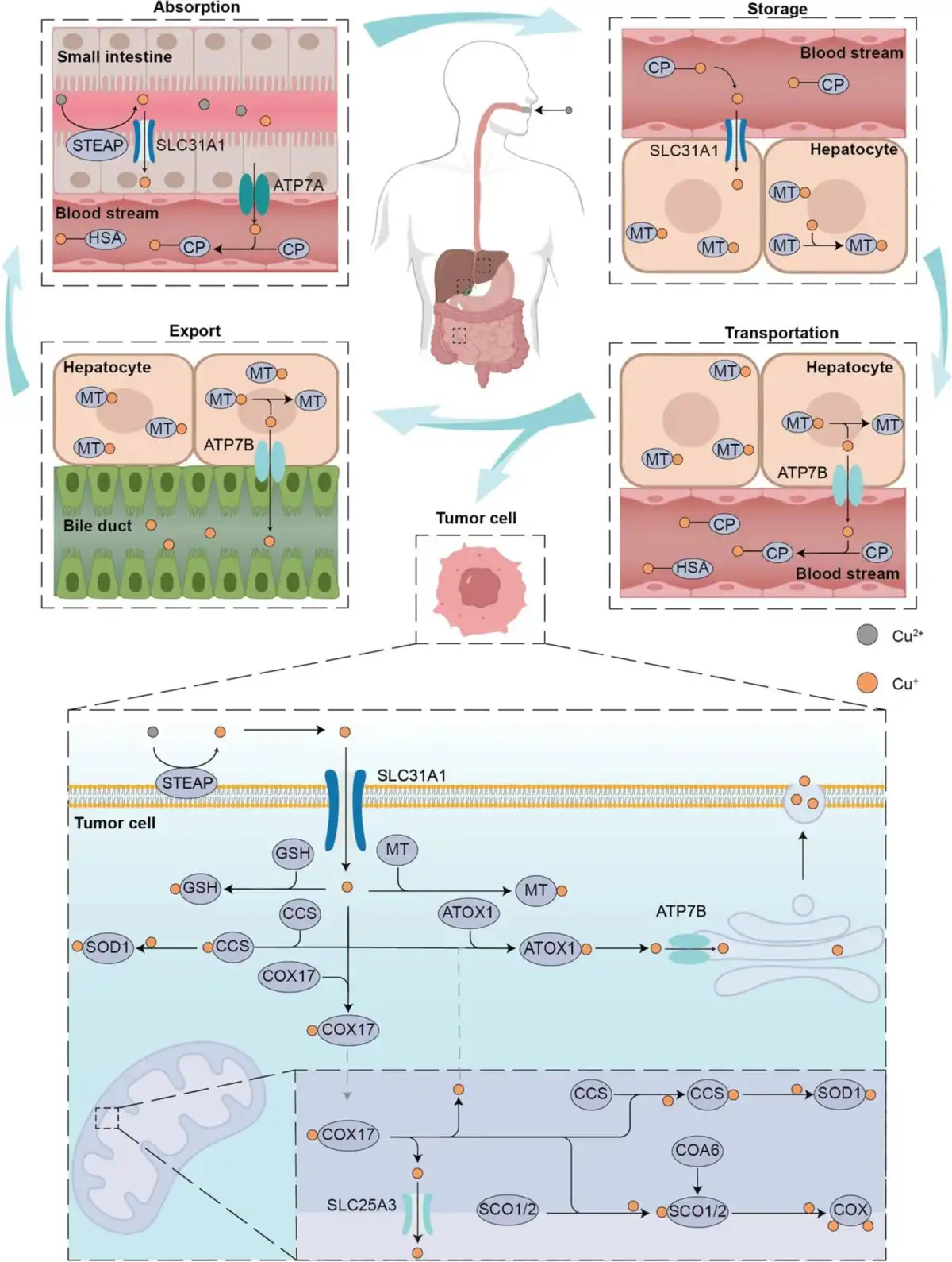
Copper is rich in a variety of foods, such as animal offal, seafood, cereals, vegetables, and fruits. Drinking water is also an important source of copper in the body. The currently recommended daily copper intake for adults is 2 to 3 mg.[23] After being ingested by the human body, copper is mainly absorbed through the intestinal epithelium, with the duodenum being the main absorption site.[24] Copper ions in food often exist in the form of divalent copper ions, but only monovalent copper ions can be stored and utilized in the body. Therefore, as shown in Figure 1, the ingested divalent copper ions are first reduced to monovalent copper ions by metalloreductases such as six-transmembrane epithelial antigen of the prostate (STEAP) on the surface of the intestinal epithelial cell membrane and then enter the intestinal epithelial cells through the copper transporter 1 (CTR1) or solute carrier family 31 member 1 (SLC31A1). Copper in the intestinal epithelial cells is transported to the other side of the intestinal epithelial cells by the copper chaperone antioxidant 1 (ATOX1) and released into the blood through the ATPase copper transporter alpha (ATP7A).[26] Most of the copper ions in the blood are bound to ceruloplasmin, and the remaining copper ions are bound to human serum albumin and amino acids.[27] Copper ions in the blood are transported to the liver through the portal vein system. The liver is the main storage organ for copper. The copper ions transported to hepatocytes through CRT1 are bound to metallothionein 1/2 (MT1/2). MT1 and MT2 are rich in sulfhydryl groups and therefore have a high affinity for copper ions. MT1 and MT2 in hepatocytes are the main storage sites for copper. When the copper content in the body exceeds the normal threshold, liver cells excrete excess copper into the bile and excrete it from the body through the action of ATPase copper transporter beta (ATP7B).[28] In summary, systemic copper metabolism is mainly regulated by the small intestine and liver.
In tumor cells, copper ion homeostasis and function depend on the interaction of different types of proteins. The first group is proteins related to transmembrane copper transport, such as STEAP and CTR1, which are responsible for transporting monovalent copper ions into tumor cells. The second group is proteins that bind and store copper ions,[29] such as MT and glutathione (GSH). MT and GSH are natural chelators of copper ions in tumor cells and play an important role in the homeostasis of intracellular copper ions. The third group is copper ion chaperones,[30] such as superoxide dismutase copper chaperone, ATOX1, and cytochrome C oxidase copper chaperone 17. Copper ions bind to copper molecular chaperones and are then delivered to specific proteins or cell compartments to exert their effects.
2.2 Cuproptosis targets
Previous studies have shown that copper can cause cell death by inducing mitochondrial oxidative damage or destroying enzymes related to the tricarboxylic acid cycle.[31] Tsvetkov et al[6] identified key targets that promote cuproptosis through genome-wide CRISPR-Cas9 screening, including ferredoxin 1 (FDX1), DLAT, lipoic acid synthase (LIAS), and dihydrolipoamide dehydrogenase (DLD). The discovery of these targets helps to deepen our understanding of the molecular mechanism of cuproptosis and provides potential targets for future drug development.
3. Construction options for copper-based nanomaterials
Activating copper-induced cell death (cuproptosis) hinges on the ability to induce a state of copper overload within tumor cells. To achieve this, the selection of appropriate copper-based nanomaterials is crucial as they serve as a “Cu ions storage pool.” These nanomaterials must be capable of delivering and releasing copper ions effectively within the tumor. Table 1 summarizes the different types of copper-based nanomaterials used for cancer treatment.
Table 1
Different types of copper-based nanomaterials for cancer treatment.
| Classification | Copper-based materials | Remarks and highlights | References |
|---|---|---|---|
| Copper-ionophores hybridized nanomaterials | Cu-TCPP nanosheets | Generation of ROS through the Russell mechanism | [32] |
| PCP@ES-Cu NPs | Extends the circulation time of ES-Cu in the body | [33] | |
| Cu2O/Cu-TCPP nanosheets | Nanoplatforms for loading enzyme-mimicking NPs, generation of ROS through Russell mechanism | [34] | |
| Metal–organic frameworks nanomaterials | Cu/ZIF-8 | Optimal carriers for storing O2 for enhanced PDT | [35] |
| Lip@Fe-Cu-MOF | Photothermal enhancement of ferroptosis and cuproptosis in tumor cells | [36] | |
| Other copper-coordinated nanoassembly | Cu2O@HKUST-1 | Inducing NO release to inhibit tumor metastasis and promote ·OH production for CDT | [37] |
| CuHPT | To overcome cancer chemoresistance by disrupting cellular redox homeostasis | [38] | |
| Inorganic nanomaterials | CuSiO3 hollow microspheres | Outstanding photothermal effects (η = 48.4%) and controlled drug release | [24] |
| CuO nanospheres | More active reaction sites to improve Fenton-like efficiency | [39] | |
| CuS nanoflowers | Ideal drug carriers for loading anticancer drug | [40] |
CDT, chemodynamic therapy; ES, elesclomol; MOF, metal–organic framework; NP, nanoparticle; ZIF-8, zeolitic imidazolate framework-8.
3.1 Copper-ionophores hybridized nanomaterials
Ionophores are small molecules that can bind with copper ions and transport those ions into the cell via transmembrane transport.[41,42] Therefore, the use of such special molecules to deliver copper ions is one of the most common approaches in the field of nanomedicine nowadays. Furthermore, several ionophores, such as disulfiram (DSF), diethyldithiocarbamate (DTC), and elesclomol (ES), have been found to increase the intracellular copper levels to activate cuproptosis.[43,44] Unfortunately, the lack of tumor targeting of these ionophores, their short blood circulation period, and easy to metabolize have led to their ineffective clinical performance. The development of nanotechnology provides a new direction for the delivery of these ionophores. Wu et al synthesized a polymer micelle (PCP) using polyethylene glycol and cinnamaldehyde. The PCP is ROS-responsive and can be used to encapsulate ES-Cu (EC) compounds, resulting in ECPCP (Figure 2A–C). This formulation extends the circulation time of ES-Cu in the body and increases its concentration in tumors. Once inside cells, the PCP coating breaks down due to high ROS levels, releasing ES and copper ions to induce cell death through cuproptosis (Figure 2D–F).[33] Similarly, Wu et al[45] fabricated a nanocomposite of CuET and copper oxide (CuO) based on ionophore DTC (CCB) to disrupt protein homeostasis for enhanced cuproptosis-mediated tumor therapy (Figure 2G–I).
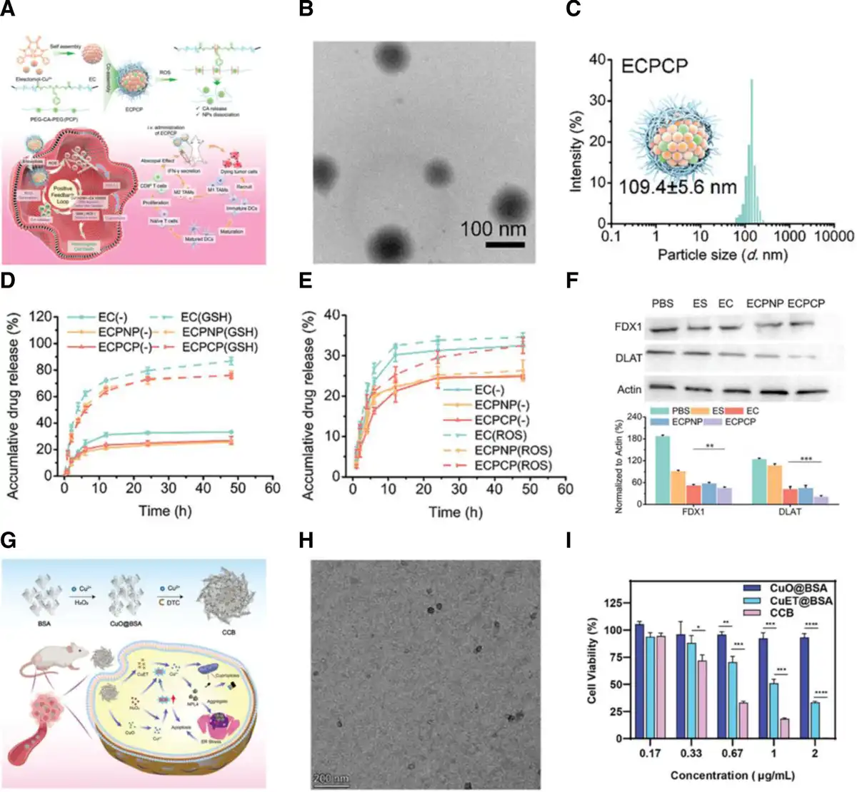
Figure 2.
(A) Schematic overview of ECPCP preparation and mechanism for cuproptosis-ICD combined therapy. TEM image (B) and size distribution (C) of ECPCP. (D) Release of ES from EC, ECPNP, and ECPCP under GSH condition. (E) Release of ES from EC, ECPNP, and ECPCP under ROS condition. (F) Western blotting of FDX1 and DLAT expression and semiquantitative analysis.[33] Copyright 2024, Wiley-VCH. (G) Schematic illustration of CCB synthesis and its therapeutic mechanism. (H) TEM image of CCB. (I) Viability of 4T1 cells after treatment with CuO@BSA, CuET@BSA, and CCB in different concentrations for 24 h.[45] Copyright 2024, Wiley-VCH.
3.2 Metal–organic frameworks nanomaterials
Metal–organic frameworks (MOFs) were built by transition metal ions with their strongly coordinating organic ligands through self-assembly to form nanoparticles with a periodic network structure.[46,47] Notably, the presence of a large number of coordination interactions between metal ions and organic groups in these MOF structures, which are weakened in the specific microenvironment of tumors, leads to the dissociation of the MOF structure and therefore can be used for the selective delivery of metal ions.[48] Much effort has been devoted to developing various copper-based MOF nanomaterials for activating cuproptosis-mediated tumor therapy. Chen et al[49] developed a system comprising bimetallic Fe-Cu MOFs as a core, which could be internalized by neutrophils to form a “cellular Trojan Horse” (Figure 3A, B). These neutrophils carried thermosensitive liposomes filled with Fe-Cu MOFs (Lip@Fe-Cu-MOFs), which released their contents at a specific temperature. The Fe-Cu MOFs exhibited robust photothermal properties (Figure 3C), whereby the generation of heat under near-infrared (NIR) light triggered drug release and enhanced cuproptosis and ferroptosis (Figure 3D). The Fenton reaction, catalyzed by Fe3+ or Cu2+, produced hydroxyl radicals that, in conjunction with Cu2+, depleted GSH in tumor cells. This process induced both ferroptosis and cuproptosis, attacking cancer cells on multiple fronts and enhancing treatment effectiveness while minimizing the risk of recurrence (Figure 3E).
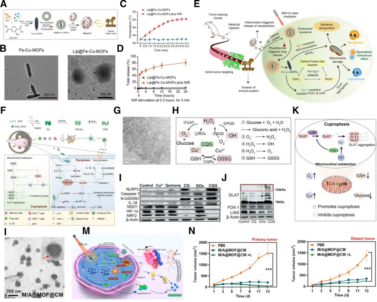
Figure 3.
(A) Synthesis of cellular Trojan Horse. (B) TEM images of Fe-Cu-MOFs and Lip@Fe-Cu-MOFs. (C) Photothermal effects of Lip@Fe-Cu-MOFs. (D) The time-dependent release of Lip@Fe-Cu-MOFs under specific conditions. (E) Schematic illustration of Lip@Fe-Cu-MOFs-induced synergistic anticancer effects.[49] Copyright 2024, American Association for the Advancement of Science. (F) Illustration of the CQG NPs synthesis and therapeutic process. (G) TEM image of CQ NPs. (H) Schematic display of multiple enzyme effects of CQG NPs. (I) Western blot analysis of several pyroptosis-related proteins levels after diverse treatments. (J) Western blot analysis of cuproptosis-related proteins levels after diverse treatments. (K) Schematic illustration of the mechanism in CQG NPs promoted cuproptosis.[50] Copyright 2023, Wiley-VCH. (L) TEM image of M/A@MOF@CM. (M) Schematic illustration of M/A@MOF@CM-based treatment for cancer. (N) Tumor growth curves of primary tumor and distant tumor.[51] Copyright 2024, Elsevier B.V.
In addition, copper-based metal-phenolic networks have likewise been extensively studied as a class of MOF materials in cuproptosis-mediated tumor therapy.[50] Qiao et al[50] designed metal-phenolic nanoparticles (CQG NPs) that integrated copper with quinone and glucose oxidase (GOx) to combat both dormant and recurring tumors by inducing a balanced pyroptosis and cuproptosis (Figure 3F, G). The multienzyme activity of CQG NPs activated NLRP3-mediated pyroptosis (Figure 3H, I), while cuproptosis was initiated by the release of copper ions from the degradation of nanoparticles (Figure 3J). Moreover, the susceptibility of cancer cells to cuproptosis was increased through several mechanisms: the depletion of copper chelators by a reaction with GSH, oxygen generation via a catalase-like reaction, and glucose scarcity due to starvation (Figure 3K).
Furthermore, due to the presence of pore structure on the surface of MOF materials, which can adsorb a variety of chemotherapeutic drugs, copper-based MOF nanomaterials can achieve more efficient cuproptosis-mediated tumor therapy with the assistance of chemotherapeutic drugs. Ji et al developed a multifunctional nanoparticle (M/A@MOF@CM) that was a copper-based MOF loaded with mitoxantrone and axitinib and coated with a tumor cell membrane for cuproptosis/ferroptosis/apoptosis synergistic cancer therapy (Figure 3L).[43] This M/A@MOF@CM showed various antitumor properties, combining photothermal therapy, chemotherapy, and chemodynamic therapy to boost ICD for feasible immunotherapy (Figure 3M). This integrated strategy resulted in an 86.45% reduction in primary tumor size and a 65.61% decrease in distant tumor growth, showcasing its potential in comprehensive cancer treatment (Figure 3N).
3.3 Other copper-coordinated nanoassembly
The coordination between metal ions and organic molecules can form multifunctional nanoassemblies different from MOF structures by tuning the organic molecule species. Metal oxides or metal sulfides can form hollow mesoporous structured core-shell nanoparticles, with the hollow mesoporous structure acting as a scaffold to load hydrophobic organic compounds within the pores of the mesoporous structure, enabling controlled release of organic drugs through the porous architecture. Furthermore, the attachment and growth of nano-sized tetrairon oxide (Fe3O4) on the core-shell structure endow the entire nanomaterial with not only high biocompatibility but also superparamagnetism and magnetothermal response, thereby achieving a precise diagnosis of tumors and effective treatment integrating chemodynamic, chemotherapy, and other multimodal therapies. Metal ions can also coordinate with chemotherapeutic drugs, small molecule inhibitors, and other organic molecules through layer-by-layer self-assembly to form tumor microenvironment (TME)-responsive nanoparticles. These nanoparticles remain stable under normal physiological conditions but rely on their responsiveness to undergo cascade reactions that eliminate tumor cells in the TME. Liang et al[52] introduced a novel copper-coordinated nanoassemblies (CCNAs) that integrated a photosensitizer (zinc phthalocyanine [ZnPc]), a prodrug of a chemotherapeutic (DOX) with a thioketal (TK) spacer and an IDO inhibitor (1-methyl tryptophan [1-MT]) (Figure 4A, B). These components self-assemble around Cu2+ ions for a combination therapy of apoptosis and cuproptosis, along with immunotherapy. Upon exposure to NIR laser irradiation, the ZnPc in CCNAs activated a photodynamic effect, producing ROS (Figure 4C, D). This led to the release of DOX, intensifying tumor cell apoptosis (Figure 4E, F). Moreover, the Cu2+ in CCNAs boosted the photodynamic process by catalyzing oxygen production and also induced the aggregation of harmful mitochondrial proteins, causing cell cuproptosis (Figure 4G).
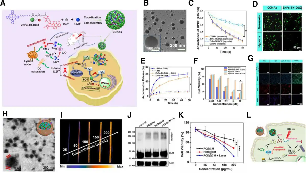
Figure 4.
(A) Schematic overview of fabrication and application in enhanced apoptosis-cuproptosis and immunotherapy from CCNAs. (B) TEM image of CCNAs. (C) The 1O2 generation of CCNAs with light irradiation measured by DPBF. (D) DCFH-DA-stained fluorescence images after treatments with ZnPc-TK-DOX and CCNAs. (E) Drug release of CCNAs in the presence of GSH. (F) The cell viabilities of PC-3 cells after different treatments. (G) Immunofluorescence of DLAT and FDX1 in PC-3 cells after different treatments.[52] Copyright 2024, Elsevier B.V. (H) TEM image of PCD@CM. (I) NIR-II PAI of PCD@CM with different polymer concentrations under 1064 nm excitation. (J) Western blot analysis of DLAT protein in 4T1 cells after various treatments. (K) Cell viability of 4T1 cells subjected to various treatments. (L) The induction mechanism of NIR-II PTT and GSH depletion dual-amplified cuproptosis.[53] Copyright 2024, Elsevier B.V. PTT, photothermal therapy.
Apart from photodynamic molecules that can be introduced, photothermal molecules can also be added to boost cuproptosis-mediated tumor therapy. Dai et al[53] developed a biomimetic cuproptosis amplifier (PCD@CM) by assembling NIR-II polymer dots and DOX drug with Cu(II), and then coating them with tumor cell membranes (Figure 4H). This system targeted tumor cells, where high levels of GSH in the TME cause the release of DOX by reducing Cu(II) to Cu(I). This enabled guided photothermal and chemotherapy using NIR-II imaging (Figure 4I). The released Cu(I) caused mitochondrial protein aggregation and proteotoxic stress, inducing cuproptosis (Figure 4J). The combination of NIR-II photothermal therapy and GSH depletion makes tumor cells more susceptible to this form of cell death (Figure 4K, L).
3.4 Inorganic nanomaterials
Inorganic nanomaterials compared with organic or polymer nanomaterials have unique physicochemical properties, stronger mechanical stability while maintaining a certain biological activity, and are easier to be compounded with a variety of carriers.[54] Metal oxides are the most common inorganic nanomaterials. The 2 main types of copper-based metal oxides are copper oxide and cuprous oxide, and both of these copper oxides have been shown to act as copper sources to activate cuproptosis. Bai et al[55] developed MitCuOHA nanozymes using copper oxide nanorods as a base, loaded with (3-carboxypropyl) triphenylphosphonium bromide, and coated with hyaluronic acid (Figure 5A, B). These nanozymes possessed multiple enzymatic activities for combined ferroptosis and cuproptosis therapy in cancer treatment. They catalyzed the depletion of cysteine and GSH while producing H2O2 (Figure 5C, D), which was converted into hydroxyl radicals to induce ferroptosis in cancer cells. Additionally, MitCuOHA nanomedicine can release copper ions in the TME, and copper ions can bind to proteins in the tricarboxylic acid cycle, causing their aggregation and loss of iron–sulfur cluster proteins, leading to proteotoxic stress and cuproptosis (Figure 5E, F). Similarly, Ning et al[56] created a biomimetic cuproptosis sensitization system by coating Cu2O with a platelet membrane to induce multiple tumor cuproptosis (Figure 5G, H). The system incorporated an AIE photosensitizer (TBP-2), which generated hydroxyl radicals to deplete GSH and hinder copper ion efflux. Cu2O broke down quickly under acidic and H2O2-rich tumor conditions, releasing copper ions (Figure 5I–K). Copper accumulation triggers the clustering of lipoylated proteins and the depletion of iron–sulfur clusters, which in turn induces protein toxicity stress, ultimately leading to cell death through cuproptosis (Figure 5L).
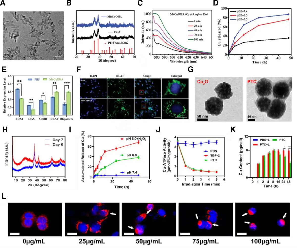
Figure 5.
(A) TEM image of MitCuOHA nanorods (scale bar: 10 nm). (B) XRD spectra of CuO and MitCuOHA nanorods. (C) Fluorescence spectrum in Cys + MitCuOHA system by using Amplex Red (H2O2 probe). (D) pH-responsive release of Cu2+ in PBS with different pH. (E) Quantification analysis of FDX1, LIAS, SDHB, DLAT, and DLAT oligomers proteins expression. (F) Immunofluorescence imagings of MCF-7 cells treated with MitCuOHA.[55] Copyright 2024, Wiley-VCH. (G) TEM images of Cu2O and PTC. (H) XRD pattern of Cu2O at 0 and 7 days after preparation. (I) Cumulative release profiles of Cu under different conditions. (J) The Cu-ATPase activity variation after indicated treatment with light irradiation. (K) The intracellular Cu content during the treatment period. (L) DLAT fluorescence images of cells after PTC + L treatment with different concentrations.[56] Copyright 2023, American Chemical Society.
Currently, an increasing number of copper-based inorganic nanomaterials are being reported to be available for efficient cuproptosis activation. Man et al[57] constructed human serum albumin-mediated defect-rich copper hydroxide nanowire (HCu nanowire) for synergetic cuproptosis and ROS-mediated apoptosis antitumor therapy (Figure 6A–C). Chan et al[58] reported a photothermic Cu2-xSe nanoparticle encapsulated with bioresponsive dimethyl maleic anhydride (DMMA@Cu2-xSe) as the copper carrier to enhance the copper accumulation in tumor for achieving cuproptosis-driven enhancement of thermotherapy (Figure 6D, E). In particular, benefiting from the fact that some of the inorganic materials can trigger a change in properties due to the realization of phase transitions in tumor-specific microenvironments, some of the copper-based nanomaterials will be expected to achieve specific activation of cuproptosis in specific TMEs. Zhao et al[59] copper hydroxyphosphate nanoparticles (Cu2(PO4)(OH) NPs) with hydrogen sulfide (H2S)-responsiveness were developed to achieve an imbalance of copper homeostasis (copper overload) in tumor cells by enhancing endocytosis of copper-based nanoparticles and inhibiting exocytosis of copper ions, which further induced cellular pyroptosis and cuproptosis. Monodisperse Cu2(PO4)(OH) NPs synthesized by a hydrothermal method and injected intravenously into H2S-overexpressing colon cancer microenvironments were able to be transformed into ultrasmall copper sulfide nanoparticles (Cu9S8 NPs) through in situ sulfation (Figure 6F, G). These ultrasmall Cu9S8 NPs can be endocytosed into colon cancer cells faster and release copper ions inside the cells, which dramatically increase the intracellular ROS level via a Fenton-like reaction. The increase in ROS not only activates NLRP3 inflammatory vesicles and Caspase-1 proteins, causing gasdermin D (GSDMD) cleavage and triggering cellular pyroptosis (Figure 6H), but also affects mitochondrial function, cuts off the supply of adenosine-5’-triphosphate (ATP), downregulates the expression of ATP7A, and reduces the efflux of copper ions. The combination of higher cellular uptake, copper ion release, and reduced copper ion efflux ultimately disrupts intracellular copper homeostasis and activates copper overload-mediated cuproptosis (Figure 6I, J).
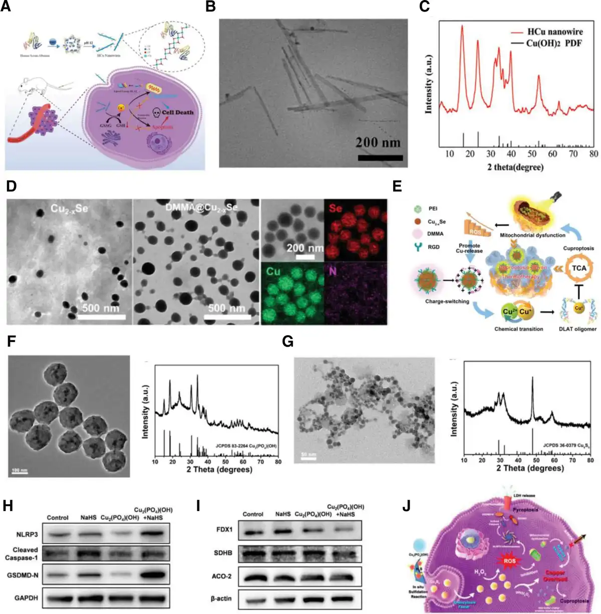
Figure 6.
(A) Schematic representation of synthetic procedure and the mechanism of enhanced antitumor efficacy by HCu nanowires. (B) TEM image of HCu nanowires. (C) XRD patterns of the HCu nanowires.[57] Copyright 2024, Wiley-VCH. (D) TEM image of Cu2-xSe and HR-TEM and mapping image of DMMA@Cu2-xSe. (E) Schematic illustration of DMMA@Cu2-xSe for cuproptosis-driven enhancement of thermotherapy.[58] Copyright 2023, Wiley-VCH. (F) TEM image and XRD pattern of Cu2(PO4)(OH) NPs. (G) TEM image and XRD pattern of Cu2(PO4)(OH) NPs after treatment with NaHS. (H) Western blot analysis of expressions in NLRP3, cleaved Caspase-1, and GSDMD-N. (I) Western blot analysis of expressions in FDX1, SDHB, and ACO-2. (J) Schematic diagram of Cu2(PO4)(OH) NPs induced copper-overload-mediated cuproptosis/pyroptosis combination therapy.[59] Copyright 2023, Wiley-VCH.
4. Cuproptosis sensitization strategies based on copper-based nanomaterials
Unfortunately, the activation efficiency of cuproptosis in tumor therapy is still not satisfactory, mainly due to the following 3 reasons. (1) It is difficult to induce copper overload effectively within the cell due to the existence of a strict copper ion transport pathway in the tumor.[12] (2) Free intracellular copper ions are susceptible to being chelated by excess GSH and lose the possibility to bind to lipid-acylated proteins.[60] (3) The hypoxic microenvironment of the tumor leads to a weak mitochondrial respiration.[61] Based on the above difficulties, the main strategies to enhance the sensitivity of tumor cells to cuproptosis with the aid of copper-based nanomaterials are summarized as follows.
4.1 Copper transporter protein regulation
Activation of cuproptosis in tumor cells is predicated on how to rapidly accumulate large amounts of copper ions within tumor cells. However, the cell has a complex network of copper-dependent transporter proteins consisting of CTR1 and ATP7A/B, to maintain its internal copper homeostasis.[62] Jointly, these proteins coordinate copper input (CTR1) and output (ATP7A/B) to maintain intracellular copper content within a specific range, and the existence of this copper homeostatic mechanism greatly prevents the occurrence of copper overload. Typically, nanomaterials enriched in tumor sites can enter into tumor cells by endocytosis, therefore the designed and constructed copper-based nanomaterials can effectively accumulate intracellular copper content by inhibiting the expression of copper efflux proteins in tumor cells. It is worth noting that the function of copper efflux proteins (ATP7A/B) needs to be energized by ATP, and once the ATP supply is insufficient, it will lead to a decrease in expression and affect the efflux process of copper ions.[63]
Based on this, Xu et al[64] developed a novel copper/iron hybrid hollow amorphous MOF with doxorubicin and hyaluronan (DOX@Fe/CuTH HaMOF) as an oxidative stress amplifier and copper/iron metabolic disrupter for anticancer therapy. In this nanomaterials system, the content of intracellular H2O2 could be promoted by the effect of DOX (Figure 7A), and combined with its Fenton catalytic properties, it can dramatically increase the intracellular level of ROS (Figure 7B), which in turn leads to mitochondrial damage and affects the production of ATP (Figure 7C), ultimately resulting in a decrease in the expression of ATP7A protein (Figure 7D). More copper ions will be locked up inside the tumor cell, enabling more effective activation of cuproptosis (Figure 7E–G). Furthermore, besides utilizing oxidative stress-induced mitochondrial damage to regulate the copper transporter proteins, it is also possible to inhibit the expression of copper efflux proteins by inhibiting glycolysis to cut off ATP supply. In addition, Yan et al[65] built an OPDEA-coated copper-based MOF system (OMP) to deliver siRNA targeting PDK1 (siPDK) for achieving antitumor effect via cuproptosis. The delivered siPDK could reduce ATP production by suppressing intracellular glycolysis (Figure 7H), leading to the blockage of the Cu efflux protein ATP7B (Figure 7I); consequently, this enhances the sensitivity of cancer cells to the cuproptotic effects triggered by OMPs (Figure 7J, K). In addition, it is also feasible to increase the activation efficiency of cuproptosis by reducing ATP7A protein expression through directly targeted delivery of siRNA targeting ATP7A.[66]
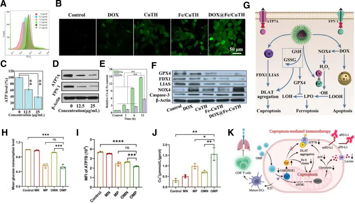
Figure 7.
(A) Flow cytometric analysis of H2O2 level in 4T1 cells after incubation with varied concentrations of DOX for 24 h. (B) CLSM images of DCFH-DA-stained cells after different treatments. Intracellular ATP (C) and ATP7A expression levels (D) after different concentrations of DOX@Fe/CuTH treatment. (E) Quantitative analysis of intracellular Cu and Fe levels after DOX@Fe/CuTH HaMOF treatment at different times. (F) Western blot analysis of the expressions in different proteins. (G) The schematic diagram of DOX@Fe/CuTH-induced cuproptosis/ferroptosis/apoptosis.[64] Copyright 2022, Wiley-VCH. Glucose consumption (H) and semiquantification of ATP7B expression (I) in B16F10 cells after various treatments. (J) Intracellular Cu+ contents in B16F10 cells after being treated with various nanoparticles. (K) Schematic overview of OMP-induced cuproptosis combined with PD-L1 for immunotherapy.[65] Copyright 2024, Elsevier B.V.
Copper transporter protein modulation strategy based on copper-based nanomaterials is focused on increasing intracellular copper levels by inhibiting the expression of copper efflux proteins (ATP7A/B). Three types of approaches are currently available: (1) increasing intracellular ROS levels through copper-based nanomaterials, which in turn causes mitochondrial dysfunction and reduces ATP production capacity, can effectively reduce ATP7A/B protein expression; (2) inhibition of intracellular glycolysis, which subsequently reduces ATP supply, similarly inhibits protein expression; and (3) delivery of siRNA for the corresponding protein directly inhibits protein expression.
4.2 GSH depletion
GSH, a naturally biological tripeptide, plays a crucial role in regulating intracellular redox balance. As the tumor cells show an overall oxidative stress state, reduced GSH is highly expressed within the tumor cells.[67] These excess reduced GSH will chelate with free intracellular copper ions, which restricts the binding of copper to the lipoylated components in the TCA, inhibiting the efficacy of cuproptosis. Thus, depletion of intracellular GSH can increase DLAT oligomerization and sensitize tumor cells to cuproptosis.
Utilizing the strong reducing property of GSH, it will be promising to deplete GSH through redox by constructing material systems containing multivalent metal ions. Chen et al[68] constructed a Cu2O@Mn3Cu3O8 (CMCO) nanozyme with a core-shell structure that served as the TME-activated copper deliverer for safe and efficient cuproptosis (Figure 8A). The redox activity of Cu+/Cu2+ and Mn2+/Mn3+/Mn4+ in CMCO reacted with the GSH in the TME, converting it to oxidized glutathione (GSSG) through a GSHOx-like mechanism (Figure 8B). This reaction reduced the ability of GSH to chelate copper ions (Figure 8C), leading to increased copper toxicity and the induction of cuproptosis in cancer cells (Figure 8D, E). Similarly, Zhang et al[69] reported CuMoO4 nanodots with multienzyme-like activities that respond to the TME for multimodal cancer treatment. The CuMoO4 nanodots, containing variable-valent metals (Cu+/Cu2+ and Mo5+/Mo6+), effectively depleted intracellular GSH through a GSHOx-like activity, sensitizing tumor cells to cuproptosis (Figure 8F).
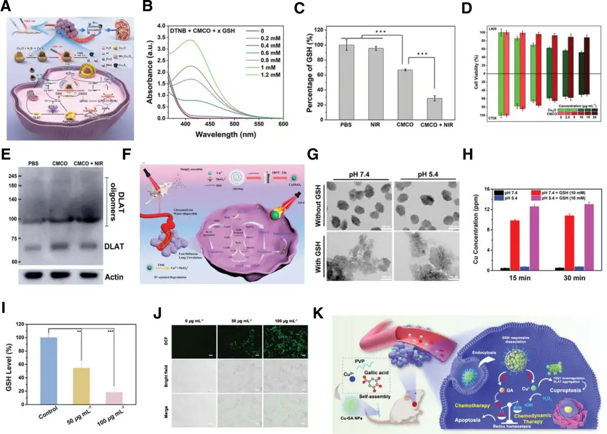
Figure 8.
(A) The potential mechanism of CMCO nanozymes-induced high-efficiency ferroptosis-boosted-cuproptosis for cancer therapy. (B) GSHOx-like activity of CMCO in PBS (pH 6.5). (C) Intracellular GSH contents in CT26 with different treatments. (D) The cell viabilities of L929 and CT26 cells treated with different concentrations of Cu2O and CMCO nanozymes. (E) Western blot analysis of DLAT after different treatments.[68] Copyright 2023, Wiley-VCH. (F) Schematic illustration of CuMoO4 nanodots synthesis and their multimodal treatment mechanism of a tumor.[69] Copyright 2023, Wiley-VCH. (G) TEM images of Cu-GA NPs after treatment with GSH. (H) The released Cu concentration from Cu-GA NPs with different treatments. (I) Intracellular GSH levels after Cu-GA NPs treatments. (J) DCFH-DA stained fluorescence images after Cu-GA NPs treatments. (K) Schematic diagram of the Cu-GA NPs mediated chemo/chemodynamic synergistic tumor therapy.[70] Copyright 2023, Wiley-VCH.
Although relying on the variable metal ion reduction strategy can efficiently consume GSH, the overall GSH depletion effect is still not satisfactory. Therefore, several strategies to assist GSH depletion have been developed for more efficient realization of intracellular GSH depletion. Zhao et al[70] utilized gallic acid (GA) with GSH-depleting properties to construct copper-based metal-phenolic network nanoparticles (Cu-GA NPs) for polyphenol drug-assisted dysregulation of intracellular redox homeostasis. The Cu-GA NPs enabled the rapid release of copper ions and GA under the conditions of the TME overexpressed GSH (Figure 8G). The released GA and Cu2+ continuously consume the remaining GSH to achieve GSH depletion and simultaneously Cu2+ will be reduced to Cu+ with higher Fenton catalytic activity (Figure 8H, I). Subsequently, ROS levels within the tumor cells are significantly increased by the Fenton-like reaction of released Cu+ (Figure 8J). Furthermore, an excessive buildup of Cu+ within tumor cells leads to the clustering of DLAT and a decrease in the expression of iron–sulfur cluster proteins, ultimately realizing the dual-wheel-driven tumor therapy of apoptosis and cuproptosis under the regulation of redox homeostasis (Figure 8K). The depletion of intracellular GSH is likewise effectively assisted by the delivery of GSH generation inhibitors. Huang et al developed a copper-based nanoplatform, MOF-199@DDM, designed to deliver buthionine-sulfoximine (BSO). BSO is an inhibitor of intracellular GSH synthesis, which makes tumor cells more susceptible to cuproptosis-inducing effects.[71]
GSH overexpressed in tumor cells has a strong ligand-chelating effect, which can effectively chelate the accumulated copper ions in the cell and then cut off the effect of copper ions and cuproptosis-related proteins, reducing the possibility of the emergence of cuproptosis. Two potential modalities have been developed to deplete GSH in order to increase the sensitivity of tumor cells to cuproptosis: (1) construction of copper-based nanomaterials with variable metal ions to regulate intracellular GSH levels by redox reaction. (2) Delivery of GSH-depleting chemotherapeutic molecules or GSH synthesis inhibitors with the assistance of copper-based nanomaterials.
4.3 Increasing intracellular oxygen content
Mitochondrial respiration involving cuproptosis necessitates the engagement of oxygen (O2), whereas the TME is typified by hypoxia, which will also restrict the impact of cuproptosis. Alleviating the hypoxic environment of the tumor (increasing intracellular oxygen content) would be expected to enhance the sensitivity of tumor cells to cuproptosis.
Peroxides are a class of inorganic materials containing peroxygen bonds (–O–O–), which can produce H2O2 stably under acidic conditions and release O2 under certain conditions, being considered as potential carriers of O2.[72] On the basis of calcium peroxide nanoparticles (CaO2), Jin et al have engineered and synthesized a nanoreactor for immunotherapy that triggers cuproptosis, designated as CCJD-FA. This nanoreactor encapsulates copper-based shell-coated CaO2 and the bromodomain-containing protein 4 inhibitor JQ-1, along with DSPE-PEG-FA (Figure 9A).[63] Within tumor cells, CCJD-FA disintegrates under the influence of GSH and an acidic milieu, exposing CaO2, which then produces H2O2 by interacting with H+ (Figure 9B–D). This H2O2, in conjunction with the liberated Cu2+, undergoes a Fenton-like reaction, releasing O2 to alleviate hypoxia and instigate cuproptosis (Figure 9E–G).
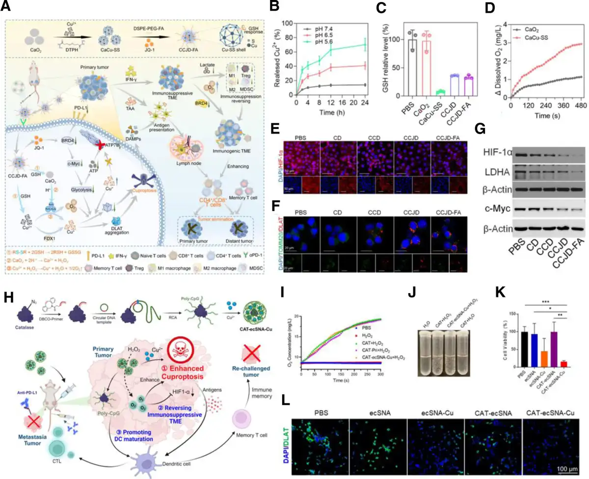
Figure 9.
(A) Schematic illustration of CCJD-FA-mediated cuproptosis for activating immune response. (B) The release curves of Cu2+ from CCJD-FA in PBS with different pH. (C) GSH consumption ability of CCJD-FA. (D) The generation of O2 with time under different treatments in pH 5.6. (E) Immunofluorescence images of HIF-1α for detection of nanoparticles-induced hypoxia reversion. (F) Immunofluorescence images of DLAT after different treatments. (G) Western blotting of cuproptosis-related protein expression.[63] Copyright 2023, Elsevier B.V. (H) Schematic illustration of the preparation of CAT-ecSNA-Cu and corresponding mechanism of enhanced cuproptosis therapy. (I) O2 generation in different media after the addition of free CAT, CAT-Pri, and CAT-ecSNA-Cu. (J) Digital photographs of O2 generation in H2O solution after various treatments. (K) Viability of CT26 cells after different treatments for 24 h. (L) Immunofluorescence images of DLAT expression in CT26 cells after various treatments.[73] Copyright 2024, American Chemical Society.
Catalase (CAT), a kind of enzyme widely existing in organism, is capable of efficiently catalyzing the release of O2 from H2O2, reversing the hypoxic TME,[74] which enhances mitochondrial respiration and promotes cuproptosis. The researchers used a technique called rolling circle amplification to combine long-chain DNA with catalase, and then obtained the enzyme-core spherical nucleic acid nanoplatform (CAT-ecSNA-Cu) through the chelation of copper ions (Figure 9H).[73] CAT can maintain its activity under the protection of long-chain DNA. In the TME, CTA catalyzes hydrogen peroxide to produce oxygen, which enhances the aerobic respiration of tumor cells and increases the sensitivity of tumor cells to copper death. Notably, some copper-based nanomaterials with CAT-like enzyme activity can likewise catalyze the production of O2 to alleviate hypoxia in the TME (Figure 9I, J), thereby promoting the therapeutic efficiency of cuproptosis (Figure 9K, L).[68]
Tumor hypoxia microenvironment can inhibit cell cuproptosis. There are 2 approaches that have been confirmed to increase intracellular oxygen content for the enhancement of cuproptosis. (1) Peroxide-based oxygen generation: design nanoreactors incorporating peroxides to release oxygen within the TME, mitigating hypoxia and boosting cuproptosis. (2) Catalase-enhanced mitochondrial respiration: utilize catalase-functionalized nanoplatforms to catalyze the conversion of H2O2 to O2, thereby sensitizing tumor cells to cuproptosis by enhancing mitochondrial respiration.
5. Copper-based nanomaterials driven cuproptosis-mediated combination cancer therapy
Due to the high heterogeneity among tumors and the existence of a protective mechanism for cuproptosis within tumors, it is difficult to achieve an ideal therapeutic effect with a single treatment modality based on cuproptosis, leading to metastasis and recurrence of tumors.[75] The combination of multiple therapeutic modalities to amplify the therapeutic effect of cuproptosis would facilitate the clinical application of cuproptosis. Accordingly, it is necessary to develop copper-based nanomaterials that can integrate cuproptosis with other therapeutic modalities to achieve efficient cancer treatment.
5.1 Cuproptosis combined chemotherapy
Chemotherapy is still the first choice for the treatment of cancers in clinical practice, and correspondingly developed chemotherapeutic drugs are mainly effective in combating cancer by activating apoptosis.[76] However, tumor cells derive multiple drug-resistant resistances to counteract the function of these chemotherapeutic drugs, resulting in unsatisfactory efficacy.[77] In addition, conventional chemotherapeutic agents without tumor selectivity require high-dose injections to achieve anticancer effects that are frequently accompanied by severe toxic side effects. These unavoidable obstacles urgently seek to develop multimechanism combination strategies to alleviate the above dilemma.
Fortunately, some studies have found that the introduction of cuproptosis can solve some of the difficulties in the application of chemotherapeutic drugs. Drug resistance, especially for drug efflux transporter-mediated chemoresistance, stands out as one of the predominant factors leading to the suboptimal outcomes in the field of contemporary cancer chemotherapy. Lu et al[78] developed a copper-based nanoplatform by coordinating with ellagic acid (EA-Cu), which effectively combated resistance to cancer chemotherapy through cuproptosis (Figure 10A). Within this system, EA curbed the self-sustaining properties of tumor cells by inhibiting the hedgehog signaling pathway. Concurrently, Cu2+ diminished the stem-like characteristics of cells by interfering with their mitochondrial function, which significantly boosted the effectiveness of chemotherapy induced by DOX. Furthermore, EA worked in tandem with copper ions to trigger mitochondrial dysfunction and cuproptosis, leading to a significant reduction in ATP levels. This reduction consequently inhibited the function of P-glycoprotein, a key player in drug resistance by facilitating the expulsion of chemotherapeutic agents, thereby increasing the sensitivity of tumor cells to chemotherapy mediated by DOX.
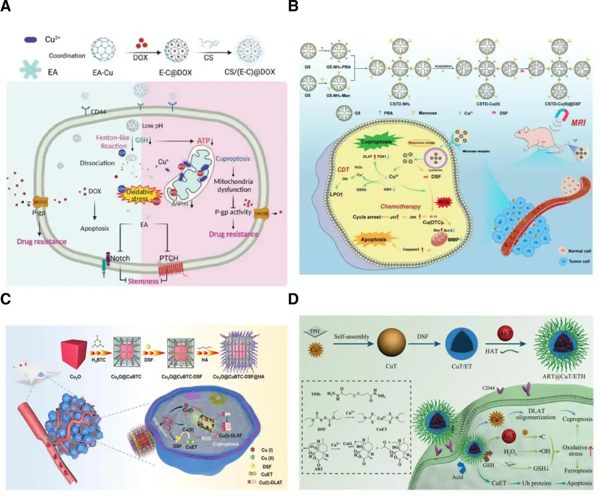
Figure 10.
(A) Preparation of CS/NPs and the underlying mechanism of CS/NPs for cuproptosis-mediated therapeutic resistance.[78] Copyright 2023, Wiley-VCH. (B) Schematic illustration of preparation and mechanism of CSTD-Cu(II)@DSF for cuproptosis-promoted chemo-chemodynamic therapy.[79] Copyright 2023, Elsevier B.V. (C) Schematic illustration of construct process of copper-enriched nanomedicine and the ROS-augmented cuproptosis without systemic toxicity.[80] Copyright 2023, Elsevier B.V. (D) Schematic illustration of the fabrication process of ART@CuT/ETH HNP and its for oxidative stress amplification-enhanced cuproptosis-based anticancer therapy.[81] Copyright 2023, Wiley-VCH.
The inefficient cytotoxic selectivity of chemotherapeutic agents has likewise been an obstacle to the further development of chemotherapy. It is worth noting that one class of chemotherapeutic drugs is of the metal ion potentiating type.[82] And one of the characteristics of cuproptosis is an overload of intracellular copper ions. Therefore, copper-based chemotherapy drugs hold great potential to achieve cuproptosis synergetic with chemotherapy. DSF, approved by the Food and Drug Administration for treating alcoholism, was found to possess antitumor activity via inducing apoptosis of tumor cells.[83] Interestingly, some recent studies suggested that the antitumor capacity of DSF could be strongly amplified once it was combined with endogenous copper ions (Cu2+), while DSF by itself exhibited low toxicity.[84] Therefore, DSF is a potential “pro-drug” with Cu2+ as an enhancer to realize in situ chemotherapy. Ni et al[79] engineered a TME-activated core-shell structured tecto dendrimer, which was laden with copper ions and DSF, designated as CSTD-Cu(II)@DSF. This formulation was intended for a dual therapeutic strategy that leverages cuproptosis to enhance chemo-chemodynamic therapy (Figure 10B). Upon systemic circulation, the CSTD-Cu(II)@DSF accumulated at the tumor site, where it released its therapeutic payload in response to the tumor's mildly acidic and elevated levels of ROS microenvironment. Once inside the cells, the elevated levels of intracellular Cu(II) ions initiated the oligomerization of lipoylated proteins, precipitating proteotoxic stress that led to cuproptosis. Additionally, these ions catalyzed the process of lipid peroxidation, a critical component of chemodynamic therapy. The CSTD-Cu(II)@DSF also induced mitochondrial dysfunction and cell cycle arrest at the G2/M phase, which, combined with DSF, amplified the apoptotic effect on the cancer cells. By combining the cytotoxic effects of chemotherapy, the selective stress induced by cuproptosis, and the oxidative stress generated through chemodynamic therapy, it offered a multifaceted approach to overcoming cancer. Likewise, researchers led by Zhong utilized Cu2O@CuBTC-DSF@HA nanocomposites, which were capable of responding to acidic conditions by dissociating to catalyze the conversion of DSF into the toxic dithiocarbamate-copper complexes (CuET). This process was designed to occur in the TME and it worked in conjunction with cuproptosis to amplify the therapeutic effects of treatment (Figure 10C).[80]
The compound artemisinin (ART), known for its antimalarial properties, has uncovered its potential in the fight against cancer.[85] Its molecular structure, featuring a sesquiterpene with an endoperoxide bridge, is susceptible to cleavage by transition metal ions, resulting in the formation of a carbon-centered radical (·C). This process significantly boosts oxidative stress within cells. Xu et al[81] introduced a novel hollow nanoplatform (HNP) that incorporated CuET and loaded with ART to enhance oxidative stress in cancer therapy (Figure 10D). This HNP was designed to be responsive to both the acidic pH and the high levels of GSH within TME. Upon exposure to these conditions, the ART@CuT/ETH HNPs disassemble, releasing Cu2+ ions. The released copper ions played a dual role: They catalyzed the formation of ·C and hydroxyl radicals (·OH) through reactions with ART and endogenous H2O2, respectively, thereby intensifying oxidative stress. Additionally, copper ions bind to the DLAT, leading to its aggregation and the induction of cuproptosis. The platform also incorporated the ligand TPH, which features disulfide bonds and is adept at depleting intracellular GSH. This GSH depletion further magnified oxidative stress and supported the induction of cuproptosis. The approach of amplifying oxidative stress through 3 distinct pathways showed promise in the realm of copper-based cancer therapies.
5.2 Cuproptosis combined chemodynamic therapy
Chemodynamic therapy (CDT) is an emerging field in cancer treatment that leverages the cytotoxic potential of ROS, which are produced from the conversion of endogenous H2O2 through Fenton or Fenton-like reactions, to provoke apoptosis in tumor cells.[86] Recognized for its precise targeting of cancer cells and minimally invasive characteristics, CDT has gained attention in the search for novel cancer therapies. Copper ions are a kind of metal ions with Fenton-like catalytic activity and have higher Fenton catalytic activity compared with conventional iron ion catalysts.[87] Once copper overload is induced to form in tumor cells, oxidative stress is also generated. When the oxidative stress exceeds the cellular threshold, it is expected to enable chemodynamic therapy in combination with cuproptosis.
Yu et al[88] designed a Cu+ and DNAzyme-driven nanocascade system (ZIF-8-Cu2O-DNA) that enhanced both cuproptosis and CDT (Figure 11A). Within the weak acidic environment of the tumor, the system was designed to release DNA, Zn2+ ions, and Cu+ ions. The Cu+ ions, which excel in mediating Fenton-like reactions and cuproptosis, were released to efficiently trigger these processes while coupled with the generation of Cu2+ ions. These Cu2+ ions were then partially recycled back into Cu+ by GSH, forming a supply loop that ensured a sustained synergistic effect. The consumption of GSH in this cycle also boosted cuproptosis and CDT. Simultaneously, the DNA and Zn2+ form DNAzymes that degrade catalase-related RNA, causing an accumulation of H2O2 and further amplifying the therapeutic effect. The GSH-depleting properties, integrated into copper-based nanomaterials that can induce CDT, open up the possibility of combining cuproptosis with ferroptosis, offering a novel and multipronged approach to cancer treatment.
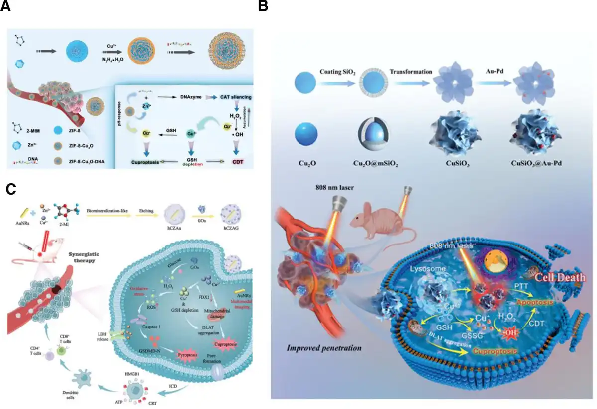
Figure 11.
(A) Schematic of ZIF-8-Cu2O-DNA synthesis and cascade reaction mechanism for cuproptosis and chemodynamic therapy.[88] Copyright 2023, Wiley-VCH. (B) Schematic illustration of the preparation scheme and therapeutic mechanism for cuproptosis-based synergistic cancer therapy of the CuSiO3@Au-Pd NMs.[89] Copyright 2024, Wiley-VCH. (C) Schematic illustration of yolk-shell nanostructured hCZAG preparation and corresponding mechanism for cuproptosis/pyroptosis-induced ICD by cascade reactions.[90] Copyright 2024, Wiley-VCH.
The efficacy of CDT agents can be substantially heightened by inducing localized hyperthermia within the tumor.[91] Photothermal therapy (PTT) is an extensively utilized technique that employs photothermal agents to convert NIR light into heat, damaging tumor cells.[92] By incorporating PTT into CDT, the therapeutic outcome is further enhanced, creating a complementary strategy that leverages the thermal effects of PTT with the oxidative mechanisms of CDT for a more potent cancer treatment. Song et al[89] developed nanomotors (CuSiO3@Au-Pd NMs) that utilized NIR light to enhance cellular uptake and tumor penetration for achieving a cuproptosis-assisted synergistic therapeutic approach combining PTT and CDT (Figure 11B). These nanomotors were capable of self-propulsion upon NIR irradiation, facilitating their entry into cells and their ability to reach deep within tumor tissues. Once internalized, the Cu2+ ions released from the nanomotors initiate the aggregation of DLAT in the TCA cycle, leading to proteotoxic stress and the induction of cuproptosis in cells. Additionally, the Cu+ ions, produced by the reduction of Cu2+ by GSH, triggered Fenton-like reactions that generated amounts of toxic ROS. The gold-palladium nanoparticles (Au-Pd NAs) within the CuSiO3@Au-Pd NMs contributed to their exceptional photothermal capabilities, further boosting the effectiveness of the cancer treatment. The CuSiO3@Au-Pd NMs-mediated synergistic therapy, which combined PTT with CDT and cuproptosis, presented a promising strategy for efficient and multifaceted cancer treatment (Figure 11C).
Recent research has verified that an increase in intracellular ROS can initiate pyroptosis, a novel form of PCD characterized by the upregulation of gasdermin family proteins. In this regard, it is expected that with the addition of CDT, simultaneous activation of cellular cuproptosis and pyroptosis to improve the efficiency of tumor treatment. Wang et al[90] constructed an integrated nanoplatform (hCZAG), which used the zeolitic imidazolate framework-8 (ZIF-8) as its basement. This platform featured Cu2+ and Zn2+ as active sites and incorporated glucose oxidase (GOx) to simultaneously induce pyroptosis and cuproptosis (Figure 11D). The GOx within the hCZAG efficiently increased intracellular H2O2 content. Meanwhile, the Cu2+ ions could be reduced to Cu+ by the overexpressed endogenous GSH, and both Cu2+ and Cu+ ions could engage in Fenton-like reactions, thereby enhancing the generation of ROS and amplifying oxidative stress. Significantly, the surge in ROS caused by the hCZAG nanoplatform activated Caspase-1 proteins, leading to the cleavage of GSDMD, and ultimately inducing pyroptosis. Furthermore, the accumulation of Cu+ ions could cause the aggregation of DLAT, which triggered cuproptosis. This dual-modality approach to cancer therapy, activating both pyroptosis and cuproptosis pathways, presented a promising strategy for enhancing the effectiveness of tumor treatments.
5.3 Cuproptosis combined immunotherapy
Cancer immunotherapy, which involves the strategic use of the immune system to identify and eliminate malignant cells, has become a pivotal and innovative method in oncological care.[93] Nevertheless, the typically weak immune reaction following immunotherapy often leads to the development of adverse effects associated with the immune system. Recently, several studies found that activation of ICD can further promote the liberation of antigens from tumors and the maturation process of dendritic cells (DCs). Hence, it amplifies the immunogenicity of the tumor and invigorates the immune system’s response.[94,95] Cuproptosis has been reported to trigger the ICD, releasing damage-associated molecular patterns (DAMPs) to promote the recruitment of T cells.[96,97] The mechanism of ICD induced by copper ions involves multiple aspects, such as copper ions and mitochondrial function, oxidative stress, DAMPs release, and activation of immune response. These mechanisms work together to cause tumor cell death and activate antitumor immune response.[98] Under normal physiological conditions, cells maintain a balance between oxidation and antioxidant defense. Copper ions can destroy the cellular antioxidant system and lead to tumor cell death.[99] On the one hand, copper ions undergo a Fenton-like reaction, catalyzing hydrogen peroxide to produce a large amount of ROS, which cause multiple damages to cells, including DNA damage, interference with mitochondrial function, and destruction of cell membrane integrity.[100] On the other hand, copper can oxidize reduced GSH to oxidized glutathione disulfide (GSSG), leading to the depletion of the antioxidant GSH, thereby interfering with the GSH-related antioxidant defense system and leading to tumor cell apoptosis.[101] In addition, the copper ion carrier elesclomol can bind extracellular divalent copper ions and transport them to intracellular compartments. FDX1 reduces divalent copper ions to monovalent copper ions, inhibiting the synthesis of Fe–S cluster proteins. LIAS is an upstream regulator of protein lipoylation and can regulate the lipoylation of mitochondrial enzymes such as DLAT. Monovalent copper ions can bind to lipoylated DLAT in the TCA cycle and induce the aggregation of lipoylated proteins.[6] These abnormal processes together lead to proteotoxic stress and mitochondrial dysfunction, ultimately leading to cell death. Hence, the immunotherapeutic effect of tumors could be enhanced by regulating the cell cuproptosis. Du et al[102] prepared a bismuth copper oxide nanomaterial enriched with copper vacancies (BCO-VCu), which possessed superior piezoelectric and ferroelectric characteristics. The incorporation of copper vacancies (VCu) led to an enhancement in the piezoelectric properties and residual ferroelectric polarization. The optimized BCO-VCu exhibited outstanding ferroelectric catalytic capabilities and a strong initial polarization, thereby efficiently generating ROS through a ferroelectric catalytic mechanism. This advanced ferroelectric nanomaterial was capable of being precisely controlled, allowing it to facilitate penetration into cells by initially increasing the permeability of the cell membrane and then inhibiting the efflux of copper ions via specific proteins. This process intensified the activation of cuproptosis, inducing ICD and provoking a robust immune response. In turn, the occurrence of ICD stimulated the maturation of DCs and the polarization of macrophages toward the M1 phenotype, promoting the differentiation of cytotoxic T cells for immunotherapy (Figure 12A). Likewise, Liu et al[105] synthesized morphology-transforming multifunctional nanotransformers (CTMF NPs) to deliver copper ions and induce cuproptosis by disrupting the TCA cycle, which yielded notable ICD and synergistic treatment by multiple anticancer mechanisms in a single therapeutic entity.
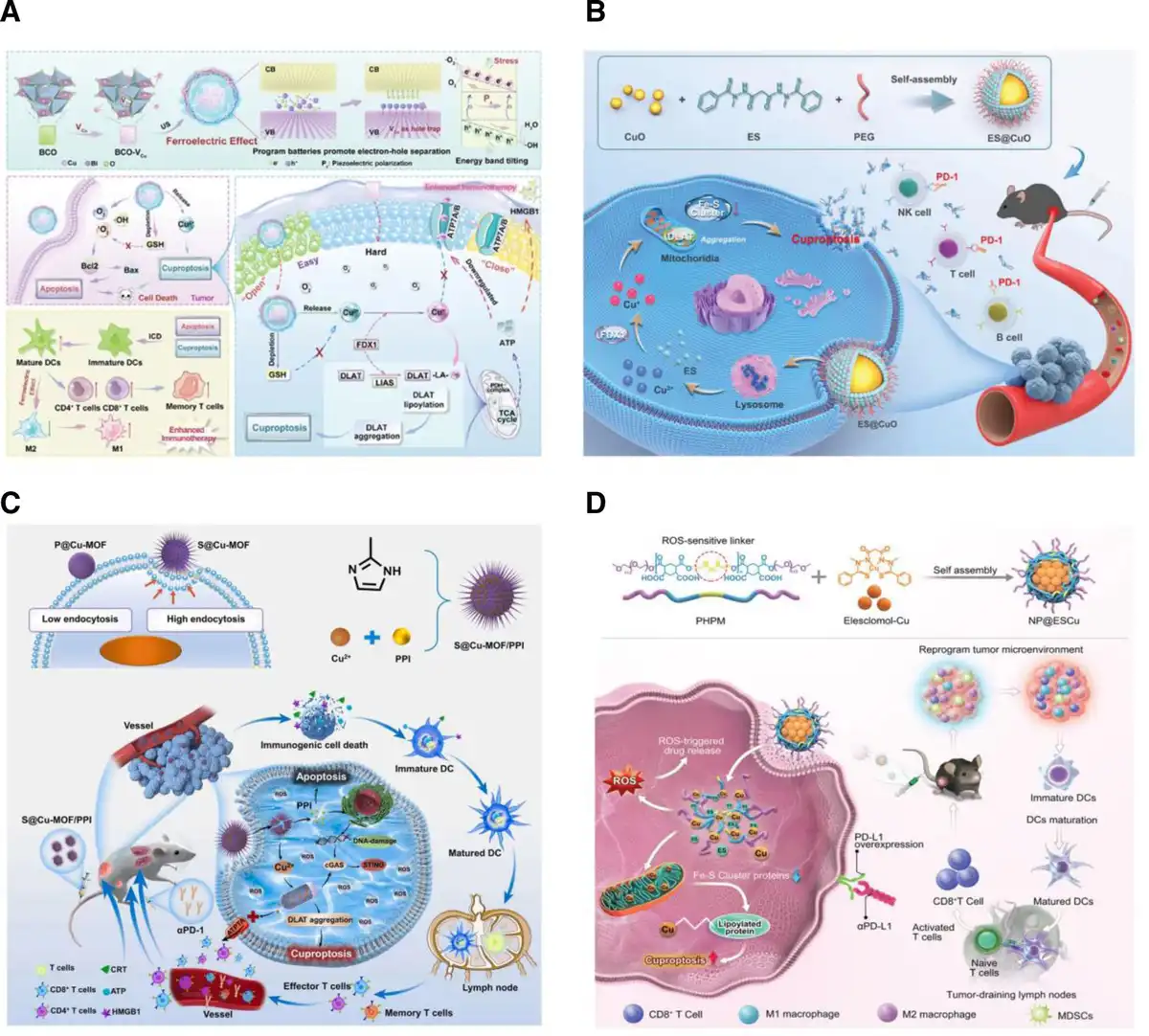
Figure 12.
(A) Schematic of BCO-VCu with ferroelectric effect induced apoptosis and cuproptosis in tumor cells under US irradiation, leading to ICD and strong immune responses.[102] Copyright 2024, Wiley-VCH. (B) A schematic representation of the synergistic mechanism for melanoma immunotherapy, where ES@CuO-induced cuproptosis was combined with PD-1 checkpoint inhibition to enhance the therapeutic effect.[103] Copyright 2024, Wiley-VCH. (C) Schematic design of the S@Cu-MOF/PPI and its therapeutic action for cuproptosis-mediated αPD-1 immunotherapy.[104] Copyright 2024, Elsevier B.V. (D) Schematic design of NP@ESCu and corresponding therapeutic mechanism by activating cuproptosis and combining with αPD-L1 for enhanced cancer therapy.[98] Copyright 2023, Wiley-VCH.
In addition, tumor cells tend to express programmed death ligand 1 (PD-L1) on their surface and achieve immune escape by binding to programmed death receptor 1 (PD-1) on T cells (immune cells), inhibiting the function of T cells, thereby leading to immune escape.[106] Hence, utilizing immune checkpoint inhibitors (ICIs), particularly those targeting the PD-1/PD-L1 axis, has demonstrated unprecedented clinical outcomes in various malignancies.[107] Moreover, on the basis of cuproptosis-mediated ICD, the combination of ICIs will be able to further improve the immunotherapeutic efficiency of tumors. Lu et al[103] developed an innovative cuproptosis-inducing nanosystem (ES@CuO) involving CuO nanoparticles with ionophore ES. Upon internalization by tumor cells, ES@CuO underwent degradation, liberating Cu2+ ions and ES to initiate cuproptosis, effectively suppressing the proliferation of murine B16 melanoma cells. Furthermore, the ES@CuO could enhance cuproptosis-mediated immune responses and alter the typically immunosuppressive TME by increasing the infiltration of lymphocytes into the tumor and promoting the secretion of inflammatory cytokines, resulting in an effective immune response against cancer. In addition, the integration of ES@CuO with PD-1 immunotherapy could significantly boost the antitumor efficacy in a murine melanoma model (Figure 12B). In a related study, Xu et al[104] constructed a spiky copper-based MOF (S@Cu-MOF) and combined it with αPD-1 for cancer therapy. The spiky structure could promote tumor cell uptake of S@Cu-MOF. Moreover, this synergistic approach has been shown to further enhance cuproptosis-mediated cancer immunotherapy. By combining the cuproptosis activation property of the S@Cu-MOF with the immune checkpoint inhibition offered by αPD-1, it could overcome the limitations of individual treatments and provide a more potent therapeutic effect against cancer (Figure 12C).
Furthermore, recent studies indicated that intratumoral Cu levels in tumors could affect PD-L1 expression, consequently affecting the responsiveness of tissue to immune therapy.[108,109] Guo et al[98] built a ROS-sensitive polymer for coencapsulating ES and Cu to create nanoparticles (NP@ESCu). Once these nanoparticles penetrate cancer cells, the high levels of ROS within the cells trigger the release of ES and Cu. The released ES would be quickly effluxed and act as a chelator, facilitating the uptake of additional extracellular Cu2+ ions into the cancer cells. This procedure established a shuttling mechanism that promoted the continuous accumulation of Cu within the mitochondria of the cancer cells, leading to the occurrence of cuproptosis. Significantly, the sustained increase in copper levels within the cells can induce the upregulation of PD-L1 expression on the cancer cell surface. This upregulation is a key factor in enhancing the effectiveness of αPD-L1 for immune therapy. In a bladder cancer mouse model, the combined antitumor efficacy of NP@ESCu and αPD-L1 was evaluated. This study marked the first instance of integrating a nanomaterial capable of inducing cuproptosis with αPD-L1 to augment the therapeutic impact on cancer (Figure 12D).
6. Conclusion and perspective
Cuproptosis initially identified in 2022 is an emerging modality of programmed cell death, which has gained increasing interest, especially in the domain of cancer therapy.[6,110,111] Afterwards, an extensive study of biological mechanisms involving cuproptosis has been deeply explored, which promoted the use of cuproptosis inducers for the elimination of cancers.[112,113] Rapid advances in nanotechnology are driving the development of cuproptosis-related therapeutic agents. Moreover, several copper-based nanomaterials have been designed and constructed to realize cuproptosis-mediated cancer therapy.[114–115–116] In this review, a list of copper-based nanomaterials that have been developed to activate cuproptosis is first outlined and categorized. The advantages of each type of nanomaterial are also explored, providing a comparison for the subsequent selection of material species. In the meantime, 3 feasible cuproptosis sensitization strategies (copper transporter protein regulation, GSH depletion, and increasing intracellular oxygen content) have been proposed to amplify the efficacy of cuproptosis. Furthermore, it is concluded that some treatments such as chemotherapy, chemodynamic therapy, and immunotherapy can be further combined with cuproptosis to maximize the therapeutic effect, which offers promising insights into the engineering of copper-based nanomaterials for cancer combination therapy.
In addition, this review summarizes the application ideas of copper-based nanomedicines in tumor treatment, which provides inspiration for the design of advanced copper-based nanotherapeutic platforms. Initially, copper-based nanomaterials can be used as carriers for targeted copper therapy, and precise tumor treatment can be achieved through chelators and copper ion carriers. Subsequently, copper-based nanomaterials can respond to the TME and enhance the therapeutic effect by changing the local chemical and physical conditions of the tumor; ultimately, copper-based nanomaterials show the potential for multimodal synergistic treatment in cancer treatment, such as PTT-chemotherapy, PTT-PDT, PTT-CDT, and PDT-immunotherapy (Table 2).
Table 2
Treatment strategies of copper-based nanomedicines.
| Classification | Remarks and highlights | Methods | References |
|---|---|---|---|
| Targeted copper therapy | Mild side effects and promising clinical application prospects | Copper ions carrier | [117] |
| Copper ion chelating agent | [118] | ||
| Tumor microenvironment response to therapy | Achieve controlled release of drugs and improve tumor-targeted delivery efficiency | H2S response | [119] |
| pH response | [103] | ||
| GSH response | [120] | ||
| ROS response | [121] | ||
| Multimodal combined treatment | Multimodal synergistic effects for enhanced therapeutic efficacy | PTT-CDT | [122] |
| PTT-chemotherapy | [24] | ||
| PTT-PDT | [123] | ||
| PTT-PDT-chemotherapy | [124] | ||
| PTT-CDT-chemotherapy | [125] | ||
| PTT-immunotherapy | [126] |
However, the research on cuproptosis is still in its infancy, and there are still many difficult issues to be resolved. (1) Facing the complex molecular mechanism of cuproptosis, the relevant regulatory approaches, such as the functional regulation of FDX1 and the regulation of proteotoxic stress induced by copper-promoted oligomerization of DLAT, have not yet been clarified.[127,128] Therefore, more fundamental studies are needed to further clarify the molecular regulatory mechanisms of cuproptosis. It is only after clarifying the regulatory mechanisms of cuproptosis at the molecular level that copper-based nanomaterials can be designed to activate cuproptosis more efficiently. (2) The central prerequisite for cuproptosis is the induction of intracellular copper overload. Unfortunately, both normal cells and tumor cells have the potential to trigger cuproptosis causing cellular damage once excessive intracellular copper content accumulates. Currently, developed copper-based nanomaterials that can be used to activate cuproptosis are based on copper ion-delivery strategies in response to the TME; however, due to the diversity and heterogeneity of tumor tissues, the differences in the microenvironment between some tumors and normal tissues are very small, and it is difficult for the existing materials to satisfy such needs.[62,129,130] Hence, on the one hand, it is necessary to design ultrasensitive copper-based nanomaterial facilities with tumor-specific responses, but also to control the injection dosage and duration of the nanomaterials as much as possible to achieve a balance between efficacy and side effects. (3) Tumor cells have a variety of protective effects against cuproptosis, for example, overexpressed GSH has a strong ligand-chelating effect, which can effectively chelate the accumulated copper ions in the cell and then cut off the interaction between copper ions and cuproptosis-associated proteins, thus reducing the possibility of the emergence of proteotoxic stress.[131,132] Moreover, once intracellular cuproptosis-associated proteotoxic stress is generated, tumor cells will increase the expression of heat shock proteins to effectively weaken the impact of proteotoxic stress, and then reduce the activation efficiency of cuproptosis.[68,133] It is the presence of these self-protective effects within tumor cells that further impedes the initiation of the cuproptosis pathway and attenuates the sensitivity of tumor cells to cuproptosis. Thus, there is a need to design and develop copper-based nanomaterials that effectively inhibit this self-protective effect to sensitize cuproptosis. (4) Single cuproptosis-mediated tumor therapy still fails to achieve satisfactory therapeutic effects.[134,135] It has been shown that copper, which is the core of cuproptosis, not only catalyzes the overexpression of H2O2 in tumors to increase the intracellular ROS level but also combines with some specific chemotherapeutic agents to potentiate chemotherapy.[136,137] In addition, the accumulation of copper in the tumor site also affects the expression of immune proteins in tumor cells, which in turn promotes the immunotherapy of tumors.[138,139] Therefore, copper-based nanomaterials can be designed to combine multiple therapeutic modalities to achieve more efficient tumor therapy. (5) It is well known that toxicity is one of the key factors in the clinical translation of nanomaterials. Since nanomaterials interact with target cells, it is important to ensure that this interaction does not cause any side effects.[140] Researchers have developed standard experimental models for in vitro (cell lines) and in vivo (experimental animals) toxicity assessment.[141] The toxicity of nanoparticles is determined by their physicochemical and biological properties, including their structure, shape, size, surface properties, catalytic activity, aggregation, and solubility.[142] Currently, a variety of strategies have been developed to improve the biosafety of copper-based nanomaterials: (1) using biosafe polymers to modify the surface of copper-based nanomaterials to improve their biocompatibility[143]; (2) by adjusting the size, shape, and surface charge and other physicochemical properties of nanomaterials, their interaction with biological systems can be affected, thereby reducing toxicity[144]; (3) designing biodegradable copper-based nanomaterials, such as copper-based silicate nanoparticles and copper-based MOFs, which can be degraded in vivo, reducing potential toxicity and adverse reactions caused by long-term accumulation[145]; (4) by surface modification or designing specific nanostructures, the targeting of copper-based nanomaterials to tumor tissues can be improved, reducing damage to normal tissues.[146] Through the above strategies, the biosafety of copper-based nanomaterials can be effectively improved and their potential risks in clinical applications can be reduced.
Cuproptosis provides new perspectives for cancer therapy. Comprehensive integration of expertise from the disciplines of metallurgy, oncology, and nanoscience, in-depth study of the biomolecular mechanism of cuproptosis, construction of efficient copper ion-delivery copper-based nanomaterials, and design of effective cuproptosis-sensitizing combined therapeutic strategies will offer the possibility of eradicating cancer.
Funding
This work was supported in part by the National Natural Science Foundation of China (No. 52073258), the Natural Science Foundation of Zhejiang Province (No. LR22E010001), and the Fundamental Research Funds for the Provincial Universities of Zhejiang (No. RF-B2022006).
Conflicts of interests
The authors declare that they have no conflict of interest.
Author contributions
Fan Zhao: Conceptualization, investigation, discussion, and writing original draft. Zhuangzhuang Zhao: Investigation, discussion, and writing original draft. Hao Gao, Jiarui Qi, and Hongyan Yu: Investigation and discussion. Yuxin Zhang: Discussion and writing original draft. Chen Wang and Junchen Xu: Discussion. Muhammad Zubair Yousaf: Discussion and supervision. Shenglei Che: Conceptualization, supervision, reviewing, and editing. Jing Yu: Conceptualization, supervision, funding, review, and editing.
References
- [1] Tsang T, Davis CI, Brady DC. Copper biology. Curr Biol. 2021;31(9):R421–R427.
- [2] Yang Y, Li M, Chen G, et al. Dissecting copper biology and cancer treatment: “activating cuproptosis or suppressing cuproplasia”. Coord Chem Rev. 2023;495:215395.
- [3] Garza NM, Swaminathan AB, Maremanda KP, et al. Mitochondrial copper in human genetic disorders. Trends Endocrinol Metab. 2023;34(1):21–33.
- [4] Gao L, Zhang A. Copper-instigated modulatory cell mortality mechanisms and progress in oncological treatment investigations. Front Immunol. 2023;14.
- [5] Han J. Copper trafficking systems in cells: insights into coordination chemistry and toxicity. Dalton Trans. 2023;52(42):15277–15296.
- [6] Tsvetkov P, Coy S, Petrova B, et al. Copper induces cell death by targeting lipoylated TCA cycle proteins. Science. 2022;375(6586):1254–1261.
- [7] Li Y, Du Y, Zhou Y, et al. Iron and copper: critical executioners of ferroptosis, cuproptosis and other forms of cell death. Cell Commun Signal. 2023;21(1):327.
- [8] Mao C, Wang M, Zhuang L, et al. Metabolic cell death in cancer: ferroptosis, cuproptosis, disulfidptosis, and beyond. Protein Cell. 2024;15:642–660.
- [9] Tang D, Kroemer G, Kang R. Targeting cuproplasia and cuproptosis in cancer. Nat Rev Clin Oncol. 2024;21(5):370–388.
- [10] Tian Z, Jiang S, Zhou J, et al. Copper homeostasis and cuproptosis in mitochondria. Life Sci. 2023;334:122223.
- [11] Zhang X, Tao T, Qiu Y, et al. Copper-mediated novel cell death pathway in tumor cells and implications for innovative cancer therapies. Biomed Pharmacother. 2023;168:115730.
- [12] Guan D, Zhao L, Shi X, et al. Copper in cancer: from pathogenesis to therapy. Biomed Pharmacother. 2023;163:114791.
- [13] Tang X, Yan Z, Miao Y, et al. Copper in cancer: from limiting nutrient to therapeutic target. Front Oncol. 2023;13.
- [14] Han Y, Xie N, Zhou W. Copper coordination-based nanomedicine for tumor theranostics. Adv Therap. 2024;7(2):2300305.
- [15] Shen Z, Qiu Y, Ding H, et al. Cuproptosis and cuproptosis-based synergistic therapy for cancer treatment. ChemMedChem. 2024;19(21):e202400216.
- [16] Zhang Z, Ding C, Sun T, et al. Tumor therapy strategies based on microenvironment-specific responsive nanomaterials. Adv Healthcare Mater. 2023;12(20):2300153.
- [17] Su T, Zhao F, Ying Y, et al. Self-monitoring theranostic nanomaterials: emerging visual agents for real-time monitoring of tumor treatment processes. Small Methods. 2024;8(5):2301470.
- [18] Jing Z, Du Q, Zhang X, et al. Nanomedicines and nanomaterials for cancer therapy: progress, challenge and perspectives. Chem Eng J. 2022;446:137147.
- [19] Dutta B, Barick KC, Hassan PA. Recent advances in active targeting of nanomaterials for anticancer drug delivery. Adv Colloid Interface Sci. 2021;296:102509.
- [20] Wei Q, Pan Y, Zhang Z, et al. Copper-based nanomaterials for biomedical applications. Chem Eng J. 2024;483:149040.
- [21] Zhong X, Dai X, Wang Y, et al. Copper-based nanomaterials for cancer theranostics. Wiley Interdiscip Rev Nanomed Nanobiotechnol. 2022;14(4):e1797.
- [22] Lu J, Miao Y, Li Y. Cuproptosis: advances in stimulus-responsive nanomaterials for cancer therapy. Adv Healthcare Mater. 2024;13(19):2400652.
- [23] Bost M, Houdart S, Oberli M, et al. Dietary copper and human health: current evidence and unresolved issues. J Trace Elem Med Biol. 2016;35:107–115.
- [24] Yu Q, Han Y, Wang X, et al. Copper silicate hollow microspheres-incorporated scaffolds for chemo-photothermal therapy of melanoma and tissue healing. ACS Nano. 2018;12:2695–2707.
- [25] Xie J, Yang Y, Gao Y, et al. Cuproptosis: mechanisms and links with cancers. Mol Cancer. 2023;22(1):46.
- [26] Lönnerdal B. Intestinal regulation of copper homeostasis: a developmental perspective. Am J Clin Nutr. 2008;88(3):846S–850S.
- [27] Lutsenko S. Dynamic and cell-specific transport networks for intracellular copper ions. J Cell Sci. 2021;134:jcs240523.
- [28] Hernandez S, Tsuchiya Y, García-Ruiz JP, et al. ATP7B copper-regulated traffic and association with the tight junctions: copper excretion into the bile. Gastroenterology. 2008;134(4):1215–1223.
- [29] van, Rensburg MJ, van, Rooy M, Bester MJ, et al. Oxidative and haemostatic effects of copper, manganese and mercury, alone and in combination at physiologically relevant levels: an ex vivo study. Hum Exp Toxicol. 2019;38:419–433.Full TextGoogle Scholar
- [30] Kim BE, Nevitt T, Thiele DJ. Mechanisms for copper acquisition, distribution and regulation. Nat Chem Biol. 2008;4:176–185.
- [31] Sheline CT, Choi DW. Cu2+ toxicity inhibition of mitochondrial dehydrogenases in vitro and in vivo. Ann Neurol. 2004;55:645–653.
- [32] Wang C, Cao F, Ruan Y, et al. Specific generation of singlet oxygen through the Russell mechanism in hypoxic tumors and GSH depletion by Cu-TCPP nanosheets for cancer therapy. Angew Chem Int Ed Engl. 2019;58:9846–9850.
- [33] Wu H, Zhang Z, Cao Y, et al. A self-amplifying ROS-responsive nanoplatform for simultaneous cuproptosis and cancer immunotherapy. Adv Sci. 2024;11(23):e2401047.
- [34] Gao L, Song Y, Zhong J, et al. Biocompatible 2d Cu-TCPP nanosheets derived from Cu2O nanocubes as multifunctional nanoplatforms for combined anticancer therapy. ACS Biomater Sci Eng. 2022;8:1074–1086.
- [35] Xie Z, Liang S, Cai X, et al. O2-Cu/ZIF-8@Ce6/ZIF-8@F127 composite as a tumor microenvironment-responsive nanoplatform with enhanced photo-/chemodynamic antitumor efficacy. ACS Appl Mater Interfaces. 2019;11:31671–31680.
- [36] Chen K, Zhou A, Zhou X, et al. Cellular Trojan Horse initiates bimetallic Fe-Cu MOF-mediated synergistic cuproptosis and ferroptosis against malignancies. Sci Adv. 2024;10(15):eadk3201.
- [37] Ren D, Cheng Y, Xu W, et al. Copper-based metal-organic framework induces no generation for synergistic tumor therapy and antimetastasis activity. Small. 2023;19:e2205772.
- [38] Liu J, Yuan Y, Cheng D, et al. Copper-based metal–organic framework overcomes cancer chemoresistance through systemically disrupting dynamically balanced cellular redox homeostasis. J Am Chem Soc. 2022;144(11):4799–4809.
- [39] Jin X, Zhao H, Chao Z, et al. Self-assembled cupric oxide nanoclusters for highly efficient chemodynamic therapy. Chem Asian J. 2022;17:e202200296.
- [40] Zhang H, Chen Y, Cai Y, et al. Paramagnetic CuS hollow nanoflowers for T2-flair magnetic resonance imaging-guided thermochemotherapy of cancer. Biomater Sci. 2019;7:409–418.
- [41] Oliveri V. Biomedical applications of copper ionophores. Coord Chem Rev. 2020;422:213474.
- [42] Oliveri V. Selective targeting of cancer cells by copper ionophores: an overview. Front Mol Biosci. 2022;9:841814.
- [43] Zheng P, Zhou C, Lu L, et al. Elesclomol: a copper ionophore targeting mitochondrial metabolism for cancer therapy. J Exp Clin Cancer Res. 2022;41(1):271.
- [44] Xie W, Guo Z, Zhao L, et al. The copper age in cancer treatment: from copper metabolism to cuproptosis. Prog Mater Sci. 2023;138:101145.
- [45] Wu X, Wu Q, Hou M, et al. Regenerating chemotherapeutics through copper-based nanomedicine: disrupting protein homeostasis for enhanced tumor therapy. Adv Healthcare Mater. 2024;13(28):e2401954.
- [46] Kudarha R, Dhas N, Mutalik S. Distinct features of iron based metal organic frameworks (MOFs) for ferroptosis mediated cancer therapy: a comprehensive review. Coord Chem Rev. 2023;494:215330.
- [47] Coluccia M, Parisse V, Guglielmi P, et al. Metal-organic frameworks (MOFs) as biomolecules drug delivery systems for anticancer purposes. Eur J Med Chem. 2022;244:114801.
- [48] Di X, Pei Z, Pei Y, et al. Tumor microenvironment-oriented MOFs for chemodynamic therapy. Coord Chem Rev. 2023;484:215098.
- [49] Chen K, Zhou A, Zhou X, et al. Cellular Trojan Horse initiates bimetallic Fe-Cu MOF-mediated synergistic cuproptosis and ferroptosis against malignancies. Sci Adv. 10(15):eadk3201.
- [50] Qiao L, Zhu G, Jiang T, et al. Self-destructive copper carriers induce pyroptosis and cuproptosis for efficient tumor immunotherapy against dormant and recurrent tumors. Adv Mater. 2024;36(8):2308241.
- [51] Ji M, Liu H, Liang X, et al. Tumor cells are under siege from all sides: tumor cell-mimic metal−organic framework nanoparticles triggering cuproptosis/ferroptosis/apoptosis for chemo-chemodynamic-photothermal-immunological synergistic antitumor therapy. Chem Eng J. 2024;485:149640.
- [52] Liang W, Han C, Zhang D, et al. Copper-coordinated nanoassemblies based on photosensitizer-chemo prodrugs and checkpoint inhibitors for enhanced apoptosis-cuproptosis and immunotherapy. Acta Biomater. 2024;175:341–352.
- [53] Dai Y, Zhu L, Li X, et al. A biomimetic cuproptosis amplifier for targeted NIR-II fluorescence/photoacoustic imaging-guided synergistic NIR-II photothermal immunotherapy. Biomaterials. 2024;305:122455.
- [54] Pei Z, Lei H, Cheng L. Bioactive inorganic nanomaterials for cancer theranostics. Chem Soc Rev. 2023;52(6):2031–2081.
- [55] Bai J, Zhang X, Zhao Z, et al. CuO nanozymes catalyze cysteine and glutathione depletion induced ferroptosis and cuproptosis for synergistic tumor therapy. Small. 2024;20(40):e2400326.
- [56] Ning S, Lyu M, Zhu D, et al. Type-I AIE photosensitizer loaded biomimetic system boosting cuproptosis to inhibit breast cancer metastasis and rechallenge. ACS Nano. 2023;17(11):10206–10217.
- [57] Man R, Xia M, Li H, et al. Human serum albumin mediated controllable synthesis of defect-rich copper hydroxide nanowire for cuproptosis-based anti-tumor therapy. Adv Healthcare Mater. 2024;13(23):e2401078.
- [58] Chan L, Liu Y, Chen M, et al. Cuproptosis-driven enhancement of thermotherapy by sequentially response Cu2-Se via copper chemical transition. Adv Funct Mater. 2023;33(33):2302054.
- [59] Zhao F, Liang L, Wang H, et al. H2S-activated ion-interference therapy: a novel tumor targeted therapy based on copper-overload-mediated cuproptosis and pyroptosis. Adv Funct Mater. 2023;33(38):2300941.
- [60] Zheng J, Ge H, Guo M, et al. Photoinduced cuproptosis with tumor-specific for metastasis-inhibited cancer therapy. Small. 2024;20(10):2304407.
- [61] Chen X, Cai Q, Liang R, et al. Copper homeostasis and copper-induced cell death in the pathogenesis of cardiovascular disease and therapeutic strategies. Cell Death Dis. 2023;14(2):105.
- [62] Wei C, Fu Q. Cell death mediated by nanotechnology via the cuproptosis pathway: a novel horizon for cancer therapy. View. 2023;4(3):20230001.
- [63] Jin X-K, Liang J-L, Zhang S-M, et al. Orchestrated copper-based nanoreactor for remodeling tumor microenvironment to amplify cuproptosis-mediated anti-tumor immunity in colorectal cancer. Mater Today. 2023;68:108–124.
- [64] Xu W, Qian J, Hou G, et al. A hollow amorphous bimetal organic framework for synergistic cuproptosis/ferroptosis/apoptosis anticancer therapy via disrupting intracellular redox homeostasis and copper/iron metabolisms. Adv Funct Mater. 2022;32(40):2205013.
- [65] Yan C, Liu Y, Zhao G, et al. Inhalable metal–organic framework-mediated cuproptosis combined with PD-L1 checkpoint blockade for lung metastasis synergistic immunotherapy. Acta Pharmaceutica Sinica B. 2024;14(5):2281–2297.
- [66] Zhang J, Han M, Zhang J, et al. Syphilis mimetic nanoparticles for cuproptosis-based synergistic cancer therapy via reprogramming copper metabolism. Int J Pharm. 2023;640:123025.
- [67] Corso CR, Acco A. Glutathione system in animal model of solid tumors: from regulation to therapeutic target. Crit Rev Oncol Hematol. 2018;128:43–57.
- [68] Chen W, Xie W, Gao Z, et al. Mild-photothermal effect induced high efficiency ferroptosis-boosted-cuproptosis based on Cu2O@Mn3Cu3O8 nanozyme. Adv Sci. 2023;10(33):2303694.
- [69] Zhang J, Peng L, Hao Y, et al. Biodegradable CuMoO4 nanodots with multienzyme activities for multimodal treatment of tumor. Adv Healthcare Mater. 2023;12(22):2300167.
- [70] Zhao F, Yu H, Liang L, et al. Redox homeostasis disruptors based on metal-phenolic network nanoparticles for chemo/chemodynamic synergistic tumor therapy through activating apoptosis and cuproptosis. Adv Healthcare Mater. 2023;12(29):2301346.
- [71] Huang Q-X, Liang J-L, Chen Q-W, et al. Metal-organic framework nanoagent induces cuproptosis for effective immunotherapy of malignant glioblastoma. Nano Today. 2023;51:101911.
- [72] Khorshidi S, Younesi S, Karkhaneh A. Peroxide mediated oxygen delivery in cancer therapy. Colloids Surf B. 2022;219:112832.
- [73] Huang Y, Liu X, Zhu J, et al. Enzyme core spherical nucleic acid that enables enhanced cuproptosis and antitumor immune response through alleviating tumor hypoxia. J Am Chem Soc. 2024;146(20):13805–13816.
- [74] Fan X, Luo Z, Chen Y, et al. Oxygen self-supplied enzyme nanogels for tumor targeting with amplified synergistic starvation and photodynamic therapy. Acta Biomater. 2022;142:274–283.
- [75] Wang W, Mo W, Hang Z, et al. Cuproptosis: harnessing transition metal for cancer therapy. ACS Nano. 2023;17(20):19581–19599.
- [76] Qin S-Y, Cheng Y-J, Lei Q, et al. Combinational strategy for high-performance cancer chemotherapy. Biomaterials. 2018;171:178–197.
- [77] Liu S, Khan AR, Yang X, et al. The reversal of chemotherapy-induced multidrug resistance by nanomedicine for cancer therapy. J Control Release. 2021;335:1–20.
- [78] Lu S, Tian H, Li B, et al. An ellagic acid coordinated copper-based nanoplatform for efficiently overcoming cancer chemoresistance by cuproptosis and synergistic inhibition of cancer cell stemness. Small. 2024;20(17):2309215.
- [79] Ni C, Ouyang Z, Li G, et al. A tumor microenvironment-responsive core-shell tecto dendrimer nanoplatform for magnetic resonance imaging-guided and cuproptosis-promoted chemo-chemodynamic therapy. Acta Biomater. 2023;164:474–486.
- [80] Zhong J, Zheng X, Wen Y, et al. In situ sacrificial growth of metastable copper-enriched nanomedicine for cuproptosis-based synergistic cancer therapy. Chem Eng J. 2023;474:145795.
- [81] Xu W, Wang Y, Hou G, et al. Tumor microenvironment responsive hollow nanoplatform for triple amplification of oxidative stress to enhance cuproptosis-based synergistic cancer therapy. Adv Healthcare Mater. 2023;12(13):2202949.
- [82] Yang B, Shi J. Developing new cancer nanomedicines by repurposing old drugs. Angew Chem Int Ed Engl. 2020;59(49):21829–21838.
- [83] Xu L, Sun Y, Li Y, et al. Disulfiram: a food and drug administration-approved multifunctional role in synergistically drug delivery systems for tumor treatment. Int J Pharm. 2022;626:122130.
- [84] McMahon A, Chen W, Li F. Old wine in new bottles: advanced drug delivery systems for disulfiram-based cancer therapy. J Control Release. 2020;319:352–359.
- [85] Ding Y, Wan J, Zhang Z, et al. Localized Fe(II)-induced cytotoxic reactive oxygen species generating nanosystem for enhanced anticancer therapy. ACS Appl Mater Interfaces. 2018;10(5):4439–4449.
- [86] Zhou Y, Fan S, Feng L, et al. Manipulating intratumoral Fenton chemistry for enhanced chemodynamic and chemodynamic-synergized multimodal therapy. Adv Mater. 2021;33(48):2104223.
- [87] Xing Y, Xiu J, Zhou M, et al. Copper single-atom jellyfish-like nanomotors for enhanced tumor penetration and nanocatalytic therapy. ACS Nano. 2023;17(7):6789–6799.
- [88] Yu Q, Zhou J, Liu Y, et al. DNAzyme-mediated cascade nanoreactor for cuproptosis-promoted pancreatic cancer synergistic therapy. Adv Healthcare Mater. 2023;12(28):2301429.
- [89] Song Y, Zhan G, Zhou S-F. Design of near infrared light-powered copper phyllosilicate nanomotors for cuproptosis-based synergistic cancer therapy. Adv Funct Mater. 2024;34(18):2314568.
- [90] Wang Y-Y, Li S-L, Zhang X-Y, et al. “Multi-in-one” yolk-shell structured nanoplatform inducing pyroptosis and antitumor immune response through cascade reactions. Small. 2024;20(30):2400254.
- [91] Tao J, Wang B, Dong Y, et al. Photothermal and acid-responsive fucoidan-CuS bubble pump microneedles for combined CDT/PTT/CT treatment of melanoma. ACS Appl Mater Interfaces. 2023;15(34):40267–40279.
- [92] Zhao L, Zhang X, Wang X, et al. Recent advances in selective photothermal therapy of tumor. J Nanobiotechnol. 2021;19(1):335.
- [93] Farooq MU, Lawrie CH, Deng N-N. Engineering nanoparticles for cancer immunotherapy: current achievements, key considerations and future perspectives. Chem Eng J. 2024;486:150356.
- [94] Zhang Z, Pan Z, Li Q, et al. Rational design of ICD-inducing nanoparticles for cancer immunotherapy. Sci Adv. 10(6):eadk0716.
- [95] Dogheim GM, El Feel NE, Abd El-Maksod EA, et al. Nanomedicines as enhancers of tumor immunogenicity to augment cancer immunotherapy. Drug Discov Today. 2024;29(3):103905.
- [96] Pei P, Wang Y, Shen W, et al. Oxygen-driven cuproptosis synergizes with radiotherapy to potentiate tumor immunotherapy. Aggregate. 2024;5(3):e484.
- [97] Chang J, Yin W, Zhi H, et al. Copper deposition in polydopamine nanostructure to promote cuproptosis by catalytically inhibiting copper exporters of tumor cells for cancer immunotherapy. Small. 2024;20(27):2308565.
- [98] Guo B, Yang F, Zhang L, et al. Cuproptosis induced by ROS responsive nanoparticles with elesclomol and copper combined with αpd-l1 for enhanced cancer immunotherapy. Adv Mater. 2023;35(22):e2212267.
- [99] Chio IIC, Tuveson DA. ROS in cancer: the burning question. Trends Mol Med. 2017;23(5):411–429.
- [100] Reczek CR, Chandel NS. The two faces of reactive oxygen species in cancer. Ann Rev Cancer Biol. 2017;1:79–98.
- [101] Ngamchuea K, Batchelor-McAuley C, Compton RG. The copper(II)-catalyzed oxidation of glutathione. Chemistry. 2016;22(44):15937–15944.
- [102] Du Y, Zhao X, He F, et al. A vacancy-engineering ferroelectric nanomedicine for cuproptosis/apoptosis co-activated immunotherapy. Adv Mater. 2024;36(30):2403253.
- [103] Lu X, Chen X, Lin C, et al. Elesclomol loaded copper oxide nanoplatform triggers cuproptosis to enhance antitumor immunotherapy. Adv Sci. 2024;11(18):e2309984.
- [104] Xu M, Chen H, Zhu G, et al. Spiky metal-organic framework nanosystem for enhanced cuproptosis-mediated cancer immunotherapy. Nano Today. 2024;56:102231.
- [105] Liu F, Guo C, Li X, et al. A versatile nano-transformer for efficient localization-specific imaging and synergistic therapy of bladder cancer. Nano Today. 2024;54:102116.
- [106] Yang J, Hu L. Immunomodulators targeting the PD-1/PD-L1 protein-protein interaction: from antibodies to small molecules. Med Res Rev. 2019;39(1):265–301.
- [107] Chen M, Hu S, Li Y, et al. Targeting nuclear acid-mediated immunity in cancer immune checkpoint inhibitor therapies. Signal Transduct Target Ther. 2020;5(1):270.
- [108] Voli F, Valli E, Lerra L, et al. Intratumoral copper modulates PD-L1 expression and influences tumor immune evasion. Cancer Res. 2020;80(19):4129–4144.
- [109] Li W, Xiao Y, Guo G, et al. Cuprous oxide nanocomposites with photothermal (PTT) and chemical dynamics (CDT) effects induce cuproptosis in breast cancer using the strategy of increasing inflow and reducing outflow. Nano Today. 2024;56:102223.
- [110] Feng Y, Yang Z, Wang J, et al. Cuproptosis: unveiling a new frontier in cancer biology and therapeutics. Cell Commun Signal. 2024;22(1):249.
- [111] Wu J, He J, Liu Z, et al. Cuproptosis: mechanism, role, and advances in urological malignancies. Med Res Rev. 2024;44(4):1662–1682.
- [112] Zhong Y, Zeng W, Chen Y, et al. The effect of lipid metabolism on cuproptosis-inducing cancer therapy. Biomed Pharmacother. 2024;172:116247.
- [113] Wang Y, Pei P, Yang K, et al. Copper in colorectal cancer: from copper-related mechanisms to clinical cancer therapies. Clin Transl Med. 2024;14(6):e1724.
- [114] Zhang N, Ping W, Rao K, et al. Biomimetic copper-doped polypyrrole nanoparticles induce glutamine metabolism inhibition to enhance breast cancer cuproptosis and immunotherapy. J Control Release. 2024;371:204–215.
- [115] Hu G, Cheng W, Gu M, et al. Vitamin E succinate-glycol chitosan modified copper ferrite nanocomposites for lung cancer: targeted oxidative stress regulation induces cuproptosis and ferroptosis. Chem Eng J. 2024;493:152408.
- [116] Hu P, Li Y, Zhang L, et al. Defect-engineered photothermal nanozyme with NIR-II absorption induces cuproptosis-apoptosis for synergized cancer immunotherapy and fast wound healing. Mater Des. 2024;237:112568.
- [117] Pan M, Zheng Q, Yu Y, et al. Seesaw conformations of Npl4 in the human p97 complex and the inhibitory mechanism of a disulfiram derivative. Nat Commun. 2021;12(1):121.
- [118] Ramchandani D, Berisa M, Tavarez DA, et al. Copper depletion modulates mitochondrial oxidative phosphorylation to impair triple negative breast cancer metastasis. Nat Commun. 2021;12:7311.
- [119] An L, Wang X, Rui X, et al. The in situ sulfidation of Cu2O by endogenous H2S for colon cancer theranostics. Angew Chem Int Ed. 2018;57:15782–15786.
- [120] He H, Du L, Guo H, et al. Redox responsive metal organic framework nanoparticles induces ferroptosis for cancer therapy. Small. 2020;16:2001251.
- [121] Wang N, Liu Y, Peng D, et al. Copper-based composites nanoparticles improve triple-negative breast cancer treatment with induction of apoptosis-cuproptosis and immune activation. Adv Healthcare Mater. 2024;13.
- [122] Wang X, Zhong H, Lei Y, et al. Hollow Cu2Se nanozymes for tumor photothermal-catalytic therapy. Chem Mater. 2019;31:6174–6186.
- [123] You Y, Cao F, Zhao Y, et al. Near-infrared light dual-promoted heterogeneous copper nanocatalyst for highly efficient bioorthogonal chemistry in vivo. ACS Nano. 2020;14:4178–4187.
- [124] Xu M, Yang G, Bi H, et al. An intelligent nanoplatform for imaging-guided photodynamic/photothermal/chemotherapy based on upconversion nanoparticles and CuS integrated black phosphorus. Chem Eng J. 2020;382:122822.
- [125] Wang S, Pang Y, Hu S, et al. Copper sulfide engineered covalent organic frameworks for pH-responsive chemo/photothermal/chemodynamic synergistic therapy against cancer. Chem Eng J. 2023;451:138864.
- [126] Sun Y, Liang Y, Dai W, et al. Peptide-drug conjugate-based nanocombination actualizes breast cancer treatment by maytansinoid and photothermia with the assistance of fluorescent and photoacoustic images. Nano Lett. 2019;19:3229–3237.
- [127] Wang X, Zhou M, Liu Y, et al. Cope with copper: from copper linked mechanisms to copper-based clinical cancer therapies. Cancer Lett. 2023;561:216157.
- [128] Huang X-Y, Shen J-Y, Huang K, et al. Cuproptosis in cancers: function and implications from bench to bedside. Biomed Pharmacother. 2024;176:116874.
- [129] Wu D, Wang S, Yu G, et al. Cell death mediated by the pyroptosis pathway with the aid of nanotechnology: prospects for cancer therapy. Angew Chem Int Ed Engl. 2021;60(15):8018–8034.
- [130] da Silva DA, De Luca A, Squitti R, et al. Copper in tumors and the use of copper-based compounds in cancer treatment. J Inorg Biochem. 2022;226:111634.
- [131] Xiao Y, Yu D. Tumor microenvironment as a therapeutic target in cancer. Pharmacol Ther. 2021;221:107753.
- [132] Zhang P, Zhou C, Ren X, et al. Inhibiting the compensatory elevation of xCT collaborates with disulfiram/copper-induced GSH consumption for cascade ferroptosis and cuproptosis. Redox Biol. 2024;69:103007.
- [133] Dubrez L, Causse S, Borges Bonan N, et al. Heat-shock proteins: chaperoning DNA repair. Oncogene. 2020;39(3):516–529.
- [134] Tian X, Xu H, Zhou F, et al. An intelligent cupreous nanoplatform with self-supplied H2O2 and Cu2+/Cu+ conversion to boost cuproptosis and chemodynamic combined therapy. Chem Mater. 2024;36(2):815–828.
- [135] Lee SY, Seo J-H, Kim S, et al. Cuproptosis-inducible chemotherapeutic/cascade catalytic reactor system for combating with breast cancer. Small. 2023;19(35):2301402.
- [136] Xia Y, Gu M, Wang J, et al. Tumor microenvironment-activated, immunomodulatory nanosheets loaded with copper(II) and 5-FU for synergistic chemodynamic therapy and chemotherapy. J Colloid Interface Sci. 2024;653:137–147.
- [137] Zhao W-N, Li H, Sun S, et al. The construction of hierarchical assemblies with in situ generation of chemotherapy drugs to enhance the efficacy of chemodynamic therapy for multi-modal anti-tumor treatments. J Mater Chem B. 2023;11(46):11044–11051.
- [138] Li Y, Liu J, Chen Y, et al. Nanoparticles synergize ferroptosis and cuproptosis to potentiate cancer immunotherapy. Adv Sci. 2024;11(23):e2310309.
- [139] Liu W-Q, Lin W-R, Yan L, et al. Copper homeostasis and cuproptosis in cancer immunity and therapy. Immunol Rev. 2024;321(1):211–227.
- [140] Lewinski N, Colvin V, Drezek R. Cytotoxicity of nanoparticles. Small. 2008;4:26–49.
- [141] Sukhanova A, Bozrova S, Sokolov P, et al. Dependence of nanoparticle toxicity on their physical and chemical properties. Nanoscale Res Lett. 2018;13:44.
- [142] Xie W, Guo Z, Zhao L, et al. Metal-phenolic networks: facile assembled complexes for cancer theranostics. Theranostics. 2021;11(13):6407–6426.
- [143] Feng W, Nie W, Cheng Y, et al. In vitro and in vivo toxicity studies of copper sulfide nanoplates for potential photothermal applications. Nanomed Nanotechnol Biol Med. 2015;11(4):901–912.
- [144] Galúcio JMP, de Souza SGB, Vasconcelos AA, et al. Synthesis, characterization, applications, and toxicity of green synthesized nanoparticles. Curr Pharm Biotechnol. 2022;23(3):420–443.
- [145] Liu C, Wang D, Zhang S, et al. Biodegradable biomimic copper/manganese silicate nanospheres for chemodynamic/photodynamic synergistic therapy with simultaneous glutathione depletion and hypoxia relief. ACS Nano. 2019;13(4):4267–4277.
- [146] Tian H, Duan J, Li B, et al. Clinical chemotherapeutic agent coordinated copper-based nanoadjuvants for efficiently sensitizing cancer chemo-immunotherapy by cuproptosis-mediated mitochondrial metabolic reprogramming. Adv Funct Mater. 2023;33(51):2306584.
This article is available under Open Access.
© 2024 The Authors