Metalloptosis: metal ions-induced programmed cell death based on nanomaterials for cancer therapy
Authors
Shuren Wang, Ran Ma, Zi Mei, Yanglong Hou*
- aBeijing Key Laboratory for Magnetoelectric Materials and Devices, School of Materials Science and Engineering, Peking University, Beijing, China, p bDepartment of Infection and Immunity, Luxembourg Institute of Health, Esch-sura Alzette, Luxembourg
- cSchool of Materials, Shenzhen Campus of Sun Yat-Sen t University, Shenzhen, China. D
* Correspondence: Address: Yanglong Hou, School of Materials, Shenzhen G Campus of Sun Yat-Sen University, Shenzhen 518107, China. Email: a hou@mail.sysu.edu.cn (Y. Hou). l
MedMat · 2024 · Vol. 1 · No. 1 · pp. 6-26
Abstract
Programmed cell death (PCD) is defined as regulated cell death controlled by an intracellular program. While apoptosis was once thought to be the only kind of PCD, current understanding has expanded to include other forms such as pyroptosis, autophagy, and necroptosis. These processes, especially apoptosis and necroptosis, serve as natural defenses that restrict cancer cells from surviving and disseminating. However, cancer cells have evolved various strategies to evade PCD, including genetic mutations and epigenetic modifications in key modulators of PCD pathways. With the continuous development of nanotechnology, emerging nanomaterials (NMs) are considered to break through this bottleneck due to their intrinsic physicochemical properties. Especially, new kinds of cell death induced by NMs, such as ferroptosis, cuproptosis, and calcium overload, show gratifying potential in cancer therapy, which is closely linked to the role of metal ions. Additionally, other metal ions-induced cell death such as sodium and zinc have also emerged in an endless stream. Hence, we propose the term “metalloptosis” to describe cell death induced by metal ions and summarize its application in cancer therapy through NMs. This review will delve into the critical design principles for engineering NMs involved in metalloptosis and provide a comprehensive summary of current metal ions-mediated cancer therapies, focusing on nanoplatforms and their mechanisms of action. We hope that this review will provide a new perspective on metal ions-mediated cancer therapy based on nanotechnology.
Translations
Long abstracts in additional languages. The English article is the version of record.
中文zh-Hans
程序性细胞死亡(PCD)是指由细胞内程序调控的受控细胞死亡过程。过去认为凋亡是唯一的 PCD 形式,但当前认知已扩展至包括焦亡、自噬和坏死性凋亡等多种形式。其中,凋亡和坏死性凋亡作为自然防御机制,能有效限制癌细胞的存活与扩散。然而,癌细胞通过关键调节因子的基因突变或表观遗传修饰等策略逃避 PCD,导致治疗困境。随着纳米技术的持续发展,具有独特理化性质的新型纳米材料(NMs)被视为突破这一瓶颈的关键手段。本文旨在提出“金属死亡”(metalloptosis)这一新概念,以描述由金属离子诱导的细胞死亡现象,并系统总结其在癌症治疗中的应用前景与机制基础。
本综述文章深入探讨了工程化参与金属死亡的纳米材料的设计原则。重点分析了新型纳米平台在介导铁死亡、铜死亡及钙超载等特定代谢途径中的构建策略。这些设计核心在于利用纳米材料的固有特性,精准调控细胞内金属离子的稳态平衡。通过整合不同金属离子(如钠、锌及其他新兴金属)的生物学效应,研究构建了多种功能化纳米载体系统。文章详细阐述了如何通过这些材料诱导特定的氧化应激或代谢紊乱,从而触发癌细胞内的程序性死亡信号通路,为设计高效抗癌纳米药物提供了理论框架和实验依据。
综述指出,由铁、铜等特定金属离子介导的细胞死亡形式在癌症治疗中展现出令人鼓舞的潜力。这些过程与金属离子的生物学作用紧密相关,且钠、锌等其他金属离子诱导的细胞死亡机制也层出不穷。研究系统梳理了当前基于纳米平台的金属离子介导疗法,揭示了其独特的分子作用机理。通过工程化设计,纳米材料能够特异性地积累于肿瘤部位并释放特定金属离子,进而破坏癌细胞内的氧化还原平衡或线粒体功能。这种策略不仅克服了传统化疗的耐药性问题,还为理解非凋亡性细胞死亡提供了新的科学视角,证实了利用金属毒性进行精准治疗的可行性与有效性。
本综述为基于纳米技术的金属离子介导癌症治疗提供了全新的理论视角和综合框架。尽管现有研究展示了显著的治疗潜力,但仍需进一步探索不同金属离子的长期生物安全性及体内代谢动力学特征。未来的工作应聚焦于优化纳米载体的靶向效率、降低对正常组织的潜在毒性,并推动从体外模型向临床前研究的转化。此外,针对多离子协同诱导的复合死亡机制仍需深入解析。通过持续创新材料设计策略,有望克服当前治疗瓶颈,开发出更安全、更高效的新型抗癌疗法,最终实现金属离子介导细胞死亡在肿瘤精准医疗中的广泛应用与突破。
Françaisfr
La mort cellulaire programmée (MCP) est définie comme une mort cellulaire régulée contrôlée par un programme intracellulaire. Bien que l'apoptose ait été longtemps considérée comme la seule forme de MCP, les connaissances actuelles s'étendent désormais à d'autres formes telles que la pyroptose, l'autophagie et la nécroptose. Ces processus, en particulier l'apoptose et la nécroptose, servent de défenses naturelles pour restreindre la survie et la dissémination des cellules cancéreuses. Cependant, les cellules cancéreuses ont évolué diverses stratégies pour échapper à la MCP, notamment par des mutations génétiques ou des modifications épigénétiques dans les modulateurs clés des voies de signalisation de la mort cellulaire. Avec le développement continu des nanotechnologies, les nouveaux matériaux nanostructurés (MN) sont considérés comme capables de franchir ce goulot d'étranglement grâce à leurs propriétés physico-chimiques intrinsèques.
Cette revue examine en détail les principes critiques pour l'ingénierie des MN impliqués dans la mort cellulaire induite par les métaux. Elle met l'accent sur le développement de nouvelles plateformes nanométriques conçues pour déclencher des formes spécifiques de mort, telles que la ferroptose, la cuproptose et la surcharge en calcium. L'approche repose sur l'utilisation stratégique d'ions métalliques comme fer, cuivre, sodium ou zinc au sein de systèmes nanostructurés fonctionnalisés. Les auteurs décrivent comment ces matériaux sont conçus pour perturber sélectivement l'équilibre ionique intracellulaire des cellules tumorales. Le cadre théorique proposé intègre les mécanismes d'action spécifiques où les propriétés physico-chimiques des nanomatériaux permettent une libération contrôlée et ciblée, maximisant ainsi l'efficacité thérapeutique tout en minimisant les effets systémiques non désirés.
Les résultats de la synthèse montrent que les nouvelles formes de mort cellulaire induites par les MN, notamment celles liées aux ions métalliques comme le fer ou le cuivre, présentent un potentiel prometteur pour la thérapie du cancer. D'autres types de mort cellulaire induits par des ions tels que le sodium et le zinc émergent également à un rythme soutenu. La revue fournit une synthèse complète des thérapies anticancéreuses médiées par les métaux actuelles, en se concentrant sur leurs mécanismes d'action au niveau moléculaire. Elle interprète ces phénomènes comme étant étroitement liés au rôle physiologique et pathologique des ions métalliques dans la régulation de l'homéostasie cellulaire. Cette compréhension approfondie permet de concevoir des stratégies thérapeutiques innovantes qui exploitent les vulnérabilités métaboliques spécifiques aux cellules cancéreuses pour induire leur élimination programmée.
Cette revue offre une nouvelle perspective sur la thérapie anticancéreuse basée sur l'interaction entre nanotechnologie et ions métalliques. Bien que le potentiel soit gratifiant, des limites subsistent concernant la complexité de la régulation biologique in vivo et les défis liés à la toxicité potentielle à long terme des accumulations d'ions. Les travaux futurs devront se concentrer sur l'amélioration de la spécificité tumorale et la compréhension approfondie des mécanismes synergiques entre différents ions métalliques. Il est essentiel de poursuivre le développement de plateformes nanométriques plus sûres et plus efficaces pour traduire ces découvertes fondamentales en applications cliniques concrètes. L'objectif final reste d'établir un paradigme thérapeutique robuste capable de surmonter les mécanismes d'évasion des cellules cancéreuses par une approche ciblée et précise basée sur la mort cellulaire programmée induite par les métaux.
Españoles
La muerte celular programada (MCP) se define como una muerte celular regulada controlada por un programa intracelular. Aunque la apoptosis fue considerada durante mucho tiempo como el único tipo de MCP, la comprensión actual se ha expandido para incluir otras formas como la piroptosis, la autofagia y la necroptosis. Estos procesos, especialmente la apoptosis y la necroptosis, sirven como defensas naturales que restringen la supervivencia y diseminación de las células cancerosas. Sin embargo, las células cancerosas han evolucionado diversas estrategias para evadir la MCP, incluyendo mutaciones genéticas y modificaciones epigenéticas en moduladores clave de las vías de MCP. Con el desarrollo continuo de la nanotecnología, los nuevos materiales nanoestructurados (NMs) se consideran capaces de superar este cuello de botella debido a sus propiedades fisicoquímicas intrínsecas. En consecuencia, proponemos el término "metaloftosis" para describir la muerte celular inducida por iones metálicos y resumimos su aplicación en la terapia del cáncer.
Esta revisión profundiza en los principios críticos de diseño para ingeniería de NMs involucrados en metaloftosis. Se proporciona un resumen exhaustivo de las terapias actuales mediadas por iones metálicos, centrándose en plataformas nanoplataformas y sus mecanismos de acción. El enfoque se basa en el desarrollo de nuevos tipos de muerte celular inducida por NMs, como la ferroptosis, cuproftosis y sobrecarga de calcio, que muestran un potencial gratificante en terapia oncológica debido a su estrecha vinculación con el papel de los iones metálicos. Además, se discuten otros tipos de muerte celular inducidos por iones como sodio y zinc que han surgido recientemente. La síntesis abarca cómo diseñar materiales nanométricos para manipular selectivamente la homeostasis iónica intracelular en células tumorales.
Los hallazgos principales indican que las nuevas formas de muerte celular, particularmente aquellas vinculadas a iones metálicos como hierro y cobre, presentan un potencial prometedor. La revisión interpreta científicamente estos fenómenos destacando cómo los NMs pueden desencadenar vías específicas de muerte programada al alterar el equilibrio redox o la función mitocondrial en células cancerosas. Se concluye que procesos como la sobrecarga de calcio y la ferroptosis son mecanismos eficaces para superar las estrategias de evasión celular. La interpretación científica subraya que la toxicidad selectiva mediada por metales, potenciada por nanotecnología, ofrece una vía viable para atacar células resistentes a tratamientos convencionales sin depender exclusivamente de los mecanismos apoptóticos tradicionales.
La importancia de esta revisión radica en ofrecer una nueva perspectiva sobre la terapia del cáncer basada en iones metálicos y nanotecnología. Aunque el potencial es gratificante, existen limitaciones relacionadas con la complejidad biológica in vivo y la necesidad de optimizar la especificidad tumoral para minimizar efectos secundarios. El trabajo futuro debe centrarse en explorar sinergias entre diferentes iones metálicos y mejorar las plataformas nanométricas existentes. Es crucial avanzar hacia aplicaciones clínicas que demuestren seguridad a largo plazo y eficacia superior frente a terapias estándar. Al superar los actuales obstáculos, la metaloftosis podría convertirse en un pilar fundamental de la oncología moderna, transformando el tratamiento del cáncer mediante mecanismos biológicos precisos y controlados por nanomateriales avanzados.
日本語ja
プログラム細胞死(PCD)は、細胞内プログラムによって制御された調節された細胞死として定義されます。かつてアポトーシスが唯一の PCD と考えられていましたが、現在の理解ではピロトーシス、オートファジー、ネクロプトーシスなどの他の形態も含まれるようになりました。これらのプロセス、特にアポトーシスとネクロプトーシスは、がん細胞の生存や転移を制限する自然な防御機構として機能しています。しかしながら、がん細胞は PCD 経路の主要な調節因子における遺伝子変異やエピジェネティック修飾など、PCD を回避するためのさまざまな戦略を進化させてきました。ナノテクノロジーの継続的な発展に伴い、固有の物理化学的性質を持つ新興のナノ材料(NMs)が、このボトルネックを突破する手段として注目されています。本稿では、「メタロプトーシス」という用語を提案し、金属イオン誘導性細胞死とそのがん治療への応用について総説します。
本レビューは、メタロプトーシスに関与するナノ材料の設計に関する重要な原則に深く立ち入ります。特に、鉄死亡(ferroptosis)、銅死亡(cuproptosis)、およびカルシウム過負荷など、金属イオンの役割と密接に関連した新しい種類の細胞死を誘導するためのナノプラットフォームとその作用機序について包括的に要約します。アプローチとしては、ナノ材料の固有特性を利用して特定の金属イオン(鉄、銅、さらにはナトリウムや亜鉛など)を標的細胞に効率的に送達し、細胞内恒常性を乱す設計原理が解説されています。これらのナノ材料は、がん細胞内の代謝経路を撹乱し、プログラムされた細胞死シグナルを引き起こすように精密にエンジニアリングされており、従来の治療法では克服できなかった課題に対する新たな解決策を提供します。
主要な知見として、新興のナノ材料が誘導する鉄死亡や銅死亡などの金属イオン関連細胞死は、がん治療において非常に有望であることが示されています。また、ナトリウムや亜鉛など他の金属イオンによる細胞死も次々と報告されるようになっています。本レビューでは、現在の金属イオン媒介型がん療法を包括的に要約し、それらの分子レベルでの作用機序を解明しています。これらのプロセスは、金属イオンの生物学的役割と密接にリンクしており、ナノ材料を用いることで特定の細胞死経路を選択的に活性化できることが科学的解釈として提示されています。これにより、がん細胞が PCD を回避するメカニズムに対する新たな攻撃戦略が可能となり、治療効果の向上が期待されます。
本レビューは、ナノ技術に基づく金属イオン媒介型がん療法に関する新しい視点を提供することを目的としています。しかしながら、現在の知見には限界もあり、特に長期的な生体安全性や体内動態の詳細についてはさらなる検討が必要です。今後の研究では、異なる金属イオンの相乗効果の解明や、正常組織への影響を最小限に抑えるためのナノ材料設計の最適化が重要となります。また、臨床応用に向けた基礎的な課題解決と併せて、より安全で効率的な治療法の開発が進められるべきです。メタロプトーシスの概念は、がん細胞による PCD 回避戦略を打破する新たなパラダイムとなり得るため、継続的な研究を通じてその可能性を実証し、最終的に臨床現場での応用を実現することが期待されます。
العربيةar
يُعرَّف موت الخلايا المبرمج (PCD) بأنه عملية موت خلوي منظم يتم التحكم فيها بواسطة برنامج داخلي. بينما كان يُعتقد سابقًا أن الاستماتة هي النوع الوحيد من PCD، فإن الفهم الحالي قد توسع ليشمل أشكالاً أخرى مثل الموت الخلوي الالتهابي والالتهام الذاتي والموت المبرمج النخري. تلعب هذه العمليات، وخاصة الاستماتة والنخرية، دور الدفاع الطبيعي الذي يحد من بقاء الخلايا السرطانية وانتشارها. ومع ذلك، طورت الخلايا السرطانية استراتيجيات متعددة للفرار من PCD، بما في ذلك الطفرات الجينية والتعديلات اللاجينية في المعدّلات الرئيسية لمسارات موت الخلايا المبرمج. مع التطور المستمر لتقنية النانو، يُنظر إلى المواد النانوية الناشئة (NMs) على أنها قادرة على كسر هذا العائق بسبب خصائصها الفيزيوكيميائية الذاتية.
تتناول هذه المقالة الاستعراضية بعمق مبادئ التصميم الحرجة لهندسة المواد النانوية المشاركة في موت الخلايا المبرمج المحفز بالمعادن. تقدم مراجعة شاملة للعلاجات الحالية للسرطان التي تتوسطها أيونات المعادن، مع التركيز على المنصات النانوية وآليات عملها. يركز النهج المطروح على تطوير أنواع جديدة من موت الخلايا الناتجة عن المواد النانوية، مثل الموت الحديدي والموت النحاسي وفرط حمل الكالسيوم، والتي تظهر إمكانات واعدة في علاج السرطان نظرًا لارتباطها الوثيق بدور أيونات المعادن. كما يتم استعراض الآليات التي تعتمد على أيونات أخرى مثل الصوديوم والزنك، مما يوفر إطاراً نظرياً لتصميم أنظمة نانوية تستهدف التوازن الأيوني داخل الخلايا.
تُظهر النتائج الرئيسية أن الأنواع الجديدة من موت الخلايا المحفزة بالمواد النانوية، وخاصة تلك المرتبطة بأيونات المعادن مثل الحديد والنحاس، تظهر إمكانات مشجعة في علاج السرطان. كما برزت أنواع أخرى من موت الخلايا الناتجة عن أيونات الصوديوم والزنك بشكل متواصل. تفسر المراجعة هذه الظواهر علمياً على أنها مرتبطة ارتباطاً وثيقًا بالدور البيولوجي لأيونات المعادن في تنظيم وظائف الخلية، حيث يمكن للمواد النانوية هندسة إطلاق محدد لهذه الأيونات لبدء مسارات موت الخلايا المبرمج داخل الخلايا السرطانية. هذا التفسير العلمي يؤكد أن استغلال سمية المعادن عبر تقنيات النانو يمثل استراتيجية فعالة للتغلب على مقاومة الأدوية التقليدية.
تقدم هذه المراجعة منظوراً جديداً لعلاجات السرطان التي تتوسطها أيونات المعادن بناءً على تقنية النانو، مما يفتح آفاقاً جديدة في مجال الأورام. ومع ذلك، لا تزال هناك قيود تتعلق بالسلامة البيولوجية طويلة المدى والحاجة إلى فهم أعمق للآليات التفاعلية داخل الجسم الحي. يجب أن تركز الأعمال المستقبلية على تحسين استهداف المواد النانوية وتقليل السمية المحتملة على الأنسجة السليمة، بالإضافة إلى دراسة الآليات التعاونية بين أيونات المعادن المختلفة. من خلال تجاوز هذه التحديات الحالية، يمكن لمفهوم موت الخلايا المبرمج المحفز بالمعادن أن يصبح ركيزة أساسية في العلاج الدقيق للسرطان، مما يعزز فعالية العلاجات ويقلل الآثار الجانبية بشكل كبير.
Keywords
Full Text
1. Introduction
Programmed cell death (PCD) is a regulated, intracellular program essential for eliminating damaged, infected, or obsolete cells, thereby maintaining tissue homeostasis and development.[1] PCDs, categorized into apoptosis, pyroptosis, autophagy, and necroptosis based on distinct mechanisms, are intricately linked to human pathologies, including inflammatory disorders and malignant tumors. Among these, apoptosis is one of the earliest discovered forms of PCD, characterized by cell shrinkage and chromosomal DNA fragmentation.[2] The classical apoptotic pathways bifurcate into extrinsic and intrinsic categories.[3] The intrinsic apoptotic pathway is induced by intracellular stimuli such as oxidative stress and DNA damage, regulated by multiple kinds of proteins. Bcl-2 family proteins such as Bax and Bcl-2 modulate mitochondrial membrane permeabilization, facilitating the release of cytochrome c. This cytochrome c, in conjunction with procaspase-9 and apoptotic protease activating factor (Apaf-1), assembles into the apoptosome complex, a crucial regulator of the intrinsic pathway. On the other hand, the extrinsic apoptotic pathway is mediated by the interaction between death ligands and death receptors. Taking Fas as an example, Fas-associated death domain protein and procaspase-8/10 form the death-inducing signaling complex (DISC). DISC then activates the downstream effector caspases through self-cutting procaspase-8 and procaspase-3 into caspase-8 and caspase-3, respectively, ultimately leading to cell apoptosis.[4] Pyroptosis, distinguished by DNA fragmentation, cell blebbing, and leakage of cellular content, represents another kind of PCD that exhibits notable differences from apoptosis.[5] Gasdermin (GSDM) family proteins play a crucial regulatory role throughout the entire process of pyroptosis. Since the discovery and identification of the GSDM D protein in 2015, which remains in an inhibitory state under normal circumstances, the regulatory pathway of pyroptosis has progressively been elucidated. Upon activation, the GSDM D protein is cleaved by caspases, clipping to generate GSDM D-N (the N-terminal domain) and GSDM D-C (the C-terminal domain).[6,7] In addition to GSDM D, other GSDM family proteins such as GSDM A-C and GSDM E are also capable of inducing pyroptosis.[8–9–10–11–12] Autophagy is a relatively conservative way of regulating cell growth and death, which is crucial for maintaining intracellular homeostasis.[13] Serving as a physiologically cellular strategy, autophagy safeguards cell survival under stress conditions. Only under certain adverse circumstances, autophagy will over-activate and result in cell death.[14] It is mediated by some nutritional metabolic pathways through multistep lysosomal degradation, in which various kinds of proteins participate, such as autophagy-related (ATG) proteins and their core complexes. These proteins run through the entire process of autophagy from initiation, fusion, and degradation.[15] Necrosis initially seen as an accidental cell death has gradually been proven to have a programmatic regulation pathway, which is termed “necroptosis.”[16] Different from necrosis caused by physical trauma, necroptosis can be affected by many receptors, such as Toll-like receptors,[17] interferon receptors,[18] tumor necrosis factor receptors,[19] and T cell receptors.[20] Besides, some chemical compounds such as necrostatin-1[21] can also induce necroptosis through metabolic toxic stress.
Intriguingly, PCD, especially apoptosis and necroptosis, is originally a natural self-protection mechanism formed by the body to prevent the survival or spread of malignant cells. However, cancer cells can successfully escape the recognition of PCD by regulating their own transcriptome and proteome. With the continuous development of nanotechnology, nanomaterials (NMs) are emerging to successfully treat cancer through inducing PCD in versatile ways thanks to their intrinsic physicochemical properties. The role of metal elements in NMs has aroused great interest among researchers. As research continually deepens, an expanding body of evidence underscores the significant role of metal elements in regulating biological processes, including cell growth and cell death. Various metal elements, including sodium (Na), potassium (K), calcium (Ca), magnesium (Mg), and trace elements such as iron (Fe), manganese (Mn), zinc (Zn), copper (Cu), cobalt (Co), and molybdenum (Mo), participate in many biological processes.[22] For instance, metal elements often act as cofactors to help some enzymes exercise their specific functions by stabilizing the structures of these enzymes and facilitating their catalytic activity. On the other hand, some metal elements such as Fe, Cu, and Zn are closely bound up with maintaining the structural integrity of proteins and other macromolecules within cells.[23] In terms of regulating cellular signaling pathways, Zn and Ca can participate in some signaling pathways of regulating some cell differentiation and gene expression[24]; Zn and Mg help the process advancement of DNA replication and repair[25]; Fe and Cu have been proven to be conducive to redox reactions through increasing the efficiency of transferring electrons between molecules.[26] However, disruption of the body’s internal metal element homeostasis can cause a series of harmful consequences, such as cytotoxicity and various forms of cell death, which can significantly contribute to the onset of numerous diseases. Specifically, the deficiency of Fe ions can easily lead to anemia and cause low oxygen-loading in the body.[27] Similarly, the deficiency of Cu ions can also cause anemia, as well as bone abnormalities, and neurological dysfunction.[28] On the other hand, excessive metal ions can trigger different kinds of PCD through multiple pathways, including inducing oxidative stress response, disrupting cell membranes, causing protein inactivation, and splitting DNA fragments, ultimately making tissues and organs damaged.[29]
While the imbalance of metal ions can lead to various health issues, an interesting turn of events is observed in the field of nanotechnology. Many NMs, ingeniously designed, have been found to induce PCD. This property opens new avenues for research, especially in the potential therapeutic exploitation of NMs to target diseased or cancerous cells by manipulating PCD mechanisms. For example, recently proposed ferroptosis and cuproptosis based on NMs exert gratifying potential in cancer therapy. On the other hand, other metal ion-induced cell death including Ca, Na, and Zn (as well as termed ion interference therapy) has emerged in an endless stream. Therefore, we propose the term “metalloptosis” to describe the unique process of cell death induced by metal ions. This new definition aims to summarize existing metal ion-mediated cancer therapy, potentially paving the way for innovative research directions. It highlights the significant role that specialized metal ions in NMs could play in advancing cancer treatment strategies. The definition of metalloptosis will include the following aspects: (1) specific metal elements; (2) induced PCD; (3) specific signal pathways; and (4) specific detection methods or markers. Based on existing research, PCDs that meet the definition of metalloptosis include ferroptosis, cuproptosis, and calcium overload because all of them have specific signal pathways and specific detection methods or markers. Considering the current research status, some other metal ions (eg, Mg ions, Zn ions, Mn ions, and Na ions) can also induce PCD, while it is through some relatively universal signaling pathways, such as destruction of mitochondria, apoptosis, pyroptosis, necrosis, etc, we temporarily classify it as the definition of metalloptosis, hoping that as research deepens, we will discover that they have their own unique signaling pathways and detection methods or markers. In detail, this review will discuss the essential points of engineering NMs design pertinent to metalloptosis, and summarize existing metal ions-mediated cancer therapy consisting of nanoplatforms. It will also elucidate the underlying mechanisms of these therapies (including ferroptosis, cuproptosis, calcium overload, Zn ions-mediated cell death, Na ions-mediated cell death, etc), offering fresh insights and perspectives on metal ions-mediated cancer therapy through the lens of nanotechnology.
2. Design of engineering NMs in metalloptosis
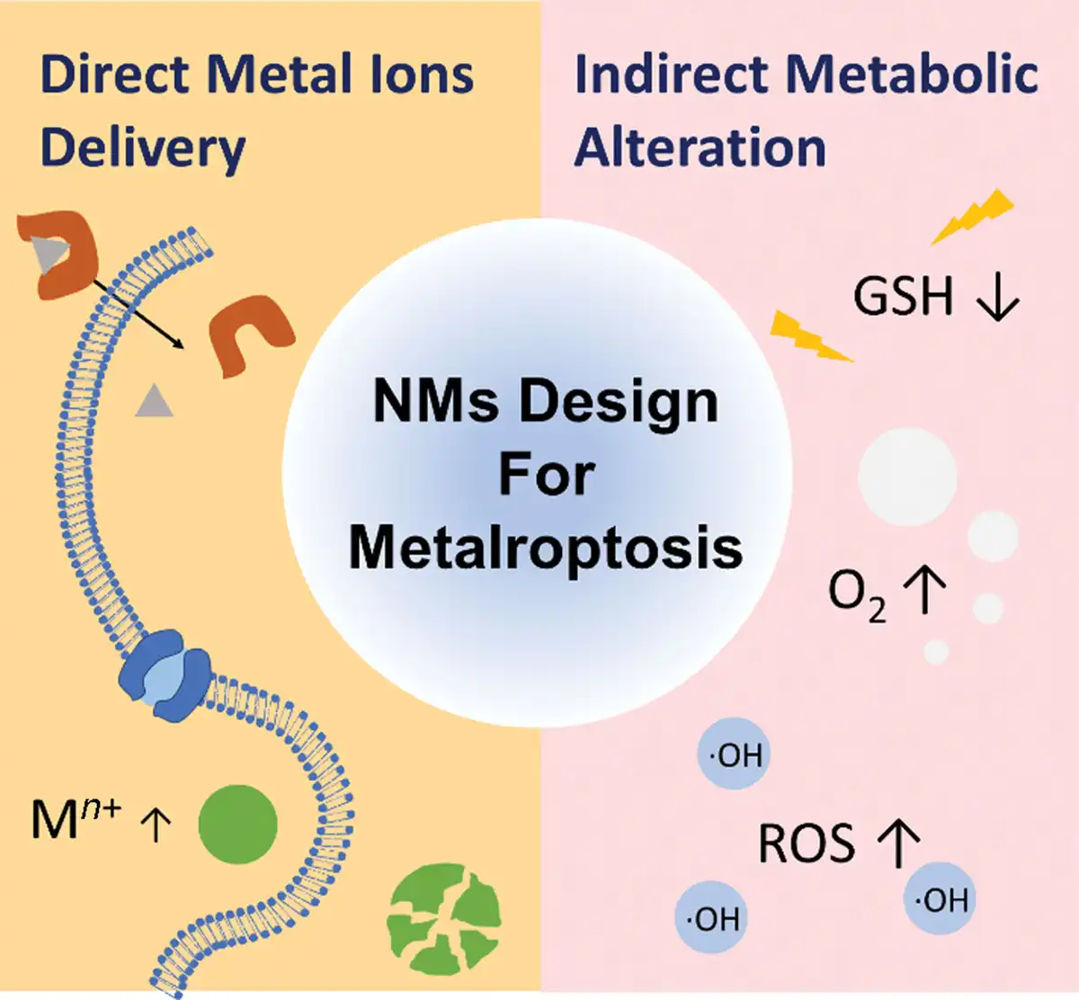
As a kind of metal-dependent cell death, metalloptosis is closely linked to intracellular ion levels. Direct or indirect delivery of metal ions into cells is a common strategy to induce metalloptosis (Figure 1). Compared to small-molecule metalloptosis inducers, engineering NMs, with their unique physicochemical properties, offer significant advantages in enhancing drug stability, increasing tumor accumulation, reducing systemic toxicity, and achieving sustained release.[30] Therefore, this section will mainly focus on discussing the design strategies and the prominent role of engineering NMs in metalloptosis.
Figure 1.
Scheme of engineering NMs in metalloptosis. Engineering NMs used for inducing metalloptosis can be categorized into 2 main types. The first type is utilized for direct metal ions delivery, such as NMs based on ionophores, inorganic NPs, and MOFs, which contain specific metal elements. The second type involves NMs used for metabolic alteration, which enhance the sensitivity of tumor cells to metal ions through pathways such as GSH depletion, ROS production, hypoxia modulation, oxygen generation, and affecting cell’s ion channels, thereby causing or facilitating the occurrence of metalloptosis.
2.1 Engineering NMs for direct delivery of metal ions
Various NMs containing specific metallic elements have been employed to directly enhance intracellular metal ion levels, thereby inducing metalloptosis. The design of such NMs often requires attention to the following aspects: (1) The proportion of metallic elements in NMs should be appropriate, ensuring sufficient metal transport efficiency while maintaining viable biosafety and avoiding severe toxic side effects; (2) The modification of surface ligands should be strategically implemented to enhance the biocompatibility and stability of NMs. Additionally, ingenious surface modification can introduce new biofunctionalities such as targeted tissue specificity, while also enhancing interactions with biological membranes and biomolecules, thereby augmenting the therapeutic potential of the NMs. (3) The designed NMs should be stimulus-responsive, capable of releasing metal ions in a predictable manner upon exposure to specific external or internal environmental stimuli. These stimuli include physical factors (such as light, temperature, magnetic fields, and electric fields), mechanical factors (such as ultrasound), chemical factors (such as pH and certain molecules like glucose), and biological factors (such as specific enzymes); (4) Through multifunctional design, additional capabilities such as imaging and synergistic therapeutic functions could be integrated into NMs.
Ionophores are well-known regulators of metal homeostasis, with many such molecules currently under clinical trials.[31] These molecules are capable of binding metal ions and facilitating their transport into cells, circumventing the standard metal ion importers.[32] The reversible nature of the binding between ionophores and metal ions lays the foundation for the targeted release of metal ions in response to the specific environment of tumor sites. However, due to their low molecular weight, ionophores are rapidly cleared and metabolized in circulation, which severely limits their uptake by tumor cells and, consequently, their therapeutic efficacy.[33] This challenge can be addressed by engineering ionophores into NMs, which has shown promising results in inducing metalloptosis. For instance, when elesclomol (ES), a well-known copper ionophore, was formulated into NMs in the form of the ES-Cu2+ complex, it overcame the limitations posed by the short half-life of small-molecule ES in the bloodstream, thereby enhancing copper accumulation in tumors and facilitating cuproptosis.[34]
Inorganic nanoparticles (NPs) containing specific metal elements are also extensively utilized for inducing metalloptosis. For example, iron oxide NPs,[35] amorphous iron NPs,[36] FePt NPs,[37] and iron carbide NPs[38] are employed in killing cancer cells through ferroptosis. Similarly, cuprous oxide NPs[39] and Cu2(PO4)(OH) NPs[40] have been employed to initiate cuproptosis, while CaO2 has been used to induce calcium overload.[41] These inorganic NPs often exhibit enhanced tumor tissue targeting, effectively reducing the adverse effects of metalloptosis on normal tissues. Additionally, they demonstrate prolonged circulation time in the blood as well, possessing a superior capability for metal ions delivery. This showcases the diverse potential of inorganic NPs in regulating metal homeostasis and advancing therapeutic applications.
Constructed from metal ions/clusters and organic linkers, metal-organic frameworks (MOFs) represent a category of highly ordered coordination polymers, distinguished by their porous crystalline structure.[42] MOFs, through ingenious design, customization, and modification of structural parameters, can achieve significant high surface areas, high-density active sites, and enhanced stability.[43] These features have led to their widespread applications in various fields including drug delivery, tissue engineering, and regenerative medicine, as well as antibacterial and anticancer treatments.[44–45–46] Furthermore, MOFs can be specifically tailored for efficient metal ions delivery to induce metalloptosis,[47,48] which will be discussed in detail in subsequent sections.
In addition to the aforementioned types, NMs based on single-atom nanozymes, hydrogels, and other coordination complexes can also be employed to directly deliver metal ions into cells, inducing metalloptosis.[49–50–51–52] This diversity in the design of NMs opens new avenues for exploring metalloptosis in cancer treatment.
2.2 Engineering NMs for indirect metabolic alteration
Metalloptosis has a strong connection with the metabolic processes within cells. By modifying the metabolic pathways specific to cancer cells, one can notably influence their responsiveness to metal ions. A combined strategy that integrates the induction of metalloptosis with metabolic reprogramming holds promising potential in the field of nanomedicine design. This method has been widely applied in various research contexts.[53,54] The design of these NMs for indirect metabolic alteration should thoroughly consider the interaction between cancer metabolism and the microenvironment, ingeniously utilizing the specific metabolic signaling pathways involved in various forms of metalloptosis for rational material design. Employing NMs to achieve glutathione (GSH) depletion,[54,55] ameliorate hypoxic conditions,[56] and provoke a burst of reactive oxygen species (ROS),[57] thereby initiating and enhancing metalloptosis, are all available strategies. For instance, by strategically employing NMs to deplete GSH, cancer cells are deprived of their primary antioxidant defense, which drastically increases their susceptibility to oxidative stress and metal ion toxicity. This depletion not only disrupts the redox homeostasis but also primes the cells for enhanced reactivity to subsequent treatments.[58] Moreover, addressing the challenge of tumor hypoxia can significantly improve the efficacy of metalloptosis. By utilizing NMs designed to improve oxygenation within the tumor microenvironment (TME), the oxygen levels are increased, which in turn facilitates the generation of ROS upon metal-ion interaction. This approach not only mitigates the limitations posed by hypoxic conditions but also synergizes with the oxidative stress induced by GSH depletion to further drive the metalloptosis process.
It is noteworthy that some engineered NMs can impact the cell’s ion channels, thereby potentially inducing metalloptosis. Previous reports have demonstrated the possibility of stimulating the opening of membrane ion channels via optogenetic approaches, leading to an influx of extracellular ions and subsequent intracellular ion overload. For instance, Ai et al have strategically localized lanthanide-doped upconversion nanocrystals (UCNs) to cell membranes.[59] The emitted blue light from these UCNs could effectively activate light-gated ion channels, inducing Ca2+ influx and triggering cell death in HeLa cells. Although research in this area is still limited, future studies are expected to clarify how NMs influence ion channel behavior. Understanding these interactions could potentially improve the use of these NMs in targeted cancer therapies and contribute to advancements in metalloptosis-based treatments.
The abovementioned strategies, when combined, offer a robust framework for designing next-generation nanomedicines that can efficiently target and eradicate tumor cells by exploiting their metabolic vulnerabilities. This comprehensive approach to cancer therapy highlights the innovative potential of integrating metalloptosis with metabolic alteration, establishing a new way for treatments in oncology.
3. Strategic NMs for metalloptosis
3.1 Metal ions-specific-induced metalloptosis
3.1.1 Ferroptosis
Ferroptosis as a novel Fe-dependent PCD has been discovered in 2012,[60] which is morphologically and mechanistically distinct from common PCDs such as apoptosis, necrosis, and autophagy. Its prominent feature is an overload of lipid peroxides on cellular membranes, mitochondria shrinkage, and reduction.[60,61] In detail, hallmarks of ferroptosis are listed as follows. (1) Morphological features: ferroptosis is accompanied by cell membrane rupture, cytoplasmic swelling, chromosome condensation, autophagosomes increasing, and mitochondrial abnormalities.[60,62–63–64] (2) Biochemical features: Fe accumulation and lipid peroxidation are the 2 most important biochemical characteristics of ferroptosis. The accumulation of Fe ions in vivo can directly induce the Fenton reaction to generate excessive ROS, ultimately leading to oxidative damage-mediated ferroptosis. In addition to the classic Fe ions-induced ferroptosis, other metal ions such as Zn ions can also achieve this process,[65] which we will discuss in detail in the following sections. On the other hand, lipid peroxidation is mainly driven by free radical, which often occurs in the cell membrane. Its mechanism is that cell membrane lipids are oxidized and produce many metabolic products such as lipid hydroperoxides and reactive aldehydes.[66,67] These metabolic products significantly increase during the process of ferroptosis, which can be the most important biochemical detection marker of ferroptosis. (3) Genetic features: ferroptosis is regulated by various kinds of relatively specific genes and proteins, thus, their abnormal expression is often considered a detection marker for ferroptosis. These genes and proteins are often associated with substance uptake and metabolic pathways, including cystine uptake, oxidant metabolism, lipid metabolism, energy metabolism, and Fe metabolism[68] (Table 1). (4) Immune features: the immunological consequences of ferroptosis on the immune cells are mainly manifested as the loss of their functions. For instance, ferroptosis that occurred in T cells led to a decrease in their antiviral or antiparasitic function.[74] More importantly, the immune system’s response is determined by ferroptosis-induced cell death. This is because various types of cell death can induce distinct immune reactions through the activation of different damage-associated molecular pattern (DAMP) signals. For instance, death cells induced by ferroptosis could activate the nuclear factor-κB pathway[75] and target lipid metabolism-related DAMP signaling,[76] eventually triggering the inflammatory response in peripheral macrophages.[77] In short, the discrimination of ferroptosis is clear and specific, which emphatically includes the following points: ROS generation, lipid peroxidation, Fe abundance, glutathione peroxidase 4 (GPX4) activity, GSH depletion, release of glutamate, and cystine uptake, SLC7A11 expression and so on. These indicators are essential for detection and study in the context of ferroptosis research.
Table 1
Ferroptosis-related genes and corresponding proteins.
| Signaling pathways | Genes and proteins | Functions |
|---|---|---|
| Cystine uptake | SLC7A11 (solute carrier family 7 membrane 11)[60] | Promotes cystine uptake |
| SLC3A2 (solute carrier family 3 membrane 2)[69] | Maintains SLC7A11 stability | |
| Energy metabolism | SLC1A5 (solute carrier family 1 member 5)[70] | Regulates glutamine uptake |
| Fe metabolism | TF (transferrin)[71] | Delivers Fe ions |
| TFR1 (transferrin receptor)[72] | Transfers Fe into cells | |
| Lipid metabolism | GPX4 (glutathione peroxidase 4)[73] | Prevents lipids hydroperoxides |
In terms of mechanisms, Fe ions-induced ferroptosis was initially discovered, and as research progressed, researchers have gradually found that the ferroptosis pathway is not solely dependent on Fe ions, some Fe-independent pathways, such as ROS metabolism (particularly lipid peroxides) and GSH metabolism, can also induce ferroptosis. Hence, the metabolic pathways of ferroptosis are mainly summarized into the following 3 pathways: Fe metabolism, ROS metabolism, and GSH metabolism (Figure 2).[78] According to different triggering mechanisms of ferroptosis, different treatment strategies based on nanotechnology can be adopted.
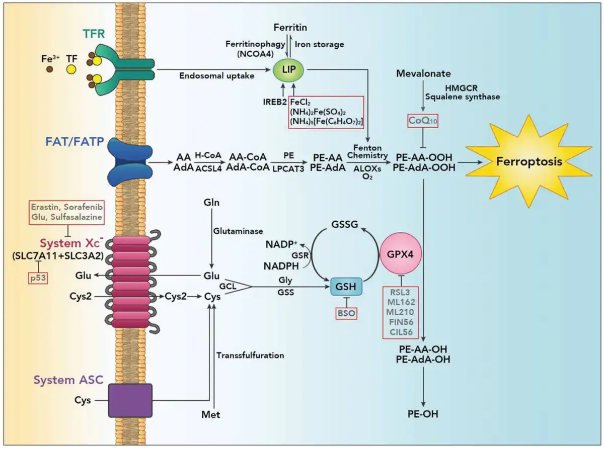
Figure 2.
The regulatory mechanism of ferroptosis. (1) Fe metabolism: the transfer of Fe ions into the cell is dependent on transferrin receptor (TFR)-mediated endocytosis, and then activates the labile iron pool (LIP), which is also can be regulated by ferritin degradation (ferritinophagy). The increased LIP further promotes the Fenton reaction, enhancing cell sensitivity to ferroptosis. (2) ROS metabolism: in the process of lipid peroxidation, the fatty acid translocase (FAT) and fatty acid transport protein (FATP) participate in the generation of phospholipid hydroperoxides through the Fenton reaction, with the occurrence of ultimately induced ferroptosis. Meanwhile, GPX4 as a central regulator of ferroptosis, can combat lipid peroxidation by transforming toxic phospholipid acid into nontoxic phospholipid alcohols. (3) GSH metabolism: cystine (Cys2) and GSH metabolism constitute the principal line in the Fe-independent pathway of regulating ferroptosis. When oxidative stress occurs under extracellular conditions, cystine is taken up by system Xc−, and the cystine is gradually reduced through GSH, which acts as an important intracellular antioxidant. This process can promote GPX4 to resist ferroptosis. Reproduced with permission from Liang et al.[78] Copyright 2019, WILEY-VCH Verlag GmbH & Co. KGaA, Weinheim.
Fe ions play an essential role in inducing ferroptosis whether through enzymatic or nonenzymatic reaction. Thanks to the Fe transport systems, intracellular Fe ions can always maintain a delicate balance under normal conditions. Extracellular Fe ions can be transferred into the cytoplasm by transferrin (TF) and its corresponding transferrin receptor (TFR) and stored in the ferritin.[61] On the other hand, intracellular Fe ions are exported outside the cell through ferroportin (FPN), which is the only known Fe ions exporter in mammal.[79] When Fe ions accumulate in the cytoplasm, disrupting the balance of Fe ions, it can trigger oxidative stress responses and the following ferroptosis, leading to cellular damage.[80] Interestingly, many studies have demonstrated that cancer cells have a higher demand for Fe than normal cells to maintain their rapid proliferation, specifically manifested as downregulation of FPN and upregulation of TFR1.[70,81] Based on the high dependence of cancer cells on Fe content, cancer cells are more susceptible to Fe overload or oxidative stress compared to normal cells, thus, inducing ferroptosis by altering the intracellular Fe ion content has become one of the emerging ways to treat cancers.[82]
At present, the majority of NMs triggering ferroptosis by changing the accumulation of Fe ions in the cytoplasm are Fe-based NMs. They can be internalized by cancer cells and degraded into ferrous (Fe2+) or ferric (Fe3+) ions in lysosomes, thereby directly boosting the intracellular Fe ions content. The excessive Fe ions can further trigger the Fenton reaction, inducing ferroptosis to kill cancer cells. For instance, the most common Fe-containing NPs are Fe3O4 NPs. Liang et al reported self-assembled ultrasmall Fe3O4 NPs with the surface modification of porphyrin-grafted lipid (PGL) to enhance ROS production and lipid peroxidation. These ultrasmall Fe3O4@PGL NPs with a diameter of ∼10 nm exhibited good biosafety (Figure 3A). In terms of treatment mechanism, the Fe3O4@PGL NPs generated abundant ROS through porphyrin-mediated photodynamic therapy (PDT), which is beneficial for the occurrence of ferroptosis. Moreover, the results indicated that the content of ROS produced was the highest only when the coincubation of NPs endocytosed by HT-29 cells and RAW264.7 cells, which suggested that Fe3O4@PGL NPs could also enhance the anticancer ability of ferroptosis through modulating macrophages in the TME (Figure 3B, C).[83] To further enhance the catalytic activity of Fe-based NPs and improve their efficiency in inducing ferroptosis, Liang et al provided an effective therapy strategy to ameliorate the low ROS production efficacy of current Fe-based NPs through synthesizing body-centered cubic-ultrasmall single-crystal Fe NPs (bcc-USINPs) (Figure 3D). These bcc-USINPs remained stable in the normal physiological environment and reacted with weak acids in the TME due to their selective acidic etching of the Fe3O4 shell and the exposure of the Fe(0) core. The results of the catalytic activity analysis showed that bcc-USINPs exhibited superior ROS generation ability than other NPs (Figure 3E), which proved that bcc-USINPs have a stronger ability to induce ferroptosis than other NPs. More importantly, bcc-USINPs also triggered immunotherapy to enhance the synergistic effect of ferroptosis treatment. In detail, bcc-USINPs could efficiently induce immunogenetic cell death at a very low concentration (1 mg/kg) by promoting the maturation of dendritic cells (DCs) and enhancing the adaptive T cell response (Figure 3F–I). The bcc-USINPs-mediated ferroptosis treatment, when combined with programmed death-ligand 1 (PD-L1) immune checkpoint blockade immunotherapy, developed a strong immune memory, thereby potentiating the immune response. This Fe(0) delivery system based on bcc-USINPs provided an effective strategy for ferroptosis-based immunotherapy.[84] Considering that the efficiency of ferroptosis induced by Fe-based NPs is related to the penetration depth of NPs at the tumor site, Wang et al prepared a kind of stimuli-responsive size-switchable nanocapsules with Au-Fe2C Janus NPs as ferroptosis agents and sorafenib (SRF) as the ferroptosis enhancer. These nanocapsules exhibited superior intratumoral accumulation compared to free Au-Fe2C NPs, subsequently penetrating deeper into tumor regions. They disrupted the intracellular redox balance, eventually leading to efficient treatment through ferroptosis.[38]
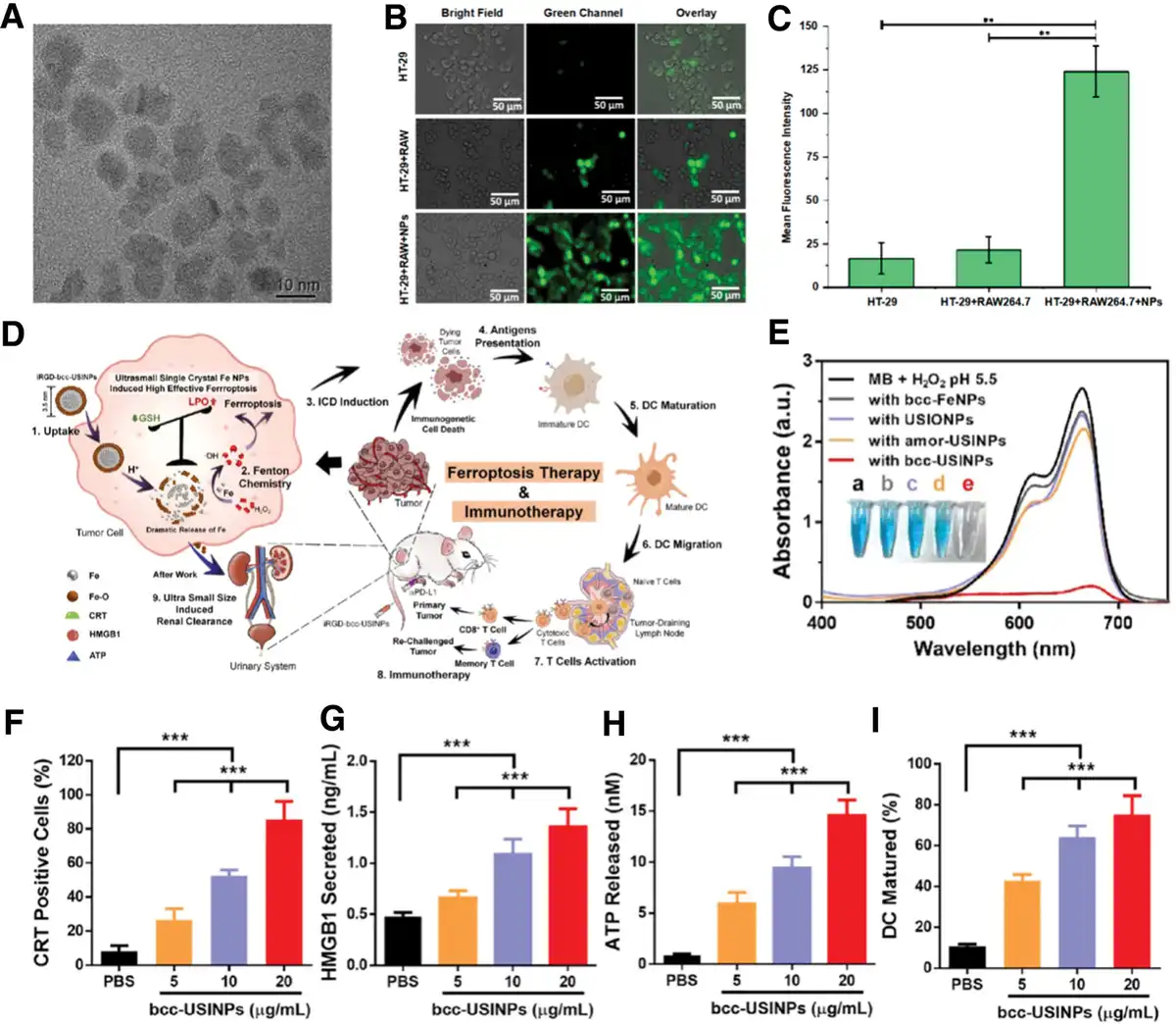
The NMs for ferroptosis via Fe metabolism. (A) Transmission electron microscopy (TEM) image of Fe3O4@PGL NPs. (B) Evaluation of intracellular ROS generation in different treatment groups for 3 days. (C) Quantification of intracellular ROS generation in (B). The values are presented as mean ± SD; **P < 0.01. (A–C) Reproduced with permission from Liang et al.[83] Copyright 2021, American Chemical Society. (D) Illustration of renal clearable bcc-USINPs for highly selective and effective ferroptosis therapy and immunotherapy. (E) UV/Vis absorption spectra of methylene blue solution in pH 5.5 buffer containing 1 mM H2O2 after adding bcc-Fe NPs, USIONPs, amor-USINPs, and bcc-USINPs at a concentration of 50 μg/mL [Fe]. (F) Flow cytometry quantification of calreticulin (CRT) on the surface of MC38 tumor cells. ***P < 0.001. (G) High mobility group box 1 (HMGB1) released from MC38 cells detected by an enzyme-linked immunosorbent assay (ELISA) kit (n = 3). ***P < 0.001. (H) Adenosine triphosphate (ATP) secretion that detected by an enhanced ATP assay kit (n = 3). ***P < 0.001. (I) Statistical analysis of DC maturation (gated on CD11c+ cells) after being incubated with PBS or bcc-USINP-treated MC38 tumor cells. ***P < 0.001. Data were expressed as means ± SD (n = 3). (D–I) Reproduced with permission from Liang et al.[84] Copyright 2021, American Chemical Society.
It is worth noting that the induction of ferroptosis is not limited to Fe ions, and many other metal ions can also achieve ferroptosis through non-Fe ions-dependent pathways, such as Mn ions and Zn ions, which will be discussed in the following sections.
Reduced GSH is the major intracellular antioxidant in mammals, whose biosynthesis is subject to the influence of cystine and cysteine. In the triggering mechanism of ferroptosis, GSH actually is a negative regulatory factor. Blocking cystine acquisition from the extracellular environment or inhibiting the biosynthesis of GSH can trigger ferroptosis.[85] Many studies have demonstrated that GSH is strongly associated with cancer aggressiveness and chemoresistance, however, the existing therapeutic drugs targeting GSH cannot effectively scavenge GSH, so the relevant treatment can only serve as a supplementary strategy of cancer treatment.[86] The emergence of nanotechnology has brought a breakthrough opportunity to solve this problem. Wang et al prepared a kind of ferroptosis-inducing agent based on arginine (Arg)-capped manganese silicate nanobubbles (AMSNs) with the efficient consumption of GSH and inactivation of GPX4. In this nanosystem, taking advantage of the argininosuccinate synthetase deficiency present in cancers, Arg as the modified surfacing ligand allowed AMSNs to have good biocompatibility and tumor homing capacity, which further dramatically enhanced the efficiency of GSH depletion and GPX4 inactivation. AMSNs were degraded to release Mn ions of higher valence (Mn3+ and Mn4+) in response to the weakly acidic conditions in TME. The released Mn3+ and Mn4+ were further reduced to Mn2+ in the presence of GSH, which precisely promoted the depletion of GSH. This depletion of GSH subsequently led to the upregulation of intracellular oxidative stress and the downregulation of GPX4 expression, collectively improving the treatment efficiency of ferroptosis. Meanwhile, the hollow structure also endowed AMSNs with the ability as drug delivery vehicles. Doxorubicin (Dox)-loaded AMSNs could suppress tumor growth more effectively compared with free Dox. In terms of bioimaging, Mn ions contributed to the T1-weighted magnetic resonance imaging (MRI), ultimately achieving imaging-guided on-demand synergistic chemotherapy and ferroptosis treatment.[87]
In addition to ameliorating the inhibitory effect of GSH on ferroptosis, targeting other antiferroptotic signaling pathways also emerges as a potent strategic approach to enhance synergistic therapeutic effects. Ferroptosis suppressor protein 1 (FSP1) can mediate detoxification of lipid peroxides through decreasing nicotinamide adenine dinucleotide phosphate (NADP+)/nicotinamide adenine dinucleotide phosphate (NADPH) ratio and catalyzing the recycling of coenzyme Q10 (CoQ10) to ubiquinol (CoQ10H2) for trapping lipid peroxyl radicals. The decreasing NADP+/NADPH ratio can further promote the conversion of cystine to cysteine and the biosynthesis of GSH and GPX4.[88,89] Consequently, devising a treatment strategy that is capable of simultaneously inhibiting both GSH- and FSP1-mediated ferroptosis-suppressing mechanisms holds considerable therapeutic potential. Li et al developed a Cu-tetra(4-carboxyphenyl)porphyrin chloride(Fe(III)) (Cu-TCPP(Fe))-based MOF nanosystem, incorporated with Au NPs via in situ nucleation and the ferroptosis inducer Ras-selective lethal small-molecule 3 (RSL3) via π–π stacking, meanwhile modified with polyethylene glycol (PEG) and internalizing RGD peptide (iRGD) for tumor-targeted drug delivery. Mechanistically, except for the classic Fe ions-induced ferroptosis, the introduction of Au NPs caused glucose depletion because of their glucose oxidase-like activities, eventually disrupting the pentose phosphate pathway and hindering reduced GSH biosynthesis. Meanwhile, Cu ions consumed GSH by oxidizing it to glutathione (GSSG), which could suppress the GSH-mediated antiferroptotic pathway in tumor cells for amplifying ferroptotic damage. Moreover, the loaded ferroptosis inducer RSL3 could deactivate the function of GPX4 to further result in pronounced ferroptotic damage. Nonetheless, Au NPs could also increase the NADP+/NADPH ratio and prevent the recycling of CoQ10 to CoQ10H2, impairing the FSP1-mediated antiferroptotic pathways (Figure 4A). Specifically, Au/Cu-TCPP(Fe) nanosheet was synthetized with a typical 2-dimensional morphology and its average size was between 100 and 200 nm (Figure 4B). The functional incorporation of the Au NPs and Cu species efficiently depleted the glucose and GSH in the tumor intracellular environment, which plays critical roles in the subsequent ferroptosis sensitization (Figure 4C). The results of evaluation experiments on the GOx-like catalytic activity demonstrated that the Au/Cu-TCPP(Fe) nanosheet consistently reduced the glucose concentration over a prolonged incubation time, with the final relative levels dropped by around 51.7% after 24 h (Figure 4D). On the other hand, as shown in Figure 4E, both Cu-TCPP(Fe) and Au/Cu-TCPP(Fe) could reduce the concentration of GSH in a dose-dependent manner. After incubation with a nanosheet (100 μg/mL) for 24 h, the concentration of GSH was decreased to around 81.4% and 82.1%, evidently suggesting the GSH-consuming ability of the Cu species in the nanosheet. Furthermore, the effect of the catalytic activity of composite nanosheets on 2 independent regulatory pathways of ferroptosis (GPX4/GSH pathway and FSP1/CoQ10H2 pathway) was systematically evaluated. In terms of GPX4/GSH pathway, the results indicated that the GSH-oxidizing capability of the Cu species in the Cu-TCPP(Fe)-PEG and Au/Cu-TCPP(Fe)-PEG nanosheets caused an evident decrease of the cellular GSH (Figure 4F) and increase of the cellular GSSG levels (Figure 4G), which was consistent with the results of reduced expression of GPX4 induced by both Cu-TCPP(Fe)-PEG and Au/Cu-TCPP(Fe)-PEG (Figure 4H, I). In the terms of FSP1/CoQ10H2 pathway, Au/Cu-TCPP(Fe)-PEG could enhance the ratio of NADP+/NADPH (Figure 4J) and weaken the ratio of CoQ10H2 to CoQ10 (Figure 4K) in a dose-dependent manner. These changes suggested that Au NPs in the Au/Cu-TCPP(Fe)-PEG nanosheets lowered the abundance of CoQ10H2 by the consumption of glucose and the decrease of NADPH content, potentially compromising the antiferroptotic capability of FSP1 proteins. In brief, according to different signaling pathways induced by ferroptosis, this nanosystem promoted the occurrence of ferroptosis and optimized the cell lethal effect of ferroptosis, providing a strong rationale for the application of ferroptosis therapy in the clinic (Figure 4L).[90]
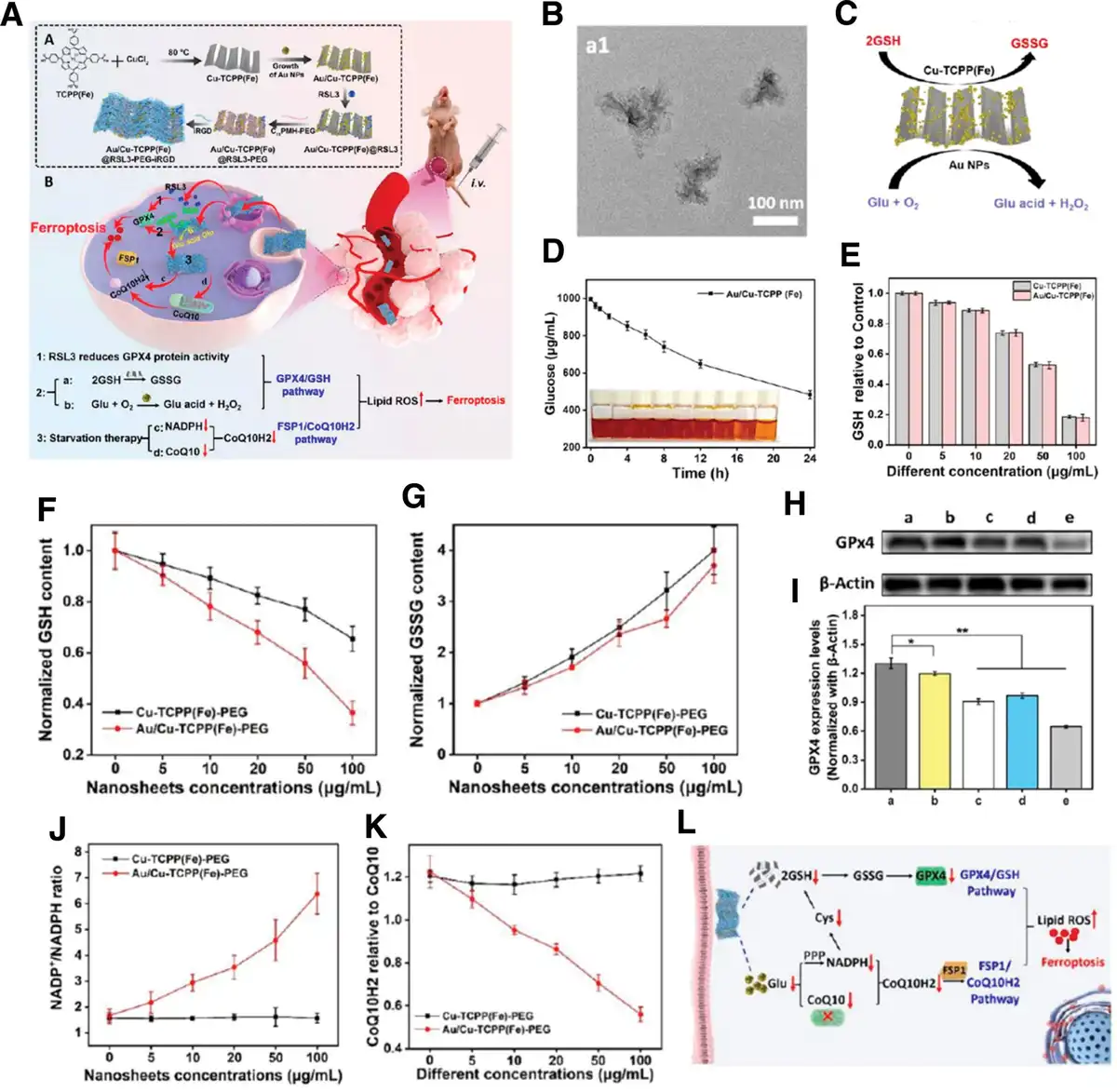
The NMs for ferroptosis via GSH metabolism and CoQ10H2 pathway. (A) Schematic illustration of the tumor-targeting composite nanosheet for sensitizing tumor ferroptosis via impairing GPX4/GSH and FSP1/CoQ10H2 pathways. (B) TEM image of Au/Cu-TCPP(Fe). (C) Schematic diagram of composite nanosheet catalysis. (D) Time-dependent GOx-like activity of Au/Cu-TCPP(Fe), n = 3. (E) Concentration-dependent GSH consumption activity of Cu-TCPP(Fe) and Au/Cu-TCPP(Fe), n = 3. (F) Relevant intracellular GSH content after Cu-TCPP(Fe)-PEG and Au/Cu-TCPP(Fe)-PEG treatment. (G) Relevant intracellular GSSG content after Cu-TCPP(Fe)-PEG and Au/Cu-TCPP(Fe)-PEG treatment. (H) Western blotting assay of the GPX4 protein expression after different treatments. (I) Relevant gray statistics of (H). (J) The ratio of intracellular NADP+/NADPH after different treatments. (K) The ratio of CoQ10H2 to CoQ10 in 4T1 cells after different treatments. (L) Mechanisms of composite nanosheets regulating 2 independent ferroptosis signaling pathways. (a) Control; (b) Cu-TCPP(Fe)-PEG (10 μg/mL); (c) Cu-TCPP(Fe)-PEG (50 μg/mL); (d) Au/Cu-TCPP(Fe)-PEG (10 μg/mL); (e) Au/Cu-TCPP(Fe)-PEG (50 μg/mL), n = 3. Reproduced with permission from Li et al.[90] Copyright 2021, American Chemical Society.
Moreover, as a central downstream regulator of ferroptosis in GSH metabolism, GPX4 combats with lipid peroxidation by transforming toxic phospholipid hydroperoxides into nontoxic phospholipid alcohols, with the generation of oxidized GSH (GSSG) as byproducts.[91] Many chemical or biological methods can intervene in the expression of GPX4 and have also shown initial effectiveness in inducing ferroptosis.[80,92,93] Therefore, targeting GPX4 is a powerful therapeutic strategy for ferroptosis-based cancer therapy[94] and various kinds of nanoplatforms have been developed for suppressing GPX4.[87,95,96] Among them, 2 common solutions are as follows: first, NMs can act as the delivery system to transport small-molecule inhibitors of GPX4 in vivo, which is capable of overcoming certain defects, such as rapid clearance from systemic circulation and poor tumor enrichment efficiency; second, NMs can reduce the expression of GPX4 by utilizing their own intrinsic physicochemical properties.[87,96] Inspired by industrial electro-Fenton technology featured with electrochemical iron cycling, Liu et al constructed a network-like corona spontaneously consisting of Fe3+ ion and naturally derived tannic acid (TA) to wrap the SRF nanocores, which is a typical small-molecule GPX4 inhibitor (Figure 5A). The results showed that the formed SRF@FeIIITA (SFT) NPs could significantly decrease the level of GSH (Figure 5B) and GPX4 (Figure 5C) for ferroptosis initiation. Meanwhile, TA chemically reduced Fe3+ to Fe2+, offering iron redox cycling to effectively sustain the Fenton reaction and produce the lipid peroxide that was required in ferroptosis (Figure 5D). In vivo results indicated that this rational material design could advance the antitumor effect of ferroptosis through the continuous internal supply of Fe ions combined with the specific GPX4 inhibitor (Figure 5E).[95] Moreover, the reconstituted NMs with docosahexaenoic acid were also proved to cause inactivation of lipid antioxidant GPX4 and accumulation of lipid hydroperoxides, with the consequence of significant cancer suppression.[96]
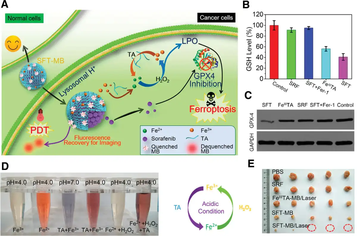
The NMs as drug delivery systems (DDSs) for ferroptosis via targeting GPX4. (A) Schematic illustration of SFT-mediated combination of ferroptosis and image-guided PDT. (B) Intracellular GSH level of 4T1 cells after treated with SRF, FeIIITA, SFT, and SFT plus Ferrostatin-1 (Fer-1, a ferroptosis inhibitor) with an equivalent SRF dosage at 20 μM. (C) Western blot analysis of GPX4 expression in 4T1 cells after the treatment with SRF, FeIIITA, SFT, and SFT plus Fer-1, respectively. (D) Observation on the iron redox cycling of SFT under the acidic condition. Fe2+ was detected using o-phenanthroline. An acidic environment triggered Fe2+ release from FeIIITA, while TA under acidic conditions can reduce Fe3+ that originates from the oxidation of Fe2+ by H2O2 into Fe2+ again. (E) Photographs of tumors that obtained on the 14th day after treatments. Reproduced with permission from Liu et al.[95] Copyright 2018, American Chemical Society.
Interestingly, as the study continues to deepen, researchers have gradually discovered some new signaling pathways of ferroptosis. The latest research indicated that supplementation with dihydroorotate or orotate, the substrate and product of dihydroorotate dehydrogenase (DHODH), could attenuate or potentiate ferroptosis induced by inhibition of GPX4, respectively, and these effects were especially pronounced in cancer cells with low expression of GPX4 (GPX4low). Inactivation of DHODH could lead to extensive mitochondrial lipid peroxidation and ferroptosis in GPX4low cancer cells and produce the same phenomenon in GPX4high cancer cells with the combination of ferroptosis inducers. All these results identified a new mechanism of ferroptosis mediated by DHODH and suggested a therapeutic strategy of targeting DHODH in cancer ferroptosis treatment.[97]
ROS, generated via various pathways, can promote the accumulation of oxidation products (particularly lipid peroxides), being acknowledged as the typical hallmark of ferroptosis.[98] Hence, it is an evident design strategy of NMs for ferroptosis to accelerate the generation of ROS. For instance, Guan et al reported a multifunctionalized nanoplatform with a ROS cascade for ferroptosis combined with chemotherapy/gas therapy in reversing multidrug resistance. Ferrocene used in this nanoplatform synergistically ameliorated the antitumor efficiency of ferroptosis through breaking the redox balance in the TME.[99] Moreover, Fe/Ni-layered double hydroxides (LDHs) were synthesized by a general and facile method to enhance apoptosis- and ferroptosis-induced cancer therapy. Compared with Fe-OH and Ni-OH, Fe/Ni-LDH exhibited exceptional catalytic activity with massive generation of ROS, •OH, and O2•− radicals, which could prominently increase intracellular oxidative stress and accumulation of lipid peroxide (LPO), ultimately optimizing ferroptosis-mediated cancer therapy.[100] However, the current research on the regulation mechanism of ROS metabolism for ferroptosis has not been sufficiently validated; most studies are limited to detecting the content of ROS and markers of ferroptosis, without verifying the direct relationship between the 2 through rational experiments. For example, appropriate inhibitors can be used to block the other signaling pathways of ferroptosis such as Fe metabolism and GSH metabolism, to further clarify the regulatory role of ROS metabolism on ferroptosis. This is also important to enhance our understanding of the ferroptosis induced by ROS metabolism.
3.1.2 Cuproptosis
Copper is an essential trace element for the human body, which is primarily obtained from food. The current recommended copper intake for adults is 0.8 to 2.4 mg/d to maintain whole-body copper homeostasis.[101] Statistically, an adult human body contains about 100 mg of copper.[102] In one regard, copper is integral to various physiological processes in the body, acting as a catalytic and structural cofactor for a range of enzymes that facilitate critical biochemical reactions. Its role is particularly prominent in enzymes such as cytochrome c oxidase and superoxide dismutase 1, which are pivotal for cellular energy production and antioxidant defense, respectively.[103] Beyond these functions, copper is also vital for the synthesis of neurotransmitters,[104] iron metabolism,[105] peptide hormone maturation, and blood clotting.[102] While an optimal amount of copper is beneficial for the organism, excessive copper levels can precipitate a sequence of cellular metabolic disturbances that ultimately lead to cell death. Early research attributed this type of death to the accumulation of copper, which disrupts the delicate balance between the generation of ROS and the body’s antioxidative defense mechanisms.[33] As of 2022, the notion of “cuproptosis” was formally published within the scientific community, representing a novel cellular demise pathway distinct from previously characterized mechanisms such as apoptosis, necrosis, and autophagy.[106]
Compared to ferroptosis, cuproptosis represents a relatively novel concept in the field of metal ions-mediated cell death research. It predominantly hinges on the intracellular accumulation of copper ions, which leads to the aggregation of lipoylated proteins and subsequent loss of iron-sulfur (Fe-S) cluster proteins. This sequence of events triggers proteotoxic stress, culminating in cell death. Specifically, researchers employed the ionophore elesclomol (ES) to facilitate the transport of Cu2+ into cells, where an excess of Cu2+ was subsequently transported to mitochondria and reduced to the more toxic Cu+ by the mammalian ferredoxin 1 (FDX1), a critical player in cuproptosis process. Besides reducing Cu2+, FDX1 also lipoylates specific mitochondrial enzymes, such as dihydrolipoamide S-acetyltransferase (DLAT), crucial for maintaining normal cellular metabolism and mitochondrial functionality. When Cu+ binds to DLAT, it triggers the aggregation of this enzyme, subsequently impeding the tricarboxylic acid cycle. Concurrently, Cu+ diminishes the synthesis of Fe-S clusters. These dual actions collectively lead to cellular demise.[106] A detailed illustration of the cuproptosis mechanism is provided in Figure 6A, offering a visual representation of how these interactions converge to induce cell death.
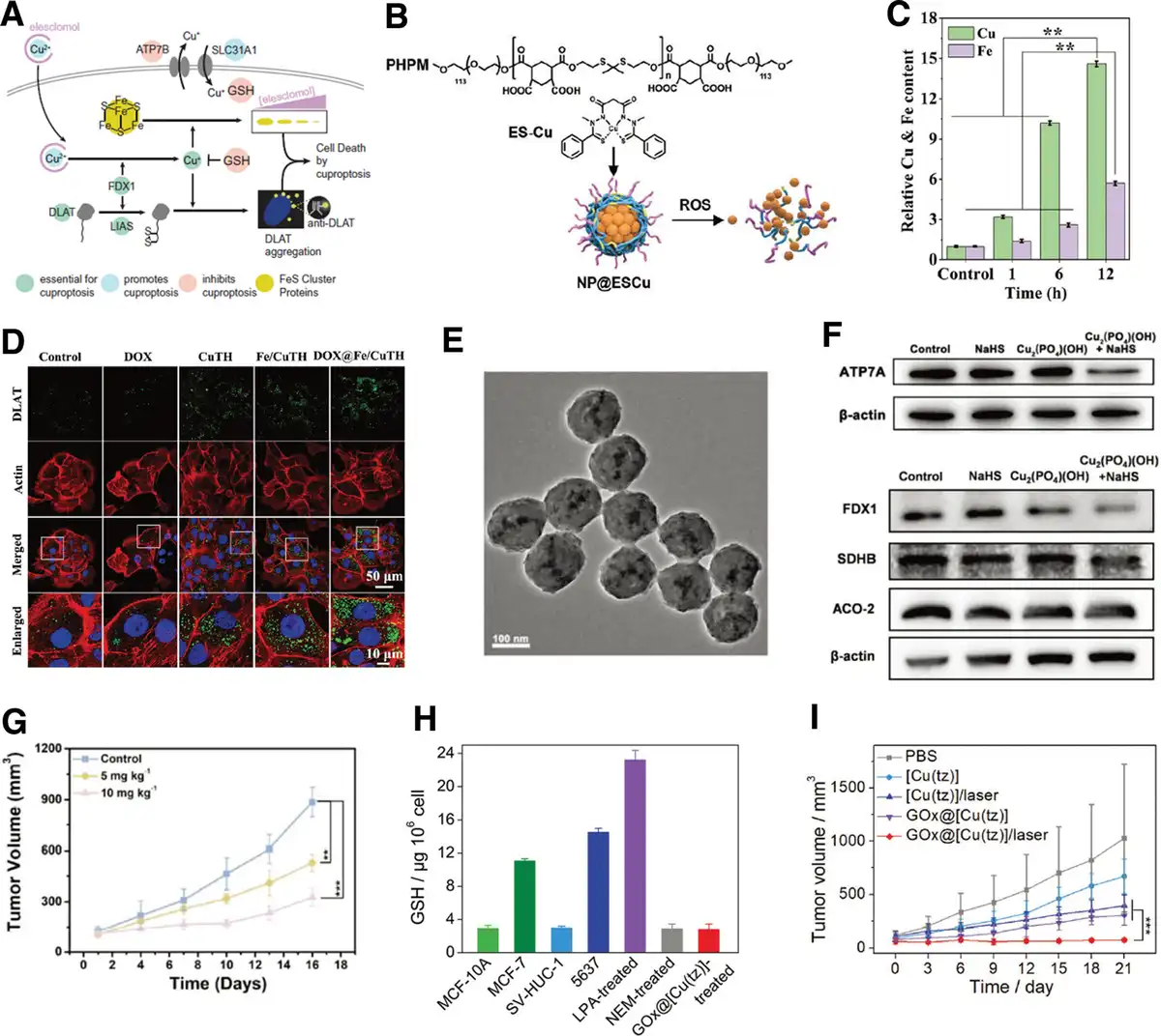
Figure 6.
Mechanisms and NMs-induced activation of cuproptosis. (A) Schematic of mechanisms of cuproptosis. Reproduced with permission from Tsvetkov et al.[106] Copyright 2022, the Authors, some rights reserved; exclusive licensee American Association for the Advancement of Science. (B) Schematic diagram of ROS-induced release of ES-Cu from NP@ESCu. Reproduced with permission from Guo et al.[34] Copyright 2023, Wiley-VCH GmbH. (C) Changes of intracellular Cu and Fe levels in 4T1 cells after treatment at different times. **P < 0.01. (D) Images of DLAT immunofluorescence in different treatment groups. (C, D) Reproduced with permission from Xu et al.[47] Copyright 2022, Wiley-VCH GmbH. (E) TEM image of Cu2(PO4)(OH) NPs. (F) Western blot analysis on the expressions of ATP7A, FDX1, SDHB, and ACO-2. (G) Tumor volume curves of different treatment groups, n = 5. **P < 0.01, ***P < 0.001. (E–G) Reproduced with permission from Zhao et al.[40] Copyright 2023, Wiley-VCH GmbH. (H) The intracellular GSH levels in the different cell lines or different treatments. (I) Tumor volume curves of different treatment groups, n = 5. ***P < 0.001. (H, I) Reproduced with permission from Xu et al.[54] Copyright 2022, Wiley-VCH GmbH.
The cuproptosis pathway is characterized by high levels of lipoylated proteins, including glycine cleavage system protein H, dihydrolipoamide branched chain transacylase E2, dihydrolipoamide S-succinyltransferase, and aforementioned DLAT.[107] These proteins’ lipoylation is controlled by enzymes such as lipolytransferase 1, lipoyl synthase (LIAS), and dihydrolipoamide dehydrogenase, and FDX1, with FDX1 playing a key role in initiating the lipoylation process.[108,109]
The utilization of ionophores such as ES and disulfiram (DSF) to transport copper ions into tumor cells represents a direct and common method to induce cuproptosis.[110] Ionophores can traverse biological membranes by forming stable complexes with ions and, upon specific stimuli, release metal ions, thereby altering intracellular and extracellular ion concentrations.[111] However, the efficacy of existing small-molecule ionophores in inducing cuproptosis in tumor cells is somewhat limited due to their short blood half-life.[112] The advancement of nanotechnology offers a solution to this issue by constructing nanomedicines that incorporate ionophores alongside other components, enhancing the properties of the drug delivery system. Such nanomedicines not only address the aforementioned challenges but also equip therapeutic agents with multifunctional features. They enhance cuproptosis effectiveness by promoting synergistic treatments with immunotherapy and chemo-chemodynamic therapy, or by enabling image-guided approaches for tumor therapy.[34,113] For instance, Guo et al developed a nanomedicine, NP@ESCu, by encapsulating elesclomol-Cu with a ROS-responsive polymer (PHPM), as shown in Figure 6B. Owing to the thioketal bonds in PHPM that were sensitive to ROS, NP@ESCu released its contents when exposed to elevated ROS levels within cancer cells, leading to cuproptosis. Furthermore, NP@ESCu enhanced PD-L1 expression on cancer cells, caused maturation of DCs, and enhanced CD8+ T cell infiltration in tumor sites. It also triggered a shift from tumor-associated macrophages from the immunosuppressive M2 phenotype to the immunostimulatory M1 phenotype, thereby augmenting immunotherapy efficacy. The combined use of NP@ESCu and αPD-L1 in a bladder cancer mouse model demonstrated superior therapeutic outcomes, illustrating the potential of NP@ESCu as a multifaceted approach to cancer treatment.[34] In another study, researchers used DSF/Cu-based nanomedicine to induce cuproptosis. By encapsulating DSF/Cu complexes within a tumor TME-responsive core-shell dendrimer, the resulting CSTD-Cu(II)@DSF achieved MRI-guided synergistic therapy by combining cuproptosis and chemo-chemodynamic treatment.[113]
In addition to using ionophores for copper delivery, alternative NMs such as MOFs,[47,49,56] inorganic NPs,[39,40,50,114] and carbon nitride[115] can also facilitate copper ions delivery into cells, effectively triggering cuproptosis. For example, Xu et al explored amorphous metal-organic frameworks (aMOF) as cuproptosis carriers due to their unique hollow structure and amorphous properties. By incorporating Cu2+ and Fe3+ during the synthesis and modifying with Dox-loaded, functionalized hyaluronan, the resultant Dox@Fe/CuTH HaMOF enhanced the intracellular copper/iron concentration in tumor cells, inducing a synergistic antitumor effect through cuproptosis, ferroptosis, and apoptosis. The inductively coupled plasma–mass spectrometry (ICP-MS) data indicated that, with prolonged incubation of the Dox@Fe/CuTH with 4T1 cells, the intracellular copper content increased 14.6 times compared to the PBS-treated control group (Figure 6C). The rise in copper levels ultimately induced the occurrence of cuproptosis in tumor cells, as evidenced by the pronounced DLAT foci in cells treated with Dox@Fe/CuTH, attributed to abnormal oligomerization of DLAT (Figure 6D). Moreover, western blot results showed a significant reduction in the levels of FDX1 and LIAS in the Dox@Fe/CuTH-treated group compared to the control group. The destabilization of these proteins further confirmed the capability of the designed nanomedicine to induce cuproptosis.[47] Using a hydrothermal method, copper hydroxyphosphate NPs (Cu2(PO4)(OH) NPs) were synthesized (Figure 6E), which could respond to endogenously overexpressed H2S to transform into ultrasmall copper sulfide NPs. This size transformation enhanced tumor cell uptake of the NPs, elevated intracellular ROS levels, and increased intracellular copper ion concentrations, ultimately leading to cuproptosis and pyroptosis. It is noteworthy that within an H2S-enriched microenvironment, Cu2(PO4)(OH) NPs were able to cause mitochondrial damage and consequently decreased the expression of copper-specific efflux proteins such as ATP7A (Figure 6F). This reduction led to decreased copper excretion, further facilitating the occurrence of cuproptosis. As shown in Figure 6F, the observed reduction in FDX1, succinate dehydrogenase B (SDHB), and aconitase-2 (ACO-2) levels following the treatment with Cu2(PO4)(OH) NPs in an H2S environment indicated the successful initiation of cuproptosis. After a 16-day treatment period, the tumors in H2S-overexpressed colon cancer-bearing mice, treated with Cu2(PO4)(OH) NPs, showed significant suppression, as depicted in Figure 6G.[40] By utilizing various NMs, these innovative approaches provide a range of mechanisms for “encapsulating” and “releasing” copper ions in cancer cells, thereby broadening the array of tools available for targeted cancer treatment via cuproptosis.
The above-discussed methods leverage nanoplatforms to elevate intracellular copper levels, inducing cuproptosis. As Figure 6A illustrates, there is a complex link between cuproptosis and cell metabolism. Modifying tumor metabolism to heighten copper sensitivity presents another viable strategy for inducing cuproptosis. Given that GSH, an endogenous copper chelator, can mitigate the therapeutic effect of cuproptosis by reducing free intracellular copper levels, incorporating components that deplete GSH or inhibitors blocking GSH synthesis into nanomedicines could potentially enhance the antitumor efficiency of cuproptosis. For example, researchers ingeniously utilized glucose oxidase (GOx) to respond to GSH in tumor cells, creating a nanomedicine (called GOx@[Cu(tz)]) based on nonporous copper(I) 1,2,4-triazolate ([Cu(tz)]) coordination polymer. This nanomedicine achieved dual depletion of glucose and glutathione, facilitating cuproptosis-based synergistic cancer therapy. From the intracellular GSH levels test in Figure 6H, it is evident that there was a significant decrease in GSH within tumor cells after treatment with GOx@[Cu(tz)]. Furthermore, in vivo animal experiments further confirmed that GOx@[Cu(tz)] exhibited excellent tumor suppression effects (Figure 6I).[54] Additionally, another study incorporated buthionine-sulfoximine (BSO) to inhibit GSH synthesis and catalase (CAT) to decompose endogenous hydrogen peroxide, constructing BSO-CAT@MOF-199@DDM nanomedicine that sensitized tumor cells to cuproptosis by alleviating hypoxia and reducing GSH synthesis.[56]
As a crucial subset of metalloptosis, research on cuproptosis is still in its infancy, with ongoing exploration into its mechanisms and the design of cuproptosis-inducing nanomedicines. Questions about the specific roles of key factors such as FDX1, specific compounds for cuproptosis, the resistance mechanisms of cuproptosis-inducing nanomedicines, the morphological features of cuproptosis, and the interaction with other cell death pathways remain open. Future designs should focus on optimizing nanomedicine transport efficiency, blood retention, tumor targeting, biocompatibility, and metabolic pathways. It is noteworthy that while the therapeutic efficacy of cuproptosis is limited when used in isolation, synergistic cancer treatments achieved through strategic NMs design can significantly enhance outcomes. Particularly, some copper-containing NMs exhibit unique properties such as exceptional photothermal conversion efficiency, catalytic activity, and MRI capabilities.[116,117] These features hold promise for utilizing imaging techniques to achieve precise spatiotemporal delivery of copper and multimodal synergistic cancer treatment.
3.1.3 Calcium overload
Calcium ions (Ca2+) signaling pathway is a fundamental molecular regulation pathway that controls many cellular biological processes such as cell proliferation and apoptosis. The endoplasmic reticulum (ER) that stores the primary intracellular Ca2+ coordinates with other organelles and the plasma membrane to maintain the normal level of Ca2+. However, the imbalance of Ca2+ homeostasis has been considered as an important driving force in the initiation and progression of cancers.[118] The Ca2+ signaling pathway is finely tuned by complex Ca2+ channels, transporters, pumps, exchangers, and calcium-binding proteins.[119–120–121–122] Under normal circumstances, the concentration of Ca2+ in the cytoplasm is lower than the level of the extracellular environment.[123,124] The significant difference between intracellular and extracellular Ca2+ concentrations precisely creates an opportunity for Ca-mediated cancer treatment strategies.[119] In recent years, calcium overload as an emerging cancer therapy has been proved to disrupt the intracellular calcium homeostasis to cause irreversible cell damage.[125,126] Hence, calcium-based nanomaterials for inducing calcium overload have received increasing attention in the interdisciplinary field of medicine and materials.[41,127]
Due to the excellent biocompatibility, bioactivity, and biodegradability, calcium-based NPs[128–129–130–131–132–133] such as calcium carbonate (CaCO3), calcium phosphate (CaP), calcium carbonate (CaC), calcium silicate (CaSi), and calcium fluoride (CaF2) can directly and effectively increase the level of cellular basal calcium.[127] Their treatment mechanisms are also diverse, including calcium overload stress, ROS production, mitochondrial dysfunction, cell calcification, and abnormal immune response, which are all induced by abnormal changes in calcium concentration, especially increased intracellular Ca2+. Among the various calcium-based materials employed for treating calcium overload in tumors, CaCO3 stands out as the most prevalent choice. For instance, Li et al fabricated a kind of therapeutic nanoplatform (M@CaCO3@KAE) consisting of CaCO3 NPs loaded with Kaempferol-3-O-rutinoside (KAE) that could effectively disrupt calcium homeostasis regulation and facilitate calcium influx. These NPs were incorporated with the cancer cell membrane (M) to ensure the targeted delivery (Figure 7A–C). Because of the response to the weak acid of TME, M@CaCO3@KAE NPs specifically released KAE and Ca2+, which could break the balance of Ca2+ homeostasis through the role of KAE in promoting the introduction of Ca2+ and the continuous replenishment of Ca2+ by CaCO3. The specific manifestation was the expression changes of calcium-regulated proteins (Figure 7D), indicating the serious consequences of calcium overload such as the magnified destruction of mitochondrial structure and functions, the increased cytoskeleton collapse and oxidative stress, and the ultimate cancerous cellular apoptosis (Figure 7E).[134] Although the currently reported Ca2+ nanogenerators,[137] including CaCO3[138] and calcium peroxide (CaO2),[41] can impair the Ca2+ homeostasis of the mitochondrion to induce the calcium overload-mediated cancer therapy, there are still some limitations that need to be broken through. For example, the intramitochondrial concentration of Ca2+ can quickly return to a normal level owing to natural Ca2+ excretion via the Ca2+ channel, resulting in an inferior anticancer effect. Hence, designing a kind of easily-prepared Ca2+ nanogenerator that could continuously release Ca2+ is an urgent problem to be solved. Based on the above issues, Zheng et al synthesized a kind of a multichannel Ca2+ nanomodulator (CaNMCUR+CDDP) encapsulated with cisplatin (CDDP) and curcumin (CUR) in hybrid CaCO3 NPs (Figure 7F). First, CaCO3 NPs released a mass of Ca2+, resulting in the excessive accumulation of Ca2+ in mitochondria; then, CUR further promoted the release of Ca2+ from the endoplasmic reticulum to the cytoplasm and inhibited Ca2+ efflux; finally, CDDP detrimentally affected the structural integrity and metabolic functionality of mitochondria by inducing DNA damage, leading to the mitochondrial dysfunction (Figure 7G).[135] In addition, calcium overload caused by CaCO3 can be effectively managed in conjunction with other treatment methods, such as immunotherapy, improving the overall efficacy of synergistic therapy. Zheng et al further fabricated acid-sensitive PEG-decorated CaCO3 NPs incorporating CUR (PEGCaCUR) to systematically explore their immune function (Figure 7H). In accordance with the schematic diagram for in vivo treatment (Figure 7I), the PEGCaCUR group, when subjected to ultrasound (US), demonstrated a preferential enhancement in the maturation of DC and activation of CD8 T cells, in comparison to the other groups. This suggested the good effect of mitochondrial Ca2+ overload-induced immunogenic cell death (ICD) induced by PEGCaCUR NPs (Figure 7J, K).[136]
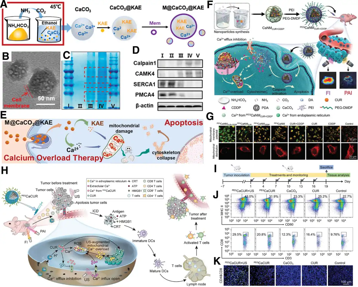
Figure 7.
The CaCO3-based NMs for calcium overload. (A) Schematic illustration of M@CaCO3@KAE NPs synthesis. (B) TEM image of the M@CaCO3@KAE NPs. (C) SDS-PAGE protein tracking of cancer cell membrane proteins. Samples from lane I to lane V are marker, CaCO3@KAE NPs, M@CaCO3@KAE NPs, cancer cell membrane, and marker, respectively. (D) Calcium-regulated proteins expression levels of A549 cells, I: control; II: CaCO3 NPs; III: KAE; IV: CaCO3@KAE NPs; V: M@CaCO3@KAE NPs. (E) Schematic illustration of M@CaCO3@KAE NP-mediated apoptosis. (A–E) Reproduced with permission from Li et al.[134] Copyright 2021, Elsevier Ltd. (F) Schematic representation of the functional pattern of multifunctional Ca2+ nanomodulator PEGCaNMCUR+CDDP. (G) Mitochondrial membrane potentials and distributions of MCF-7 cells treated with PEGCaNMCUR+CDDP or other Ca2+ nanomodulators for 12 hours. (F, G) Reproduced with permission from Zheng et al.[135] Copyright 2021, Wiley-VCH GmbH. (H) Schematic illustration of photoacoustic/fluorescence dual-mode imaging-guided synergistic cancer therapy through US-augmented mitochondrial Ca2+ overload induced by calcium nanomodulator PEGCaCUR. (I) Treatment scheme of Ca2+ nanomodulators in vivo. (J) Populations of immune cells in draining lymph nodes after different treatments. (K) CD4 (red fluorescence) and CD8 (green fluorescence) T cells in tumor tissues after different treatments. (H–K) Reproduced with permission from Zheng et al.[136] Copyright 2021, American Chemical Society.
In addition to CaCO3, calcium oxide (eg, CaO2) is also an available Ca2+ nanogenerator. Zhang et al prepared a kind of pH-sensitive sodium-hyaluronate-modified calcium peroxide nanoparticles (SH-CaO2 NPs) to induce an abnormal cytoplasm accumulation of free Ca2+. Interestingly, these NPs created an artificial calcium-overloading stress in tumor cells, while normal cells were more tolerant of the adverse influence of NPs than tumor cells. Meanwhile, the enrichment of Ca2+ showed the potential to cause tumor calcification, which may further inhibit the growth of tumor tissue.[41]
3.2 Metal ions-nonspecific-induced metalloptosis
3.2.1 Zinc ions-induced cell death
Zinc (Zn) is a necessary trace element for the daily activities of the body, ranking second in abundance. It is closely associated with various biological functions, including cell cycle progression, signal transduction, gene expression, immune functions, meiosis, and numerous other physiological processes.[139] Statistical data indicate that the activity of around 300 enzymes and the structural integrity of approximately 2000 transcription factors are zinc dependent.[140] Women typically have around 1.5 g of zinc in their bodies, while men have about 2.5 g, predominantly located in the bone and skeletal muscle.[141] Zinc homeostasis dysregulation is closely linked to cancer, with zinc ions not only induce ferroptosis or calcium overload but also directly cause apoptosis, necrosis, etc. Although zinc ions-mediated cell death has not yet been classified as a new type of PCD, it possesses distinct characteristics and will be the focus of discussion in this section.
Recent research increasingly highlights the significant role of zinc in ferroptosis. Zinc oxide NPs (ZnO NPs), approved as a safe substance by the United States Food and Drug Administration, exhibit good biocompatibility and cost-effectiveness, offering broad application prospects in the biomedical field.[142] A study by Zhang et al in 2020 demonstrated that ZnO NPs could trigger ferroptosis in human umbilical vein endothelial cells (HUVECs) by increasing ROS, lipid peroxidation, and depleting glutathione (Figure 8A–D).[143] Further investigations revealed that the zinc chelator N,N,N′,N′-Tetrakis(2-pyridylmethyl)ethylenediamine (TPEN) could protect cells from erastin-induced cell death; supplementation with zinc alone (ZnCl2) or together with the iron chelator deferoxamine significantly heightened the sensitivity of MDA-MB-231 (Figure 8E) and HT-1080 cells to ferroptosis.[144] Additional studies identified that ZIP7 plays a crucial role by regulating zinc transport from the ER to the cytosol, essential for ferroptosis. Blocking ZIP7’s function genetically or chemically safeguarded cells against ferroptosis, a protection that was negated with the addition of zinc (Figure 8F).[144] These findings confirmed that Zn2+ and ZnO NPs are capable of inducing ferroptosis, suggesting that zinc administration could serve as a promising approach to promote the death of cancer cells.
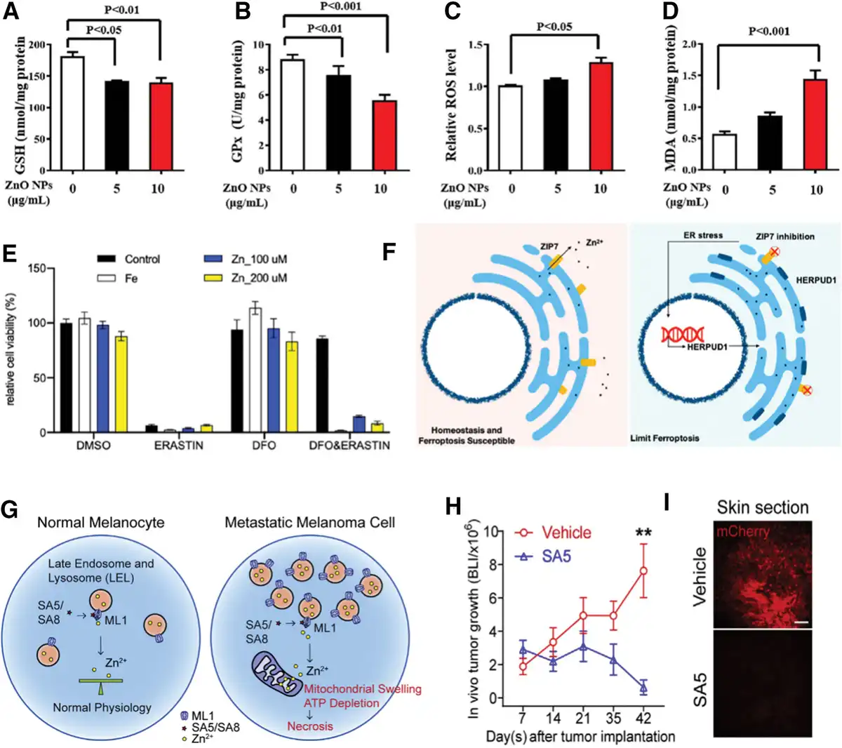
Figure 8.
Illustration of zinc ion-mediated cell death and related nNMs. (A) GSH, (B) GPx, (C) ROS, and (D) MDA levels of HUVECs after the treatment with ZnO NPs at various concentrations. (A–D) Reproduced with permission from Zhang et al.[143] Copyright 2020, the author(s). (E) Relative cell viability of MDA-MB-231 cells after different treatments for 24 hours. (F) Illustration of ferroptosis resistance through ZIP7 knockdown. (E, F) Reproduced with permission from Chen et al.[144] Copyright 2021, the author(s). (G) Illustration of lysozincrosis in metastatic melanoma cells. (H) Tumor volume curves of different treatment groups, n = 5. **P < 0.01. (I) Illustrative mCherry fluorescence images showcasing tumor cells (red) in skin sections from different treatment groups. (G–I) Reproduced with permission from Du et al.[145] Copyright 2021, the author(s).
Moreover, zinc overload can impair mitochondrial function and promote organelle death, coordinating with Ca2+-induced damage to disrupt intracellular calcium homeostasis, ultimately leading to cell death.[146,147] While Zn2+ and Ca2+ are known to share many transport pathways, the precise nature of their interplay in the context of PCD remains to be clearly defined.
Lysosomes are key organelles in cells, fundamental for metabolism and protein breakdown, and crucial for cell growth, division, and differentiation.[148] Unstable Zn2+ within cells is partly stored in lysosomes. Just as with iron, Zn2+ can also be released from its unstable reserve, influencing mitochondrial and lysosomal functions, thereby inducing zinc ions-mediated cancer cell death.[149] Mucolipin TRP channel 1 (TRPML1/MCOLN1; ML1) is a cation channel that allows the passage of Ca2+ and Zn2+/Fe2+, mainly found on the membranes of late endosomes and lysosomes in all mammalian cells.[150] It is essential for several lysosomal activities, such as transport, exocytosis, biogenesis, and heavy metal homeostasis. In metastatic melanoma cells with maladaptive upregulation of the ML1 lysosomal channel, Du et al discovered a kind of lysosomal Zn2+ release triggering nonapoptotic cell death, which they termed as lysozincrosis.[145] Upon treating these cells with TRPML-specific synthetic agonist (ML-SA), lysosomal Zn2+ release was triggered, leading to mitochondrial swelling/damage and rapid depletion of cellular ATP. This process induced necrosis, as illustrated in Figure 8G. As expected, administering ML-SA5, a specific type of ML-SA, significantly reduced tumor volume in MeWo-FmC-bearing mice over a 42-day treatment period without any relapse, demonstrating a more pronounced antitumor effect compared to the control group (Figure 8H, I).[145]
It is worth noting that the influence of zinc ions on cancer is notably varied, depending on the specific type of cancer involved.[140] Besides the aforementioned lysozincrosis in metastatic melanoma, studies indicate that ZnO NPs particularly induce cancer cell death through the apoptotic pathway in certain cancer cell lines, such as cervical carcinoma, lung carcinoma, head and neck squamous cell carcinoma, urinary bladder carcinoma, and head and neck squamous cell carcinoma.[151,152] Additionally, reports indicate that ZnFe2O4-based nanosystems could programmatically initiate and amplify the cyclic guanosine monophosphate-adenosine monophosphate synthase (cGAS)/stimulator of interferon genes (STING) signaling pathway in tumor cells, thereby inhibiting tumor progression and postoperative recurrence.[153]
In summary, current studies on zinc ions-mediated cell death mainly align with established PCD pathways, with no specific cell death mechanisms attributed to zinc ions induction yet identified. Research into the anticancer mechanisms of zinc ions-based nanomedicine, especially focusing on ZnO, remains relatively underexplored. Moreover, given the varying dependence of different cancer cells on zinc ions, establishing a universal principle for zinc ions-mediated cell death requires further investigation. However, it is undeniable that disrupting the zinc homeostasis in cancer cells represents a promising anticancer strategy.
3.2.2 Sodium ions-induced cell death
Sodium ions (Na+), a kind of common physiological ion, are crucial for normal cell function, typically existing at concentrations an order of magnitude higher extracellularly (145 mM) than intracellularly (12 mM).[154] Conversely, intracellular potassium (K+) levels are maintained at a higher concentration. This asymmetry in Na+/K+ concentration gradients is vital for amino acid transport, pH maintenance, and cell volume regulation.[155,156] Disrupting sodium homeostasis in tumor cells can induce significant osmotic changes, leading to apoptosis or necrosis. However, targeting tumor cells with excess Na+ is challenging due to the strict cellular ion transport regulation of live cells and the short circulation lifetime of single-ion delivery. Ingeniously designed sodium-based NMs can bypass these hurdles, as they enter cells via endocytosis and release Na+ internally, increasing osmotic pressure and causing selective tumor cell death while sparing normal cells due to their lower sodium levels.[157]
However, ionic compounds such as sodium chloride (NaCl) and sodium bicarbonate (NaHCO3), selected for their availability and excellent biocompatibility, are challenging to encapsulate in NMs due to their high solubility in water.[158] This makes the delivery of sodium ions to tumor cells using NMs a complex task. It is able to overcome these challenges through kinds of preparation strategies, ensuring stable encapsulation and effective delivery within the TME. Jiang et al used sodium oleate and molybdenum chloride as precursors, with oleylamine as a surfactant, to synthesize pure-phase NaCl nanoparticles (SCNPs) via a microemulsion reaction in a hexane/ethanol mixed solvent.[157] Subsequently, the obtained SCNPs surface was modified with a layer of PEGylated phospholipid to enhance its water solubility, resulting in the phospholipid-coated PSCNPs (Figure 9A). PSCNPs could enter tumor cells via endocytosis, releasing a significant amount of Na+/Cl−, leading to increased osmolarity and rapid cell lysis. Interestingly, PSCNPs showed minimal toxicity toward normal cells like human prostate epithelial cell line (HPrECs) and mouse spermatogonial stem cells (C18-4). This selective cytotoxicity was attributed to the higher uptake by rapidly proliferating tumor cells and their inherently higher intracellular sodium concentrations ([Na+]int), making them more susceptible to PSCNPs-induced osmotic shock. Figure 9B illustrates the varying cytotoxic effects on different cells and their correlation with cellular [Na+]int, indicating that [Na+]int plays a vital role in determining the susceptibility of cancer cells to PSCNPs. In vivo experiments demonstrated that PSCNPs suppressed tumor growth (Figure 9C) and induced apoptosis and necroptosis in cancer cells, with an increase in survival (Figure 9D). Moreover, the study also revealed that PSCNPs could induce ICD, simultaneously killing cancer cells and enhancing anticancer immunity. To further enhance the blood circulation time and sodium ions responsive release capability of NaCl-based nanomedicines, Li et al designed and synthesized a kind of GSH-responsive nanomedicines, termed NaCl@ssss-VHMS (Figure 9E), which comprised NaCl nanocrystals internally and a virus-mimicking hollow mesoporous tetrasulfide-organosilica (ssss-VHMS) externally.[158] The morphological advancements in NaCl@ssss-VHMS facilitated its rapid internalization into tumor cells via spike surface-assisted adhesion and invasion. Once inside, it degraded under the action of excessively produced GSH, releasing substantial amounts of Na+/Cl−, disrupting the ionic homeostasis of tumor cells, and inducing apoptosis, ferroptosis, as well as pyroptosis. The intracellular Na+ and Cl− ion levels were monitored using Na+ (sodium-binding benzofuran isophthalate acetoxymethyl ester [SBFI-AM]) and Cl− indicator probe (MQAE), respectively. The enhanced fluorescence signals of SBFI-AM and MQAE in NaCl@ssss-VHMS-treated HepG2 cells, compared to control groups, underscored its superior ion delivery performance (Figure 9F). In vivo imaging experiments also demonstrated the efficient accumulation of NaCl@ssss-VHMS within tumors (Figure 9G). In addition to NaCl, sodium citrate NPs have also been employed for sodium ion delivery, resulting in increased intracellular osmotic pressure and mediating tumor cell death.[160]
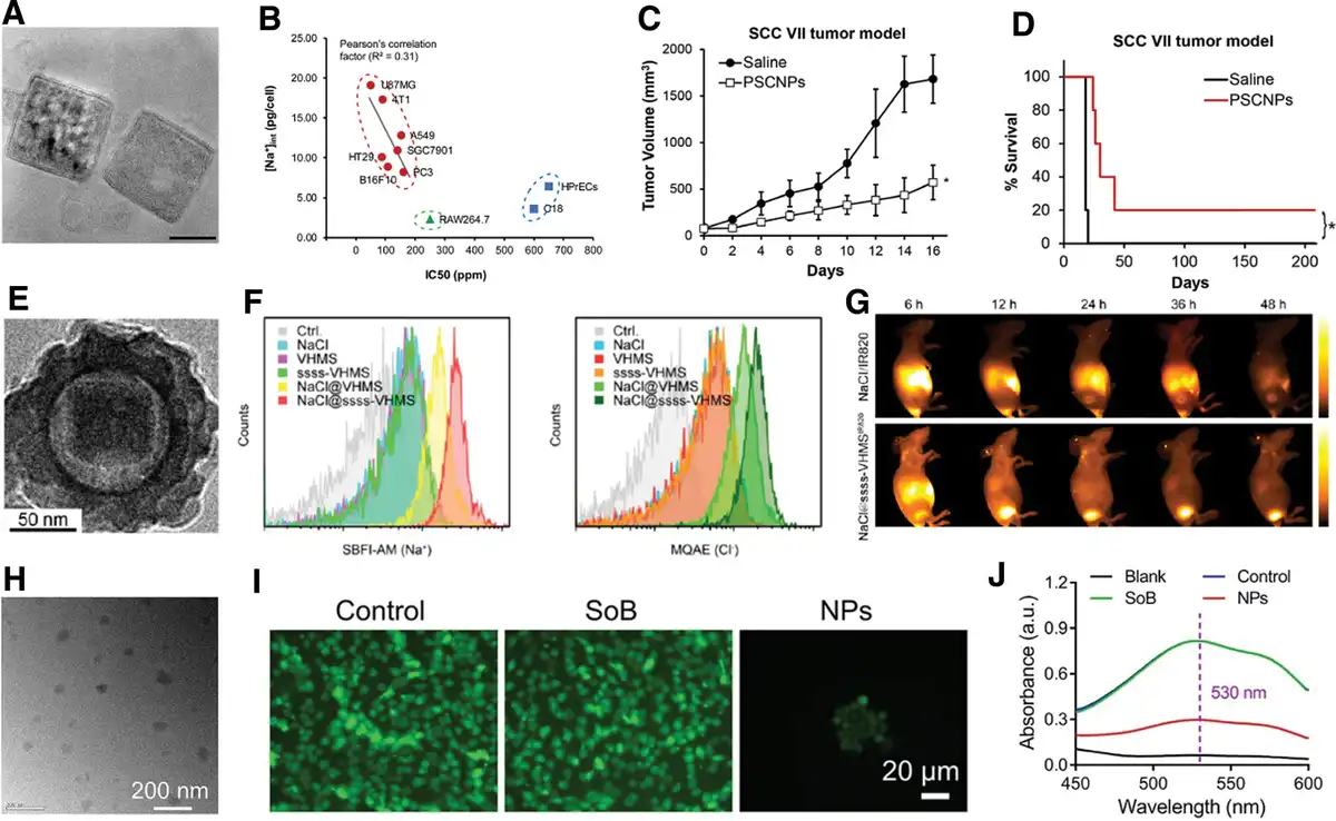
Figure 9.
Illustration of sodium ions-mediated cell death and related NMs. (A) TEM image of PSCNPs. Scale bar, 40 nm. (B) The correlation between [Na+]int and IC50. (C) Tumor growth curves of different treatment groups. *P < 0.05. (D) Survival curves of different treatment groups. *P < 0.05. (A–D) Reproduced with permission from Jiang et al.[157] Copyright 2019, WILEY-VCH Verlag GmbH & Co. KGaA, Weinheim. (E) TEM image of NaCl@ssss-VHMS. (F) Flow cytometric analysis of intracellular Na+ and Cl− levels. (G) Time-lapsed near-infrared (NIR) fluorescence imaging of tumor-bearing mice after different treatments. (E–G) Reproduced with permission from Li et al.[158] Copyright 2022, American Chemical Society. (H) TEM image of NaHCO3 NPs. (I) Intracellular pH detection after different treatments. (J) Extracellular lactic acid detection after different treatments. (H–J) Reproduced with permission from Ding et al.[159] Copyright 2023, Wiley-VCH GmbH.
As another type of sodium-containing inorganic salt, sodium bicarbonate (NaHCO3) NPs are also employed in sodium ions-mediated cell death. Distinct from the aforementioned NaCl NPs, NaHCO3 NPs possess weak alkalinity, which allows them to release a substantial amount of Na+ inside tumor cells while concurrently rectifying the acidosis of the TME.[161] In a recent study, Ding et al synthesized NaHCO3 NPs using sodium oleate and ammonium hydrogen carbonate as precursors through a rapid microemulsion method (Figure 9H).[159] NaHCO3 NPs were subsequently modified with distearoylphosphatidylethanolamine-poly(ethylene glycol) (DSPE-PEG) for enhanced biocompatibility. The resultant weakly alkaline NaHCO3 NPs could modulate lactic acid metabolism through acid-base neutralization. Treatment with NaHCO3 NPs significantly increased intracellular pH levels (Figure 9I) and markedly reduced the lactic acid concentration in the culture medium (Figure 9J), demonstrating the substantial potential of NaHCO3 NPs in regulating lactic acid metabolism and maintaining cellular pH stability. Moreover, like the aforementioned NaCl NPs, NaHCO3 NPs could also release significant quantities of Na+ within tumor cells, leading to an increase in intracellular osmolarity. This process could trigger the pyroptosis pathway and ICD, thereby enhancing immune responses.
Currently, NMs employed in sodium ions-mediated cell death are known for their good biocompatibility and simple raw material components. They induce tumor cell death by disrupting the ionic homeostasis of tumor cells, leading to a sharp increase in osmotic pressure. Additionally, some of them can also regulate lactic acid metabolism through acid-base neutralization, thereby reversing the weakly acidic, immunosuppressive TME, offering broad application prospects. In this therapeutic strategy, the controllable preparation of NMs is crucial, ensuring prolonged blood circulation while preventing the off-target leakage and degradation of water-soluble sodium-containing compounds. Future developments could involve designing more comprehensive theranostic nanoplatforms, using imaging techniques to investigate the specific mechanisms and metabolic pathways in sodium ions-mediated cell death, such as fluorescent Na+ reporter SBFI-AM or 23Na MRI. Finally, sodium ions-mediated cell death holds significant research value in improving immune responses and achieving tumor immunotherapy, with current studies making beneficial explorations in this area.[157,159,160]
3.2.4 Other ions-induced cell death
Other essential metal ions, including magnesium (Mg), cobalt (Co), molybdenum (Mo), manganese (Mn), and potassium (K) ions, are also crucial for the growth and development of humans,[22] showing potential in inducing metalloptosis.
Magnesium ion (Mg2+) as an essential trace element for the human body, participates in various physiological activities in cardiovascular, neurological systems, and muscles.[162] More importantly, Mg2+ can act as an activator involved in the synthesis of at least 200 enzymes[163] and regulate other momentous ion channels such as K+ and Ca2+.[164] Hence, abnormal levels of Mg2+ in the body can trigger various diseases, and some nanotheranostics based on Mg2+ have shown initial effectiveness in treating cancer. For instance, MgO NPs exerted a strong inhibitory effect on cancer cells[165] through the production of ROS because of the release of Mg2+ or that of ligands as well as byproducts.[166–167–168] The generated ROS further caused oxidative damage to DNA, protein denaturation, and lipid peroxidation, leading to the eventual cell death.[169] However, the detailed molecular mechanism of Mg2+-mediated cell death still needs to be clarified.[170,171] Meanwhile, the biocompatibility and cytotoxicity of MgO NPs is also a key issue that potentially hinders the translation of MgO NPs in the field of cancer therapy,[172] which may be solved through the green synthesis routes of MgO NPs.[171,173] For instance, compared with Co oxide NPs, MgO NPs synthesized by the green method showed less toxicity and great bioavailability.[174] Moreover, the researchers have also found that introducing Mg2+ into NMs can promote the rolling circle amplification of DNA, ultimately achieving the antitumor effect of the functionalized DNA.[175]
Co and Mo have been reported to induce ferroptosis. Studies indicate that Co NPs significantly downregulated the expression of GSH, GPX4, and SLC7A11 proteins in Balb/3T3 mouse fibroblast cells, while leading to a notable increase in intracellular levels of ROS, MDA, cobalt, and iron.[176] However, ferrostatin-1 showed rescue effects on cells after treatment of Co NPs, suggesting a close association between Co NPs-induced cytotoxicity and ferroptosis.[176] In another study, cobaltous oxide nanodots were encapsulated within dendritic mesoporous silica NPs, followed by further surface modifications to yield iCoDMSNs. iCoDMSNs treatment increased transferrin receptor levels and reduced solute carrier family 40 member 1 (SLC40A1), leading to Fe2+ accumulation and subsequent ferroptosis.[177] Similarly, molybdenum sulfide NPs were able to generate ROS, thereby triggering lipid peroxidation and ferroptosis in cancer cells.[178] Additionally, the simultaneous integration of Co and Mo into NMs could enhance the induction of ferroptosis more effectively. Wu et al introduced a strategy of nonferrous ferroptosis, utilizing a CoMoO4-phosphomolybdic acid nanosheet hybrid. This approach leveraged accelerated Mo(V)-Mo(VI) conversion to increase lipid peroxide levels, enhanced GSH consumption for GPX4 enzyme deactivation, and initiated a ROS surge, effectively inducing ferroptosis.[57]
Manganese-based NPs can induce ferroptosis in tumors through mechanisms such as GSH depletion, increased oxygen and ROS production, and enhanced lipid peroxidation. For example, when oral squamous cell carcinoma cells (Cal-27) were treated with MnO2 nanoflowers under near-infrared radiation, there was an increase in cellular Fe2+, an accumulation of LPO, and a downregulation of GPX4, effectively inducing ferroptosis.[179] Similarly, manganese silicate nanobubbles demonstrated a high capacity for depleting GSH, inducing ferroptosis through the inactivation of GPX4.[180] Furthermore, Mn can influence iron homeostasis by altering iron uptake and the expression of circulating transport and regulatory proteins; alterations in iron levels can, in turn, impact Mn balance and its biological roles.[181] Mn2+ may also contend with essential ions such as Ca2+ and Mg2+ for cellular entry via different transport mechanisms, suggesting its potential to trigger PCD by unbalancing ion equilibrium.[182] Moreover, Mn-containing NPs can stimulate innate and adaptive immunity through the cGAS-STING pathway, playing an important role in the immunotherapy of tumors.[183,184] As an illustration, hollow mesoporous silica-coated MnO NPs have been utilized in MRI-guided immune-chemodynamic therapy, where MnO NPs served in activating the cGAS-STING pathway for immunotherapy, upregulating ROS through Fenton-like reactions, and enhancing T1-weighted MRI.[184] This research effectively leveraged the unique properties of the Mn element for antitumor therapy.
As a vital physiological ion, potassium ions are integral to numerous crucial cellular functions, including metabolism, growth, and apoptosis.[185] Analogous to NaCl, KCl NPs are also applicable in cancer therapy. The intracellular release of a substantial amount of K+ and Cl− could alter the osmotic pressure within cancer cells, subjecting them to a hypertonic state. This change induced the rupture and subsequent death of the cancer cells.[186] Furthermore, potassium ionophores have demonstrated anticancer capability. For example, valinomycin, a natural ionophore, is able to pump intracellular potassium ions out of the cytosol, leading to mitochondrial damage and inducing apoptosis.[187] Additionally, a helical polypeptide-based potassium ionophore was shown to induce ER stress-mediated apoptosis in vivo.[188] However, it is important to note that the research on the antitumor applications of NPs constructed based on potassium ionophores remains comparatively limited.
Although the discussed metal elements in this section can mediate cell death, the exact pathways of their cancer-killing effects need more research. There is a current gap in definitive studies confirming these pathways. Future work must elucidate how these metals exert specific molecular interactions within cancer cells to maximize their therapeutic value. Unraveling these mechanisms will illuminate the details of metalloptosis and guide the development of targeted cancer treatments leveraging these metals’ unique attributes.
4. Conclusion and perspective
Metalloptosis as a new form of cell death, is worthy of defining cautiously, because the present reported several types of metalloptosis, in addition to ferroptosis, cuproptosis, and calcium overload, the downstream signaling pathway of other types of metalloptosis (such as Mg ions-, Zn ions-, Mn ions-, and Na ions-mediated cell death) is still attributed to conventional PCD. Table 2 summarizes the representative NMs mentioned in this review that induce metalloptosis. Hence, it is urgent to excavate more in-depth and specific cell death signaling pathways on metalloptosis. While the concept of “metalloimmunotherapy” has previously been introduced to the academic community, highlighting the inherent immunomodulatory properties of metal ions to enhance anticancer immune responses,[189] it is crucial to distinguish this from “metalloptosis.” Metalloptosis specifically refers to a type of cell death mediated by metal ions, characterized by cellular damage and subsequent death triggered by specific metal ions. In contrast, metalloimmunotherapy leverages these immunomodulatory properties to bolster immune responses against cancers, focusing on enhancing immunological function rather than directly inducing cell death as observed in metalloptosis. Although both concepts underscore the critical role of metal ions in cancer research, they diverge fundamentally in their primary focus and application, highlighting the diverse therapeutic potentials of metals in oncology. Meanwhile, this is also inseparable from the advancements in imaging, artificial intelligence, analytical chemistry, and other fields, an increasing array of novel characterization and detection methods are available to directly or indirectly detect metalloptosis in cancers. Such interdisciplinary collaborations enhance our understanding of metal’s anticancer mechanisms, offering new breakthroughs and progress in cancer treatment.
Table 2
NMs inducing metalloptosis mentioned in this review.
| Types of metalloptosis | Engineering NMs | Reference |
|---|---|---|
| Ferroptosis | Fe3O4@PGL | [83] |
| bcc-USINPs | [84] | |
| Au-Fe2C | [38] | |
| AMSNs | [87] | |
| Au/Cu-TCPP(Fe)-PEG | [90] | |
| SRF@FeIIITA | [95] | |
| Fe/Ni-LDH | [100] | |
| Cuproptosis | NP@ESCu | [34] |
| CSTD-Cu(II)@DSF | [113] | |
| Dox@Fe/CuTH HaMOF | [47] | |
| Cu2(PO4)(OH) NPs | [40] | |
| GOx@[Cu(tz)] | [54] | |
| BSO-CAT@MOF-199@DDM | [56] | |
| Calcium overload | M@CaCO3@KAE | [134] |
| CaNMCUR+CDDP | [135] | |
| PEGCaCUR | [136] | |
| Zinc ions-induced cell death | ZnO | [143,151,152] |
| Sodium ions-induced cell death | PSCNPs | [157] |
| NaCl@ssss-VHMS | [158] | |
| NaHCO3 NPs | [159] | |
| Magnesium ions-induced cell death | MgO NPs | [165] |
| Cobalt ions-induced cell death | Co NPs | [176] |
| iCoDMSNs | [177] | |
| Manganese ions-induced cell death | Silica-coated MnO NPs | [184] |
| Potassium ions-induced cell death | KCl NPs | [186] |
Furthermore, how to innovate the regulation signaling pathways of metalloptosis from a mechanistic perspective is a very challenging issue. Hence, in-depth studies on the interplay among different NMs components and their interactions with cancer cells are essential for a comprehensive understanding of metalloptosis. For instance, NMs with different sizes and morphologies often have varying cell phagocytic efficiency, which can directly influence their interaction with cancer cells. Composite NMs have synergistic effects between multiple elements, thus the affected signaling pathways always exhibit a networked pattern rather than a single pathway, which is beneficial for enhancing the anticancer effect of NMs, but poses certain challenges in accurately understanding the role of each element. Therefore, to delve deeper into the mechanism, the NMs designed often tend to be simple and controllable. For the composite NMs, the interactions among various metal ions in metalloptosis are intricate; for instance, zinc ions can induce lysozincrosis, and there are also reports suggesting their role in ferroptosis and calcium overload, as well as traditional PCD. Utilizing cell death inhibitors and ion chelators can further clarify different types of metalloptosis. Moreover, when exploring the anticancer mechanisms of NMs-induced metalloptosis, special attention should be paid to the metal degradation and release properties of the NMs. It is crucial to determine the specific metal concentrations, incubation time, and their impacts on cells, including morphology, metabolism, intracellular and extracellular ion content, and cell death markers.
Beyond oncology, metal homeostasis also plays a vital role in other diseases, such as neurodegenerative, cardiovascular, and metabolic disorders. Research into the mechanisms of metalloptosis can also provide new therapeutic targets for these diseases.
Funding
This work was financially supported by the National Natural Science Foundation of China (52201198, 52027801, and 92263203), the Beijing Natural Science Foundation-Haidian Primitive Innovation Joint Fund (L232015), the National Key R&D Program of China (2022YFA1203902), the China-Germany Collaboration Project (M-0199), the Clinical Medicine Plus X-Young Scholars Project of Peking University, and the Fundamental Research Funds for the Central Universities (PKU2024LCXQ014).
Conflicts of interest
The authors declare that they have no conflicts of interest.
Author contributions
Shuren Wang, Ran Ma, Zi Mei, and Yanglong Hou proposed the overall concept. Shuren Wang and Ran Ma wrote the paper. All authors contributed to the general discussion.
References
- [1] Nagata S, Tanaka M. Programmed cell death and the immune system. Nat Rev Immunol. 2017;17:333–340.
- [2] Burgess DJ. Apoptosis refined and lethal. Nat Rev Cancer. 2013;13:79–79.
- [3] Dai XF, Wang DJ, Zhang JY. Programmed cell death, redox imbalance, and cancer therapeutics. Apoptosis. 2021;26:385–414.
- [4] Verbrugge I, Johnstone RW, Smyth MJ. SnapShot: extrinsic apoptosis pathways. Cell. 2010;143:1192, 1192.e1–1192, 1192.e2.
- [5] Wu D, Wang S, Yu GC, et al. Cell death mediated by the pyroptosis pathway with the aid of nanotechnology: prospects for cancer therapy. Angew Chem Int Ed. 2021;60:8018–8034.
- [6] Shi JJ, Zhao Y, Wang K, et al. Cleavage of GSDMD by inflammatory caspases determines pyroptotic cell death. Nature. 2015;526:660–665.
- [7] Kayagaki N, Stowe IB, Lee BL, et al. Caspase-11 cleaves gasdermin D for non-canonical inflammasome signalling. Nature. 2015;526:666–671.
- [8] Wang YP, Gao WQ, Shi XY, et al. Chemotherapy drugs induce pyroptosis through caspase-3 cleavage of a gasdermin. Nature. 2017;547:99–103.
- [9] Rogers C, Fernandes-Alnemri T, Mayes L, et al. Cleavage of DFNA5 by caspase-3 during apoptosis mediates progression to secondary necrotic/pyroptotic cell death. Nat Commun. 2017;8:14128.
- [10] Orning P, Weng D, Starheim K, et al. Pathogen blockade of TAK1 triggers caspase-8-dependent cleavage of gasdermin D and cell death. Science. 2018;362:1064–1069.
- [11] Sarhan J, Liu BC, Muendlein HI, et al. Caspase-8 induces cleavage of gasdermin D to elicit pyroptosis during infection. Proc Natl Acad Sci USA. 2018;115:E10888–E10897.
- [12] Chen KW, Demarco B, Heilig R, et al. Extrinsic and intrinsic apoptosis activate pannexin-1 to drive NLRP3 inflammasome assembly. EMBO J. 2019;38:e101638.
- [13] Boya P, Reggiori F, Codogno P. Emerging regulation and functions of autophagy. Nat Cell Biol. 2013;15:713–720.
- [14] Amaravadi RK, Kimmelman AC, Debnath J. Targeting autophagy in cancer: recent advances and future directions. Cancer Discov. 2019;9:1167–1181.
- [15] Li XH, He SK, Ma BY. Autophagy and autophagy-related proteins in cancer. Mol Cancer. 2020;19:12.
- [16] Linkermann A, Green DR. Necroptosis. N Engl J Med. 2014;370:455–465.
- [17] He SD, Liang YQ, Shao F, et al. Toll-like receptors activate programmed necrosis in macrophages through a receptor-interacting kinase-3-mediated pathway. Proc Natl Acad Sci USA. 2011;108:20054–20059.
- [18] Liang SJ, Qin XB. Critical role of type I interferon-induced macrophage necroptosis during infection with serovar Typhimurium. Cell Mol Immunol. 2013;10:99–100.
- [19] Lu JV, Chen HC, Walsh CM. Necroptotic signaling in adaptive and innate immunity. Semin Cell Dev Biol. 2014;35:33–39.
- [20] Ch’en IL, Tsau JS, Molkentin JD, et al. Mechanisms of necroptosis in T cells. J Exp Med. 2011;208:633–641.
- [21] Degterev A, Huang ZH, Boyce M, et al. Chemical inhibitor of nonapoptotic cell death with therapeutic potential for ischemic brain injury. Nat Chem Biol. 2005;1:112–119.
- [22] Zoroddu MA, Aaseth J, Crisponi G, et al. The essential metals for humans: a brief overview. J Inorg Biochem. 2019;195:120–129.
- [23] Kim JK, Lee C, Lim SW, et al. Elucidating the role of metal ions in carbonic anhydrase catalysis. Nat Commun. 2020;11:4557.
- [24] Maret W. Metals on the move: zinc ions in cellular regulation and in the coordination dynamics of zinc proteins. Biometals. 2011;24:411–418.
- [25] Puig S, Ramos-Alonso L, Romero AM, et al. The elemental role of iron in DNA synthesis and repair. Metallomics. 2017;9:1483–1500.
- [26] Giese B, Karamash M, Fromm KM. Chances and challenges of long-distance electron transfer for cellular redox reactions. FEBS Lett. 2023;597:166–173.
- [27] Ganz T, Nemeth E. Hepcidin and iron homeostasis. Biochim Biophys Acta. 2012;1823:1434–1443.
- [28] Madsen E, Gitlin JD. Copper deficiency. Curr Opin Gastroenterol. 2007;23:187–192.
- [29] Xue Q, Kang R, Klionsky DJ, et al. Copper metabolism in cell death and autophagy. Autophagy. 2023;19:2175–2195.
- [30] Shen Z, Song J, Yung BC, et al. Emerging strategies of cancer therapy based on ferroptosis. Adv Mater. 2018;30:1704007.
- [31] Weekley CM, He C. Developing drugs targeting transition metal homeostasis. Curr Opin Chem Biol. 2017;37:26–32.
- [32] Lee VJ, Janisse SE, Heffern MC. Plant-derived chelators and ionophores as potential therapeutics for metabolic diseases. Chem Soc Rev. 2023;52:3927–3945.
- [33] Wang W, Mo W, Hang Z, et al. Cuproptosis: harnessing transition metal for cancer therapy. ACS Nano. 2023;17:19581–19599.
- [34] Guo B, Yang F, Zhang L, et al. Cuproptosis induced by ROS responsive nanoparticles with elesclomol and copper combined with αPD-L1 for enhanced cancer immunotherapy. Adv Mater. 2023;35:2212267.
- [35] Zanganeh S, Hutter G, Spitler R, et al. Iron oxide nanoparticles inhibit tumour growth by inducing pro-inflammatory macrophage polarization in tumour tissues. Nat Nanotechnol. 2016;11:986–994.
- [36] Zhang C, Bu W, Ni D, et al. Synthesis of iron nanometallic glasses and their application in cancer therapy by a localized Fenton reaction. Angew Chem Int Ed Engl. 2016;55:2101–2106.
- [37] Yue L, Dai Z, Chen X, et al. Development of a novel FePt-based multifunctional ferroptosis agent for high-efficiency anticancer therapy. Nanoscale. 2018;10:17858–17864.
- [38] Wang J, Fang Z, Zhao C, et al. Intelligent size-switchable iron carbide-based nanocapsules with cascade delivery capacity for hyperthermia-enhanced deep tumor ferroptosis. Adv Mater. 2024;36:2307006.
- [39] Ning S, Lyu M, Zhu D, et al. Type-I AIE photosensitizer loaded biomimetic system boosting cuproptosis to inhibit breast cancer metastasis and rechallenge. ACS Nano. 2023;17:10206–10217.
- [40] Zhao F, Liang L, Wang H, et al. H2S-activated ion-interference therapy: a novel tumor targeted therapy based on copper-overload-mediated cuproptosis and pyroptosis. Adv Funct Mater. 2023;33:2300941.
- [41] Zhang M, Song R, Liu Y, et al. Calcium-overload-mediated tumor therapy by calcium peroxide nanoparticles. Chem. 2019;5:2171–2182.
- [42] Wang K, Li Y, Xie L-H, et al. Construction and application of base-stable MOFs: a critical review. Chem Soc Rev. 2022;51:6417–6441.
- [43] Moharramnejad M, Ehsani A, Shahi M, et al. MOF as nanoscale drug delivery devices: synthesis and recent progress in biomedical applications. J Drug Delivery Sci Technol. 2023;81:104285.
- [44] Chen J, Zhang Z, Ma J, et al. Current status and prospects of MOFs in controlled delivery of Pt anticancer drugs. Dalton Trans. 2023;52:6226–6238.
- [45] Shyngys M, Ren J, Liang XQ, et al. Metal-organic framework (MOF)-based biomaterials for tissue engineering and regenerative medicine. Front Bioeng Biotechnol. 2021;9:603608.
- [46] Yang F, Dong J, Li Z, et al. Metal–organic frameworks (MOF)-assisted sonodynamic therapy in anticancer applications. ACS Nano. 2023;17:4102–4133.
- [47] Xu W, Qian J, Hou G, et al. A hollow amorphous bimetal organic framework for synergistic cuproptosis/ferroptosis/apoptosis anticancer therapy via disrupting intracellular redox homeostasis and copper/iron metabolisms. Adv Funct Mater. 2022;32:2205013.
- [48] Li J, Dai J, Zhao L, et al. Bioactive bacteria/MOF hybrids can achieve targeted synergistic chemotherapy and chemodynamic therapy against breast tumors. Adv Funct Mater. 2023;33:2303254.
- [49] Liu Y, Niu R, Zhao H, et al. Single-site nanozymes with a highly conjugated coordination structure for antitumor immunotherapy via cuproptosis and cascade-enhanced T lymphocyte activity. J Am Chem Soc. 2024;146:3675–3688.
- [50] Zhang W, Wang M, Liu B, et al. Glutathione induced in situ synthesis of Cu single-atom nanozymes with anaerobic glycolysis metabolism interference for boosting cuproptosis. Angew Chem Int Ed. 2024;63:e202402397.
- [51] Tan J, Zhou X, Zhang S. Iron-doped cross-linked lipoic acid nano-aggregates for ferroptosis-mediated cancer treatment. Acta Biomater. 2023;159:289–299.
- [52] Chen K, Gu L, Zhang Q, et al. Injectable alginate hydrogel promotes antitumor immunity through glucose oxidase and Fe3+ amplified RSL3-induced ferroptosis. Carbohydr Polym. 2024;326:121643.
- [53] Kim SE, Zhang L, Ma K, et al. Ultrasmall nanoparticles induce ferroptosis in nutrient-deprived cancer cells and suppress tumour growth. Nat Nanotechnol. 2016;11:977–985.
- [54] Xu Y, Liu SY, Zeng L. An enzyme-engineered nonporous copper(I) coordination polymer nanoplatform for cuproptosis-based synergistic cancer therapy. Adv Mater. 2022;34:2204733.
- [55] Tang H, Chen D, Li C, et al. Dual GSH-exhausting sorafenib loaded manganese-silica nanodrugs for inducing the ferroptosis of hepatocellular carcinoma cells. Int J Pharm. 2019;572:118782.
- [56] Huang Q-X, Liang J-L, Chen Q-W, et al. Metal-organic framework nanoagent induces cuproptosis for effective immunotherapy of malignant glioblastoma. Nano Today. 2023;51:101911.
- [57] Wu C, Liu Z, Chen Z, et al. A nonferrous ferroptosis-like strategy for antioxidant inhibition–synergized nanocatalytic tumor therapeutics. Sci Adv. 2021;7:eabj8833.
- [58] Li Y, Lin J, He Y, et al. Tumour-microenvironment-responsive Na2S2O8 nanocrystals encapsulated in hollow organosilica–metal–phenolic networks for cycling persistent tumour-dynamic therapy. Exploration. 2024;4:20230054.
- [59] Ai X, Lyu L, Zhang Y, et al. Remote regulation of membrane channel activity by site-specific localization of lanthanide-doped upconversion nanocrystals. Angew Chem Int Ed Engl. 2017;56:3031–3035.
- [60] Dixon SJ, Lemberg KM, Lamprecht MR, et al. Ferroptosis: an iron-dependent form of nonapoptotic cell death. Cell. 2012;149:1060–1072.
- [61] Stockwell BR, Angeli JPF, Bayir H, et al. Ferroptosis: a regulated cell death nexus linking metabolism, redox biology, and disease. Cell. 2017;171:273–285.
- [62] Dolma S, Lessnick SL, Hahn WC, et al. Identification of genotype-selective antitumor agents using synthetic lethal chemical screening in engineered human tumor cells. Cancer Cell. 2003;3:285–296.
- [63] Yagoda N, von Rechenberg M, Zaganjor E, et al. RAS-RAF-MEK-dependent oxidative cell death involving voltage-dependent anion channels. Nature. 2007;447:864–868.
- [64] Angeli JPF, Schneider M, Proneth B, et al. Inactivation of the ferroptosis regulator Gpx4 triggers acute renal failure in mice. Nat Cell Biol. 2014;16:1180–U1120.
- [65] Ayala A, Muñoz MF, Argüelles S. Lipid peroxidation: production, metabolism, and signaling mechanisms of malondialdehyde and 4-hydroxy-2-nonenal. Oxid Med Cell Longevity. 2014;2014:360438.
- [66] Kagan VE, Mao GW, Qu F, et al. Oxidized arachidonic and adrenic PEs navigate cells to ferroptosis. Nat Chem Biol. 2017;13:81–90.
- [67] Wenzel SE, Tyurina YY, Zhao JM, et al. PEBP1 wardens ferroptosis by enabling lipoxygenase generation of lipid death signals. Cell. 2017;171:628–641.
- [68] Wang HY, Cheng Y, Mao C, et al. Emerging mechanisms and targeted therapy of ferroptosis in cancer. Mol Ther. 2021;29:2185–2208.
- [69] Koppula P, Zhang YL, Zhuang L, et al. Amino acid transporter SLC7A11/xCT at the crossroads of regulating redox homeostasis and nutrient dependency of cancer. Cancer Commun (Lond). 2018;38:1.
- [70] Gao MH, Monian P, Quadri N, et al. Glutaminolysis and transferrin regulate ferroptosis. Mol Cell. 2015;59:298–308.
- [71] Kawabata H. Transferrin and transferrin receptors update. Free Radic Biol Med. 2019;133:46–54.
- [72] Gammella E, Buratti P, Cairo G, et al. The transferrin receptor: the cellular iron gate. Metallomics. 2017;9:1367–1375.
- [73] Eaton JK, Furst L, Ruberto RA, et al. Selective covalent targeting of GPX4 using masked nitrile-oxide electrophiles. Nat Chem Biol. 2020;16:497–506.
- [74] Matsushita M, Freigang S, Schneider C, et al. T cell lipid peroxidation induces ferroptosis and prevents immunity to infection. J Exp Med. 2015;212:555–568.
- [75] Jang EJ, Kim DH, Lee B, et al. Activation of proinflammatory signaling by 4-hydroxynonenal-Src adducts in aged kidneys. Oncotarget. 2016;7:50864–50874.
- [76] Kang R, Chen RC, Zhang QH, et al. HMGB1 in health and disease. Mol Aspects Med. 2014;40:1–116.
- [77] Wen QR, Liu J, Kang R, et al. The release and activity of HMGB1 in ferroptosis. Biochem Biophys Res Commun. 2019;510:278–283.
- [78] Liang C, Zhang XL, Yang MS, et al. Recent progress in ferroptosis inducers for cancer therapy. Adv Mater. 2019;31:e1904197.
- [79] Trujillo-Alonso V, Pratt EC, Zong HL, et al. FDA-approved ferumoxytol displays anti-leukaemia efficacy against cells with low ferroportin levels. Nat Nanotechnol. 2019;14:616–622.
- [80] Yang WS, Stockwell BR. Synthetic lethal screening identifies compounds activating iron-dependent, nonapoptotic cell death in oncogenic-RAS-harboring cancer cells. Chem Biol. 2008;15:234–245.
- [81] Torti SV, Torti FM. Winning the war with iron. Nat Nanotechnol. 2019;14:499–500.
- [82] Hassannia B, Vandenabeele P, Vanden Berghe T. Targeting ferroptosis to iron out cancer. Cancer Cell. 2019;35:830–849.
- [83] Liang XL, Chen M, Bhattarai P, et al. Complementing cancer photodynamic therapy with ferroptosis through iron oxide loaded porphyrin-grafted lipid nanoparticles. ACS Nano. 2021;15:20164–20180.
- [84] Liang H, Wu XY, Zhao GZ, et al. Renal clearable ultrasmall single-crystal Fe nanoparticles for highly selective and effective ferroptosis therapy and immunotherapy. J Am Chem Soc. 2021;143:15812–15823.
- [85] Shah R, Margison K, Pratt DK. The potency of diarylamine radical-trapping antioxidants as inhibitors of ferroptosis underscores the role of autoxidation in the mechanism of cell death. ACS Chem Biol. 2017;12:2538–2545.
- [86] Fan HH, Yan GB, Zhao ZL, et al. A smart photosensitizer-manganese dioxide nanosystem for enhanced photodynamic therapy by reducing glutathione levels in cancer cells. Angew Chem Int Ed. 2016;55:5477–5482.
- [87] Wang SF, Li FY, Qiao RR, et al. Arginine-rich manganese silicate nanobubbles as a ferroptosis-inducing agent for tumor-targeted theranostics. ACS Nano. 2018;12:12380–12392.
- [88] Bersuker K, Hendricks JM, Li ZP, et al. The CoQ oxidoreductase FSP1 acts parallel to GPX4 to inhibit ferroptosis. Nature. 2019;575:688–692.
- [89] Doll S, Freitas FP, Shah R, et al. FSP1 is a glutathione-independent ferroptosis suppressor. Nature. 2019;575:693–698.
- [90] Li K, Lin CC, Li MH, et al. Multienzyme-like reactivity cooperatively impairs glutathione peroxidase 4 and ferroptosis suppressor protein 1 pathways in triple-negative breast cancer for sensitized ferroptosis therapy. ACS Nano. 2022;16:2381–2398.
- [91] Ursini F, Maiorino M, Gregolin C. The selenoenzyme phospholipid hydroperoxide glutathione-peroxidase. Biochim Biophys Acta. 1985;839:62–70.
- [92] Woo JH, Shimoni Y, Yang WS, et al. Elucidating compound mechanism of action by network perturbation analysis. Cell. 2015;162:441–451.
- [93] Seiler A, Schneider M, Förster H, et al. Glutathione peroxidase 4 senses and translates oxidative stress into 12/15-lipoxygenase dependent- and AIF-mediated cell death. Cell Metab. 2008;8:237–248.
- [94] Seibt TM, Proneth B, Conrad M. Role of GPX4 in ferroptosis and its pharmacological implication. Free Radic Biol Med. 2019;133:144–152.
- [95] Liu T, Liu WL, Zhang MK, et al. Ferrous-supply-regeneration nanoengineering for cancer-cell-specific ferroptosis in combination with imaging-guided photodynamic therapy. ACS Nano. 2018;12:12181–12192.
- [96] Ou WJ, Mulik RS, Anwar A, et al. Low-density lipoprotein docosahexaenoic acid nanoparticles induce ferroptotic cell death in hepatocellular carcinoma. Free Radic Biol Med. 2017;112:597–607.
- [97] Mao C, Liu XG, Zhang YL, et al. DHODH-mediated ferroptosis defence is a targetable vulnerability in cancer. Nature. 2021;593:586–590.
- [98] D’Herde K, Krysko DV. Ferroptosis oxidized PEs trigger death. Nat Chem Biol. 2017;13:4–5.
- [99] Guan YP, Lei HQ, Xing CY, et al. Multimodal nanoplatform with ROS amplification to overcome multidrug resistance in prostate cancer via targeting P-glycoprotein and ferroptosis. Adv Healthcare Mater. 2024;13:2301345.
- [100] Pan QW, Lin F, Liu RQ, et al. Fe/Ni layered double hydroxide biocatalysts inhibit tumor growth through ROS and ferroptosis signaling pathway. Chem Eng J. 2023;466:142962.
- [101] Bost M, Houdart S, Oberli M, et al. Dietary copper and human health: current evidence and unresolved issues. J Trace Elem Med Biol. 2016;35:107–115.
- [102] Kim B-E, Nevitt T, Thiele DJ. Mechanisms for copper acquisition, distribution and regulation. Nat Chem Biol. 2008;4:176–185.
- [103] Tsang T, Davis CI, Brady DC. Copper biology. Curr Biol. 2021;31:R421–R427.
- [104] Xiao T, Ackerman CM, Carroll EC, et al. Copper regulates rest-activity cycles through the locus coeruleus-norepinephrine system. Nat Chem Biol. 2018;14:655–663.
- [105] Marston HR, Allen SH. Function of copper in the metabolism of iron. Nature. 1967;215:645–646.
- [106] Tsvetkov P, Coy S, Petrova B, et al. Copper induces cell death by targeting lipoylated TCA cycle proteins. Science. 2022;375:1254–1261.
- [107] Rowland EA, Snowden CK, Cristea IM. Protein lipoylation: an evolutionarily conserved metabolic regulator of health and disease. Curr Opin Chem Biol. 2018;42:76–85.
- [108] Solmonson A, DeBerardinis RJ. Lipoic acid metabolism and mitochondrial redox regulation. J Biol Chem. 2018;293:7522–7530.
- [109] Yang X, Deng L, Diao X, et al. Targeting cuproptosis by zinc pyrithione in triple-negative breast cancer. iScience. 2023;26:108218.
- [110] Zheng P, Zhou C, Lu L, et al. Elesclomol: a copper ionophore targeting mitochondrial metabolism for cancer therapy. J Exp Clin Cancer Res. 2022;41:271.
- [111] Steinbrueck A, Sedgwick AC, Brewster JT, et al. Transition metal chelators, pro-chelators, and ionophores as small molecule cancer chemotherapeutic agents. Chem Soc Rev. 2020;49:3726–3747.
- [112] Hu FZ, Huang J, Bing TJ, et al. Stimulus-responsive copper complex nanoparticles induce cuproptosis for augmented cancer immunotherapy. Adv Sci. 2024;11:2309388.
- [113] Ni C, Ouyang Z, Li G, et al. A tumor microenvironment-responsive core-shell tecto dendrimer nanoplatform for magnetic resonance imaging-guided and cuproptosis-promoted chemo-chemodynamic therapy. Acta Biomater. 2023;164:474–486.
- [114] Zhou J, Yu Q, Song J, et al. Photothermally triggered copper payload release for cuproptosis-promoted cancer synergistic therapy. Angew Chem Int Ed. 2023;62:e202213922.
- [115] Ding H, Ren F, Liu P, et al. Cu2+-anchored carbon nano-photocatalysts for visible water splitting to boost hydrogen cuproptosis. Angew Chem Int Ed. 2023;62:e202311549.
- [116] Wang S, Wang Z, Li Z, et al. Amelioration of systemic antitumor immune responses in cocktail therapy by immunomodulatory nanozymes. Sci Adv. 2022;8:eabn3883.
- [117] Wang Z, Wang S, Zhang X, et al. Temperature and tumor microenvironment dual responsive mesoporous magnetic nanospheres for magnetothermal effect-induced cancer theranostics. CCS Chem. 2022;5:469–485.
- [118] Zheng SL, Wang XW, Zhao D, et al. Calcium homeostasis and cancer: insights from endoplasmic reticulum-centered organelle communications. Trends Cell Biol. 2023;33:312–323.
- [119] Clapham DE. Calcium signaling. Cell. 2007;131:1047–1058.
- [120] Berridge MJ. The inositol trisphosphate/calcium signaling pathway in health and disease. Physiol Rev. 2016;96:1261–1296.
- [121] De Stefani D, Rizzuto R, Pozzan T. Enjoy the trip: calcium in mitochondria back and forth. Annu Rev Biochem. 2016;85:161–192.
- [122] Giorgi C, Danese A, Missiroli S, et al. Calcium dynamics as a machine for decoding signals. Trends Cell Biol. 2018;28:258–273.
- [123] Bootman MD, Bultynck G. Fundamentals of cellular calcium signaling: a primer. Cold Spring Harbor Perspect Biol. 2020;12:a038802.
- [124] Raffaello A, Mammucari C, Gherardi G, et al. Calcium at the center of cell signaling: interplay between endoplasmic reticulum, mitochondria, and lysosomes. Trends Biochem Sci. 2016;41:1035–1049.
- [125] Choi W, Clemente N, Sun WN, et al. The structures and gating mechanism of human calcium homeostasis modulator 2. Nature. 2019;576:163–167.
- [126] Ge CT, Huang HM, Huang FY, et al. Neurokinin-1 receptor is an effective target for treating leukemia by inducing oxidative stress through mitochondrial calcium overload. Proc Natl Acad Sci USA. 2019;116:19635–19645.
- [127] Qi C, Lin J, Fu LH, et al. Calcium-based biomaterials for diagnosis, treatment, and theranostics. Chem Soc Rev. 2018;47:357–403.
- [128] Habraken W, Habibovic P, Epple M, et al. Calcium phosphates in biomedical applications: materials for the future? Mater Today. 2016;19:69–87.
- [129] Zhang MZ, Kataoka K. Nano-structured composites based on calcium phosphate for cellular delivery of therapeutic and diagnostic agents. Nano Today. 2009;4:508–517.
- [130] Epple M, Ganesan K, Heumann R, et al. Application of calcium phosphate nanoparticles in biomedicine. J Mater Chem. 2010;20:18–23.
- [131] Sharma S, Verma A, Teja BV, et al. An insight into functionalized calcium based inorganic nanomaterials in biomedicine: trends and transitions. Colloids Surf B. 2015;133:120–139.
- [132] Zhu YJ, Sham TK. The potential of calcium silicate hydrate as a carrier of ibuprofen. Expert Opin Drug Deliv. 2014;11:1337–1342.
- [133] Zhu YJ, Guo XX, Sham TK. Calcium silicate-based drug delivery systems. Expert Opin Drug Deliv. 2017;14:215–228.
- [134] Li YL, Zhou S, Song HZ, et al. CaCO3 nanoparticles incorporated with KAE to enable amplified calcium overload cancer therapy. Biomaterials. 2021;277:121080.
- [135] Zheng P, Ding BB, Shi R, et al. A multichannel Ca2+ nanomodulator for multilevel mitochondrial destruction-mediated cancer therapy. Adv Mater. 2021;33:2007426.
- [136] Zheng P, Ding BB, Jiang ZY, et al. Ultrasound-augmented mitochondrial calcium ion overload by calcium nanomodulator to induce immunogenic cell death. Nano Lett. 2021;21:2088–2093.
- [137] Yi ZG, Luo ZC, Barth ND, et al. In vivo tumor visualization through MRI off-on switching of NaGdF-CaCO3 nanoconjugates. Adv Mater. 2019;31:1901851.
- [138] Zhang MK, Li CX, Wang SB, et al. Tumor starvation induced spatiotemporal control over chemotherapy for synergistic therapy. Small. 2018;14:1803602.
- [139] Chen B, Yu P, Chan WN, et al. Cellular zinc metabolism and zinc signaling: from biological functions to diseases and therapeutic targets. Signal Transduct Target Ther. 2024;9:6.
- [140] Bendellaa M, Lelièvre P, Coll J-L, et al. Roles of zinc in cancers: from altered metabolism to therapeutic applications. Int J Cancer. 2024;154:7–20.
- [141] Marriott BP, Birt DF, Stallings VA, Yates AA (eds). Present Knowledge in Nutrition. 11th ed. Cambridge, MA: Wiley-Blackwell; 2020:393–408.
- [142] Liu J, Feng X, Wei L, et al. The toxicology of ion-shedding zinc oxide nanoparticles. Crit Rev Toxicol. 2016;46:348–384.
- [143] Zhang C, Liu Z, Zhang Y, et al. “Iron free” zinc oxide nanoparticles with ion-leaking properties disrupt intracellular ROS and iron homeostasis to induce ferroptosis. Cell Death Dis. 2020;11:183.
- [144] Chen P-H, Wu J, Xu Y, et al. Zinc transporter ZIP7 is a novel determinant of ferroptosis. Cell Death Dis. 2021;12:198.
- [145] Du W, Gu M, Hu M, et al. Lysosomal Zn2+ release triggers rapid, mitochondria-mediated, non-apoptotic cell death in metastatic melanoma. Cell Rep. 2021;37:109848.
- [146] Sensi SL, Yin HZ, Weiss JH. Glutamate triggers preferential Zn flux through Ca permeable AMPA channels and consequent ROS production. Neuroreport. 1999;10:1723–1727.
- [147] Sensi SL, Yin HZ, Carriedo SG, et al. Preferential Zn influx through Ca-permeable AMPA/kainate channels triggers prolonged mitochondrial superoxide production. Proc Natl Acad Sci USA. 1999;96:2414–2419.
- [148] Lawrence RE, Zoncu R. The lysosome as a cellular centre for signalling, metabolism and quality control. Nat Cell Biol. 2019;21:133–142.
- [149] Maret W. Zinc coordination environments in proteins as redox sensors and signal transducers. Antioxid Redox Signal. 2006;8:1419–1441.
- [150] Dong X-P, Cheng X, Mills E, et al. The type IV mucolipidosis-associated protein TRPML1 is an endolysosomal iron release channel. Nature. 2008;455:992–996.
- [151] Wiesmann N, Kluenker M, Demuth P, et al. Zinc overload mediated by zinc oxide nanoparticles as innovative anti-tumor agent. J Trace Elem Med Biol. 2019;51:226–234.
- [152] Pandurangan M, Enkhtaivan G, Kim DH. Anticancer studies of synthesized ZnO nanoparticles against human cervical carcinoma cells. J Photochem Photobiol B. 2016;158:206–211.
- [153] Yang J, He Y, Zhang M, et al. Programmed initiation and enhancement of cGAS/STING pathway for tumour immunotherapy via tailor-designed ZnFe2O4-based nanosystem. Exploration (Beijing). 2023;3:20230061.
- [154] Leslie TK, James AD, Zaccagna F, et al. Sodium homeostasis in the tumour microenvironment. Biochim Biophys Acta Rev Cancer. 2019;1872:188304.
- [155] Hoffmann EK, Pedersen SF. Cell volume homeostatic mechanisms: effectors and signalling pathways. Acta Physiol (Oxf). 2011;202:465–485.
- [156] Okada Y. Ion channels and transporters involved in cell volume regulation and sensor mechanisms. Cell Biochem Biophys. 2004;41:233–258.
- [157] Jiang W, Yin L, Chen H, et al. NaCl nanoparticles as a cancer therapeutic. Adv Mater. 2019;31:1904058.
- [158] Li Y, Lin J, Wang P, et al. Tumor microenvironment-responsive yolk–shell NaCl@virus-inspired tetrasulfide-organosilica for ion-interference therapy via osmolarity surge and oxidative stress amplification. ACS Nano. 2022;16:7380–7397.
- [159] Ding B, Zheng P, Tan J, et al. Sodium bicarbonate nanoparticles for amplified cancer immunotherapy by inducing pyroptosis and regulating lactic acid metabolism. Angew Chem Int Ed. 2023;62:e202307706.
- [160] Li J, Ding B, Tan J, et al. Sodium citrate nanoparticles induce dual-path pyroptosis for enhanced antitumor immunotherapy through synergistic ion overload and metabolic disturbance. Nano Lett. 2023;23:10034–10043.
- [161] Wang Y, Zhou X, Wang W, et al. Sodium bicarbonate, an inorganic salt and a potential active agent for cancer therapy. Chin Chem Lett. 2021;32:3687–3695.
- [162] de Baaij JHF, Hoenderop JGJ, Bindels RJM. Magnesium in man: implications for health and disease. Physiol Rev. 2015;95:1–46.
- [163] Bairoch A. The ENZYME database in 2000. Nucleic Acids Res. 2000;28:304–305.
- [164] Costello RB, Rosanoff A, Dai Q, et al. Perspective: characterization of dietary supplements containing calcium and magnesium and their respective ratio-is a rising ratio a cause for concern? Adv Nutr. 2021;12:291–297.
- [165] Nejati M, Rostami M, Mirzaei H, et al. Green methods for the preparation of MgO nanomaterials and their drug delivery, anti-cancer and anti-bacterial potentials: a review. Inorg Chem Commun. 2022;136:109107.
- [166] Leung YH, Ng AMC, Xu XY, et al. Mechanisms of antibacterial activity of MgO: non-ROS mediated toxicity of MgO nanoparticles towards Escherichia coli. Small. 2014;10:1171–1183.
- [167] Chibli H, Carlini L, Park S, et al. Cytotoxicity of InP/ZnS quantum dots related to reactive oxygen species generation. Nanoscale. 2011;3:2552–2559.
- [168] Jiang YB, Jiang L, Yeung FSY, et al. All-inorganic quantum-dot light-emitting diodes with reduced exciton quenching by a MgO decorated inorganic hole transport layer. ACS Appl Mater Interfaces. 2019;11:11119–11124.
- [169] Manke A, Wang LY, Rojanasakul Y. Mechanisms of nanoparticle-induced oxidative stress and toxicity. Biomed Res Int. 2013;2013:942916.
- [170] Verma SK, Nisha K, Panda PK, et al. Green synthesized MgO nanoparticles infer biocompatibility by reducing molecular nanotoxicity in embryonic zebrafish through arginine interaction elicited apoptosis. Sci Total Environ. 2020;713:136521.
- [171] Rostami M, Sharafi P, Mozaffari S, et al. A facile preparation of ZnFeO-CuO-N/B/RGO and ZnFeO-CuO-CN ternary heterojunction nanophotocatalyst: characterization, biocompatibility, photo-Fenton-like degradation of MO and magnetic properties. J Mater Sci Mater Electron. 2021;32:5457–5472.
- [172] MubarakAli D, Manzoor MAP, Sabarinathan A, et al. An investigation of antibiofilm and cytotoxic property of MgO nanoparticles. Biocatal Agric Biotechnol. 2019;18:101069.
- [173] Abinaya S, Kavitha HP, Prakash M, et al. Green synthesis of magnesium oxide nanoparticles and its applications: a review. Sustainable Chem Pharm. 2021;19:100368.
- [174] Naderi S, Zare H, Taghavinia N, et al. Cadmium telluride quantum dots induce apoptosis in human breast cancer cell lines. Toxicol Ind Health. 2018;34:339–352.
- [175] Zhao HR, Yuan XX, Yu JT, et al. Magnesium-stabilized multifunctional DNA nanoparticles for tumor-targeted and pH-responsive drug delivery. ACS Appl Mater Interfaces. 2018;10:15418–15427.
- [176] Zhang W, Wang C, Zhu W, et al. Ferrostatin-1 alleviates cytotoxicity of cobalt nanoparticles by inhibiting ferroptosis. Bioengineered. 2022;13:6163–6172.
- [177] Zhao J, Chen Y, Xiong T, et al. Clustered cobalt nanodots initiate ferroptosis by upregulating heme oxygenase 1 for radiotherapy sensitization. Small. 2023;19:2206415.
- [178] Xu Y, Yang W, Han Y, et al. Biomimetic molybdenum sulfide-catalyzed tumor ferroptosis and bioimaging. Small. 2023;19:2207544.
- [179] Liu X, Sui B, Camargo PHC, et al. Tuning band gap of MnO2 nanoflowers by alkali metal doping for enhanced ferroptosis/phototherapy synergism in cancer. Appl Mater Today. 2021;23:101027.
- [180] Wang S, Li F, Qiao R, et al. Arginine-rich manganese silicate nanobubbles as a ferroptosis-inducing agent for tumor-targeted theranostics. ACS Nano. 2018;12:12380–12392.
- [181] Xu Y, Chen R, Zeng Q. Ferroptosis as a mechanism for health effects of essential trace elements and potentially toxic trace elements. Biol Trace Elem Res. 2023;201:4262–4274.
- [182] Gu J, Guo CZ, Ruan JC, et al. From ferroptosis to cuproptosis, and calcicoptosis, to find more novel metals-mediated distinct form of regulated cell death. Apoptosis. 2024;29:586–604.
- [183] Lv M, Chen M, Zhang R, et al. Manganese is critical for antitumor immune responses via cGAS-STING and improves the efficacy of clinical immunotherapy. Cell Res. 2020;30:966–979.
- [184] Sun Z, Wang Z, Wang T, et al. Biodegradable MnO-based nanoparticles with engineering surface for tumor therapy: simultaneous Fenton-like ion delivery and immune activation. ACS Nano. 2022;16:11862–11875.
- [185] Pardo LA, Stühmer W. The roles of K+ channels in cancer. Nat Rev Cancer. 2014;14:39–48.
- [186] Huang Z, Zhang X, Luo Y, et al. KCl nanoparticles as potential inducer of immunogenic cell death for cancer immunotherapy. ACS Appl Bio Mater. 2023;6:2404–2414.
- [187] Iacobazzi RM, Annese C, Azzariti A, et al. Antitumor potential of conjugable valinomycins bearing hydroxyl sites: in vitro studies. ACS Med Chem Lett. 2013;4:1189–1192.
- [188] Lee D, Lee S-H, Noh I, et al. A helical polypeptide-based potassium ionophore induces endoplasmic reticulum stress-mediated apoptosis by perturbing ion homeostasis. Adv Sci (Weinh). 2019;6:1801995.
- [189] Sun X, Zhang Y, Li J, et al. Amplifying STING activation by cyclic dinucleotide–manganese particles for local and systemic cancer metalloimmunotherapy. Nat Nanotechnol. 2021;16:1260–1270.
This article is available under Open Access.
© 2024 The Authors