Mechanism and design of organic afterglow luminescent probes for cancer theranostics
Authors
Zhiyuan Gao, Yifei Zhang, Qian Liu, Dan Ding*
- aFrontiers Science Center for Cell Responses, State Key Laboratory of Medicinal Chemical Biology, Key Laboratory of Bioactive Materials, Ministry of Education, and College of Life Sciences, Nankai University, Tianjin, China
- bDepartment of Urology, Tianjin First Central Hospital, Tianjin, China.
* Correspondence: Address: Dan Ding, College of Life Sciences, Nankai University, Tianjin 300071, China. Email: dingd@nankai.edu.cn (D. Ding).
MedMat · 2024 · Vol. 1 · No. 1 · pp. 27-39
Abstract
Organic afterglow luminescent probes (OALPs), characterized by their long-lasting luminescence after irradiation (by light, ultrasound, or X-rays) cessation, are pivotal tools in autofluorescence-free optical imaging. They exhibit ultra-low background noise interference, enhancing imaging sensitivity and ensuring clearer, more reliable imaging results. Moreover, they offer deeper tissue penetration compared to traditional optical imaging modalities, providing various information from deep tissues. Recently developed sonoafterglow and radioafterglow further enhance tissue penetration depth. This review outlines 2 design approaches for OALPs: coencapsulation and conjugation, which are derived from their luminescent mechanism. Guided by these strategies, researchers have designed 3 types of OALPs: near-infrared OALPs, responsive OALPs, and ratiometric OALPs. Additionally, we also provided examples of how OALPs are integrated with therapy and applied in the field of cancer theranostics. Finally, we discuss certain challenges encountered in the advancement of the next generation of OALPs, aiming to broaden their scope of applications.
Translations
Long abstracts in additional languages. The English article is the version of record.
中文zh-Hans
有机余辉发光探针(OALPs)是一类在光、超声或X射线照射停止后仍能持续发光的新型生物材料,已成为无自发荧光光学成像领域的关键工具。传统光学成像技术常受组织自体荧光的严重干扰,导致信噪比低且深层组织信息获取困难。尽管近红外发光有所改善,但穿透深度和背景噪声问题依然制约着其在复杂肿瘤微环境中的应用效果。本文旨在系统综述OALPs的发光机制与设计策略,探讨其如何解决上述成像瓶颈,并评估其在癌症诊疗一体化中的潜力与未来发展方向。
本综述基于OALPs独特的长余辉物理化学机制,提出了两种核心设计路径:共封装(coencapsulation)与共价偶联(conjugation)。这两种方法旨在优化发光中心与环境分子的相互作用,以延长激发态寿命并提高量子产率。在此基础上,研究者们构建了三种主要类型的探针体系:近红外OALPs、环境响应型OALPs以及比率型OALPs。特别是近年来发展的声致余辉和放射后发(sonoafterglow and radioafterglow)技术,通过利用高能激发源进一步突破了生物组织的光学穿透深度限制,为深层肿瘤成像提供了新的材料基础。
综述深入分析了不同设计策略下的探针性能表现及其科学解释。共封装与偶联技术的结合有效降低了非辐射跃迁损耗,显著提升了信号强度并实现了超低背景噪声干扰,从而大幅增强了成像的灵敏度与可靠性。近红外发射波段的选择使得光子能够穿透更厚的生物组织层;响应型探针则能特异性识别肿瘤微环境中的特定生化标志物,实现精准定位;比率型设计通过双通道信号校正,有效消除了激发源波动带来的误差。声致余辉和放射后发机制的引入,更是将成像深度从表层推进至深层器官,提供了传统光学手段无法获取的组织内部多维信息。
OALPs在癌症诊疗一体化中的应用展示了其巨大的临床转化前景,包括与光动力、化疗等治疗模式的整合及实时疗效监测。然而,当前技术仍面临余辉强度随时间衰减过快、体内长期生物安全性数据不足以及复杂制备工艺难以标准化等挑战。未来的工作需聚焦于开发具有更长持久性发光寿命的新型有机材料体系,优化其在体内的代谢行为与清除机制,并建立标准化的评价体系以推动其从实验室研究向临床前及临床应用迈进,从而进一步拓宽OALPs在精准医疗领域的应用范围。
Françaisfr
Les sondes luminescentes à après-éclat organiques (SALO), caractérisées par leur émission lumineuse persistante après l'arrêt de l'irradiation (par lumière, ultrasons ou rayons X), constituent des outils pivots pour l'imagerie optique sans autofluorescence. L'imagerie optique traditionnelle souffre souvent d'un bruit de fond élevé dû à la fluorescence tissulaire intrinsèque, ce qui réduit la sensibilité et limite la profondeur de pénétration dans les tissus profonds. Bien que certaines approches aient amélioré ces aspects, le besoin de méthodes offrant un contraste exceptionnel pour l'imagerie tumorale profonde demeure critique. Cet article vise à passer en revue les mécanismes fondamentaux des SALO et leurs stratégies de conception, tout en évaluant leur potentiel spécifique dans le domaine du theranostic du cancer.
Cette synthèse expose deux approches principales de conception dérivées directement des mécanismes d'émission : la co-encapsulation et la conjugaison. Ces méthodes permettent de stabiliser les états excités et de minimiser l'extinction non radiative, favorisant ainsi une émission prolongée. Guidées par ces principes, trois types majeurs de SALO ont été développés : des sondes émettant dans le proche infrarouge (NIR), des sondes sensibles aux stimuli environnementaux, et des sondes rationnelles à double signal. De plus, l'introduction récente des concepts d'après-éclat sonochimique et radiochimique a considérablement étendu la profondeur de pénétration tissulaire en utilisant respectivement les ultrasons et les rayons X comme sources d'excitation.
Les résultats présentés démontrent que l'utilisation de ces sondes permet une interférence par le bruit de fond ultra-faible, améliorant ainsi considérablement la sensibilité et garantissant des résultats d'imagerie plus clairs et fiables. Les émissions dans le proche infrarouge offrent une pénétration tissulaire supérieure à celle des modalités optiques traditionnelles, permettant l'acquisition d'informations provenant de tissus profonds. L'intégration avec les thérapies existantes a été illustrée par divers exemples montrant comment ces sondes peuvent être couplées à des agents thérapeutiques pour le suivi en temps réel du traitement tumoral. Les mécanismes rationnels et sonochimiques permettent d'obtenir une imagerie précise même dans des environnements biologiques complexes où les signaux faibles sont souvent masqués.
L'intégration des SALO avec diverses modalités thérapeutiques ouvre de nouvelles perspectives pour le theranostic du cancer, combinant diagnostic précis et traitement ciblé. Cependant, l'avancement vers une nouvelle génération de ces sondes se heurte à certains défis majeurs, notamment la nécessité d'améliorer la durée de vie de l'émission et les profils de biodégradation in vivo. Les limitations actuelles incluent également des contraintes liées aux protocoles de synthèse complexes qui peuvent entraver leur standardisation pour une utilisation clinique large. Des travaux futurs sont nécessaires pour surmonter ces obstacles, élargir le champ d'application de ces technologies et garantir leur sécurité à long terme avant leur adoption généralisée en milieu hospitalier.
Españoles
Las sondas de luminiscencia con después-resplandor orgánico (SLO), caracterizadas por su emisión lumínica persistente tras la cesación de la irradiación (mediante luz, ultrasonido o rayos X), son herramientas fundamentales en el campo de la imagen óptica libre de autofluorescencia. La imagenología óptica tradicional a menudo enfrenta interferencias significativas debido al ruido de fondo elevado y una penetración tisular limitada, lo que compromete la sensibilidad y la claridad de los resultados, especialmente para tejidos profundos. A pesar de los avances en modalidades como el infrarrojo cercano, persiste la necesidad de superar las barreras de contraste y profundidad para aplicaciones oncológicas precisas. Este artículo revisa sistemáticamente los mecanismos subyacentes a las SLO y sus estrategias de diseño, evaluando su potencial específico en el ámbito del teranóstico del cáncer.
Esta revisión detalla dos enfoques principales de diseño derivados directamente de sus mecanismos luminescentes: la co-encapsulación y la conjugación. Estas metodologías buscan optimizar la interacción entre los centros luminiscentes y las moléculas ambientales para prolongar el tiempo de vida del estado excitado y mejorar la eficiencia cuántica. Basándose en estas estrategias, se han diseñado tres tipos principales de SLO: sondas que emiten en el infrarrojo cercano (NIR), sondas responsivas a estímulos específicos y sondas ratiométricas para corrección de señales. Además, las recientes innovaciones en después-resplandor sónico y radiológico han ampliado significativamente la profundidad de penetración tisular al utilizar ultrasonidos y rayos X como fuentes de excitación.
Los hallazgos principales indican que el uso de estas sondas resulta en una interferencia por ruido de fondo ultra-baja, mejorando drásticamente la sensibilidad de imagen y garantizando resultados más claros y confiables. La capacidad de penetrar tejidos profundos supera a las modalidades ópticas tradicionales, proporcionando información valiosa desde regiones internas del cuerpo que antes eran inaccesibles. Las técnicas de co-encapsulación y conjugación reducen eficazmente la pérdida por transiciones no radiativas, maximizando el contraste. Los ejemplos presentados demuestran cómo estas sondas pueden integrarse con diversas terapias para monitorear en tiempo real la respuesta al tratamiento tumoral mediante señales estables y específicas.
La integración de las SLO con modalidades terapéuticas abre nuevas perspectivas prometedoras para el teranóstico del cáncer, combinando diagnóstico preciso con intervenciones dirigidas. Sin embargo, el avance hacia una nueva generación de estas sondas enfrenta ciertos desafíos críticos, incluyendo la necesidad de mejorar la duración de la emisión y comprender mejor su seguridad biológica a largo plazo in vivo. Las limitaciones actuales abarcan también dificultades en los procesos de síntesis complejos que pueden obstaculizar su estandarización para uso clínico generalizado. El trabajo futuro debe centrarse en superar estos obstáculos, ampliar el alcance de aplicaciones y garantizar la viabilidad clínica antes de una adopción más amplia en entornos hospitalarios.
日本語ja
有機余輝発光プローブ(OALPs)は、光、超音波またはX線照射の停止後も持続的な発光を示す特性を有し、自己蛍光のない光学イメージングにおける中核的ツールとして注目されています。従来の光学イメージング法では組織由来の背景ノイズが強く、感度が低下したり深部組織からの情報取得が困難であったりするという課題があります。特に、深い部位にある腫瘍の詳細な情報を得るためには、より高い透過性と低いバックグラウンドノイズを兼ね備えた新たなアプローチが必要とされています。本稿は、OALPsの発光メカニズムに基づいた設計戦略を体系的にレビューし、がん治療診断(セラノスティクス)におけるその応用可能性と将来の展望について論じます。
このレビューでは、発光機構から導き出された2つの主要な設計アプローチである共封入法および共有結合法について詳述します。これらの手法は、励起状態寿命を延長し量子収率を向上させるために不可欠です。これらを基盤として、近赤外域で発光するOALPs、環境応答型OALPs、比率測定型のOALPsの3種類のプローブが設計・開発されました。さらに近年では、超音波やX線を利用したソノアフターグローおよびラジオアフターグローの開発により、生体組織への透過深度を大幅に向上させることに成功しており、深部腫瘍イメージングのための新たな材料基盤を提供しています。
得られた知見によると、OALPsは超低い背景ノイズ干渉を示し、感度を高めるとともに、より明確で信頼性の高い画像結果をもたらすことが確認されました。近赤外発光および新しい励起源の採用により、従来の光学イメージング法よりも深い組織層への浸透が可能となり、深部からの多様な情報を取得できます。共封入と結合戦略は非放射遷移を抑制し信号強度を増大させます。応答型プローブは腫瘍微小環境の特異的マーカーに反応し、比率測定型は外部擾乱の影響を受けない安定した定量分析を実現します。これらの特性により、複雑な生体内での高精度イメージングが実現されています。
OALPsを治療法と統合しがんセラノスティクスに応用する事例を示すことで、その臨床的価値が強調されました。しかしながら、次世代のOALPs開発においては、発光持続時間の短さや体内動態に関する不確実性など、いくつかの課題に直面しています。これらの限界は、材料の安定性と生体適合性の向上を急務としています。今後の研究では、これらの課題に対処し、応用範囲を広げるための新たな有機後輝発光物質の開発と評価基準の確立が求められています。これにより、OALPsの臨床利用の可能性が大きく拡大され、がん治療診断における精度と有効性がさらに高まることが期待されます。
العربيةar
تُعدّ المجسات المضيئة العضوية ذات التوهج اللاحق (OALPs)، التي تتميز بإصدار ضوء مستمر بعد توقف الإشعاع (بالضوء، أو الموجات فوق الصوتية، أو الأشعة السينية)، أدوات محورية في التصوير البصري الخالي من الفلورة الذاتية. تواجه تقنيات التصوير البصرية التقليدية تحديات كبيرة تتمثل في الضوضاء العالية الناتجة عن التداخل الذاتي للأنسجة، مما يقلل من حساسية الكشف ويحدّ من القدرة على الحصول على معلومات واضحة من الأنسجة العميقة. ورغم التطورات الحالية، لا تزال هناك حاجة ماسة إلى حلول توفر اختراقاً أعمق وتقليلًا جذريًا للخلفية الضوئية لضمان دقة التشخيص في البيئات المعقدة مثل الأورام السرطانية. يهدف هذا الاستعراض إلى تغطية الآليات الأساسية لتصميم هذه المجسات واستراتيجياتها، مع تقييم إمكاناتها في مجال العلاج والتشخيص المتكامل للسرطان.
يستعرض هذا البحث منهجيتين رئيسيتين للتصميم مشتقتين مباشرة من آليتها الفيزيائية والكيميائية: التغليف المشترك (coencapsulation) والاقتران التساهمي (conjugation). تهدف هذه الأساليب إلى تحسين تفاعل مراكز الإضاءة مع الجزيئات المحيطة لإطالة عمر الحالة المثارة وزيادة كفاءة الكم. استنادًا إلى هاتين الاستراتيجيتين، تم تصميم ثلاثة أنواع رئيسية من المجسات: مجسات تعمل في نطاق الأشعة تحت الحمراء القريبة (NIR)، ومجسات مستجيبة للمحفزات البيئية، ومجسات ذات نسبة إشارة مزدوجة للتصحيح الدقيق. علاوة على ذلك، فإن التطورات الحديثة في تقنيات التوهج اللاحق بالموجات فوق الصوتية والإشعاع قد عززت بشكل كبير من عمق اختراق الأنسجة الحيوية.
تُظهر النتائج الرئيسية أن استخدام هذه المجسات يؤدي إلى تداخل ضوضاء خلفية منخفض للغاية، مما يعزز حساسية التصوير ويضمن نتائج أكثر وضوحًا وموثوقية. توفر الانبعاثات في نطاق الأشعة تحت الحمراء القريبة قدرة على اختراق الأنسجة أعمق مقارنة بالطرق البصرية التقليدية، مما يوفر معلومات متنوعة من داخل الأعضاء العميقة التي يصعب الوصول إليها بطرق أخرى. تسمح استراتيجيات التغليف والاقتران بتقليل الفقد غير الإشعاعي وزيادة شدة الإشارة بشكل كبير. كما توضح الأمثلة المقدمة كيفية دمج هذه المجسات مع علاجات مختلفة لمراقبة فعالية العلاج في الوقت الفعلي، مما يفتح آفاقًا جديدة للتشخيص الدقيق.
إن تكامل مجسات OALPs مع العلاجات المختلفة يظهر إمكانات هائلة لتطبيقاتها السريرية في مجال علاج وتشخيص الأورام. ومع ذلك، لا تزال هناك تحديات تواجه تطوير الجيل القادم من هذه المجسات، بما في ذلك الحاجة إلى تحسين مدة استمرار التوهج وفهم أفضل لسلامتها البيولوجية على المدى الطويل داخل الجسم الحي. تشمل القيود الحالية أيضًا تعقيدات عمليات التصنيع التي قد تعيق توحيد المعايير للاستخدام السريري الواسع النطاق. يجب أن يركز العمل المستقبلي على التغلب على هذه العقبات، وتوسيع نطاق التطبيقات المتاحة، وضمان الفعالية والسلامة قبل اعتمادها بشكل أوسع في الممارسات الطبية الروتينية.
Keywords
Full Text
1. Introduction
Optical imaging technology presents prominent advantages in both biological and medical fields.[1,2] Compared to tomographic imaging techniques such as magnetic resonance imaging, computed tomography (CT), and positron emission tomography, optical imaging boasts high spatiotemporal resolution, enabling real-time investigation of molecular and biological processes.[3,4] Its cost-effectiveness facilitates widespread adoption in clinical and research settings, while its noninvasive nature and real-time imaging capabilities render it a safe and efficient medical tool.[5] However, most optical imaging techniques rely on real-time fluorescence signals generated by light excitation, where background noise from endogenous molecules within biological specimens is unavoidable.[6] This leads to the minimization of signal-to-background ratio (SBR) and limited penetration depth, thereby affecting imaging sensitivity and ultimately reducing the reliability of the signal and image quality.[7] The emergence of bioluminescent and chemiluminescent self-luminescent probes has indeed partially addressed the background noise issue in optical imaging technology.[8] However, bioluminescent imaging and chemiluminescent imaging rely on enzyme-catalyzed or chemical reactions to trigger the luminescence of specific materials.[9,10] While bioluminescent and chemiluminescent probes do not need real-time excitation, they still require activation through interactions with specific endogenous enzymes or chemical stimuli.[11,12] Therefore, their application in vivo is inevitably restricted. Afterglow imaging probes possess the characteristic of emitting long-lasting light after irradiation (by light, ultrasound, or X-rays) ceases.[13] Afterglow imaging relies on external irradiation, with the irradiation and signal acquisition processes separated.[14] This segregation effectively eliminates background and autofluorescence interference, significantly enhancing SBR and the sensitivity of imaging.[15,16]
Afterglow imaging probes can be categorized into 2 types: inorganic afterglow imaging probes and organic afterglow luminescent probes (OALPs). Inorganic afterglow imaging probes are typically based on doping rare-earth elements or other transition metal ions.[17,18] However, due to the potential risk of leakage of toxic heavy metal ions, the use of inorganic materials in biomedical applications may be impacted.[19,20] In contrast, OALPs are safer and offer advantages such as flexible synthesis and lower production costs.[21] They can be easily modified with targeting groups to enhance their targeting capability toward specific lesion sites.[22] OALPs used for theranostics need to possess both diagnostic and therapeutic effects simultaneously. The combined diagnostic and therapeutic functionality allows for early disease detection and immediate initiation of therapy, reducing the delay between diagnosis and therapy.[23] Additionally, it can provide real-time information to assist physicians in adjusting therapeutic parameters, facilitating personalized therapy and avoiding ineffective or excessive therapy.[24]
In this review, we primarily summarize the mechanism and design strategies of existing OALPs. Subsequently, we introduce their applications in surgical therapy, photodynamic therapy, photothermal therapy, radiodynamic therapy, chemotherapy, and immunotherapy. Finally, we share our suggestions and opinions regarding the current issues and challenges of organic afterglow imaging in biomedical applications. To the best of our knowledge, there is currently no review of the design strategies of OALPs. Therefore, it is essential to provide a detailed summary to assist researchers interested in designing OALPs.
2. Luminescent mechanism
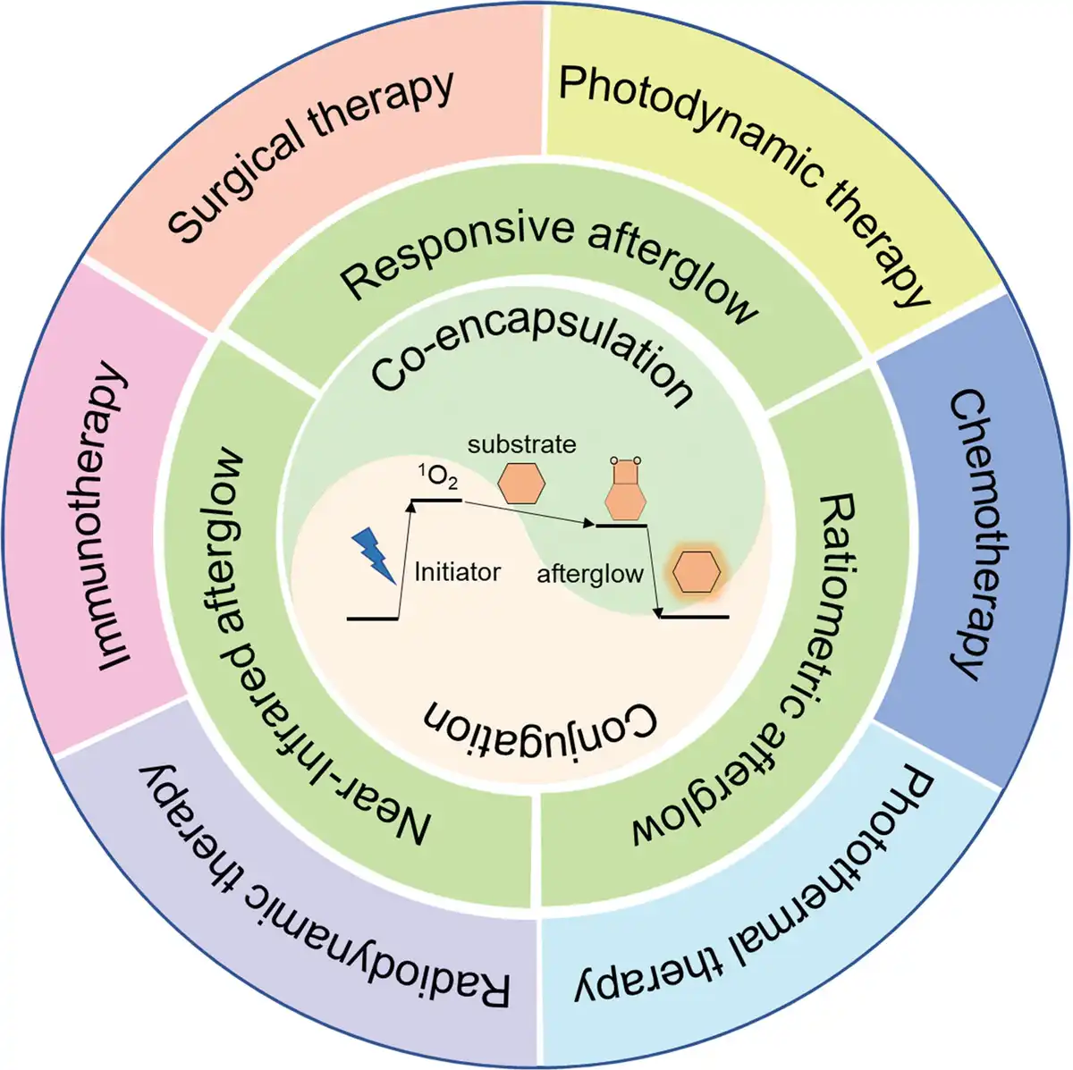
Currently, the luminescence mechanism of OALPs used in in vivo imaging can be summarized as follows. Under the mediation of singlet oxygen (1O2), radiation energy is stored in the afterglow substrate, forming the high-energy and unstable 1,2-dioxetane structure. Subsequently, the 1,2-dioxetane gradually decomposes and transfers the energy released from the bond cleavage within or between molecules. Finally, the energy receptor with fluorescence properties releases the energy in the form of photons, known as “afterglow.” According to the luminescence mechanism of OALPs, 3 components are required: (1) afterglow initiator, responsible for absorbing and converting irradiation energy into 1O2; (2) afterglow substrate, responsible for reacting with 1O2 to form the high-energy and unstable afterglow intermediate (1,2-dioxetane); (3) fluorophore acts as the afterglow relay unit, responsible for accepting energy from 1,2-dioxetane and gradually releasing it in the form of photons (Fig. 1). Based on these 3 components summarized for OALPs, we can use them to guide the design of OALPs.
Figure 1.
Scheme illustrating the design strategy and luminescent mechanism of OALPs for cancer theranostics.
3. Design strategy
3.1 Coencapsulation
The coencapsulation strategy involves encapsulating 2 or more molecules together using self-assembly of amphiphilic compounds. This strategy offers high flexibility, allowing for easy adjustment of the proportions between molecules to achieve synergistic effects. Applying this strategy to the development of OALPs enables the easy concentration of the 3 components: afterglow initiator, afterglow substrate, and afterglow relay unit through π–π interactions, facilitating chemical reactions and energy transfer processes between them.
3.1.1 Near-Infrared OALPs
Near-infrared (NIR) imaging is a noninvasive imaging technique known for its strong tissue penetration and high imaging resolution.[25,26] Combining afterglow imaging with NIR imaging can further enhance imaging effectiveness, enabling more accurate and sensitive detection of biological tissues. However, the variety of OALPs and their afterglow spectra is quite limited, which hampers their widespread application in biomedical imaging. To address this issue, Jiang et al proposed an innovative strategy for converting common fluorophores into afterglow luminescent nanoparticles (Fig. 2A).[27] The advent of this universal strategy allows for the utilization of nearly all existing fluorophores to obtain afterglow probes of different wavelengths, greatly enriching the material resources for afterglow imaging. This strategy involves coencapsulating the afterglow initiator, afterglow substrate, and afterglow relay unit within the same nanoparticle, achieving precise control over the luminescence of afterglow probes. By adjusting the doping ratio of each component within the nanoparticles, the emission wavelength of afterglow can be finely tuned, covering the entire spectrum from visible light to the NIR region. Additionally, Ni et al coencapsulated aggregation-induced emission (AIE) luminogens and afterglow substrate within the same nanoparticle, leading to the development of NIR-emitting afterglow AIE dots (Fig. 2B).[28] Leveraging the high fluorescence quantum yield and singlet oxygen generation efficiency of AIE luminogens in confined spaces (inside nanoparticles), the luminescence brightness and afterglow time of NIR-emitting afterglow AIE dots are significantly higher than those of pure afterglow substrate yellow-green emitting dots. This further confirms the importance of the AIE effect in enhancing afterglow imaging.
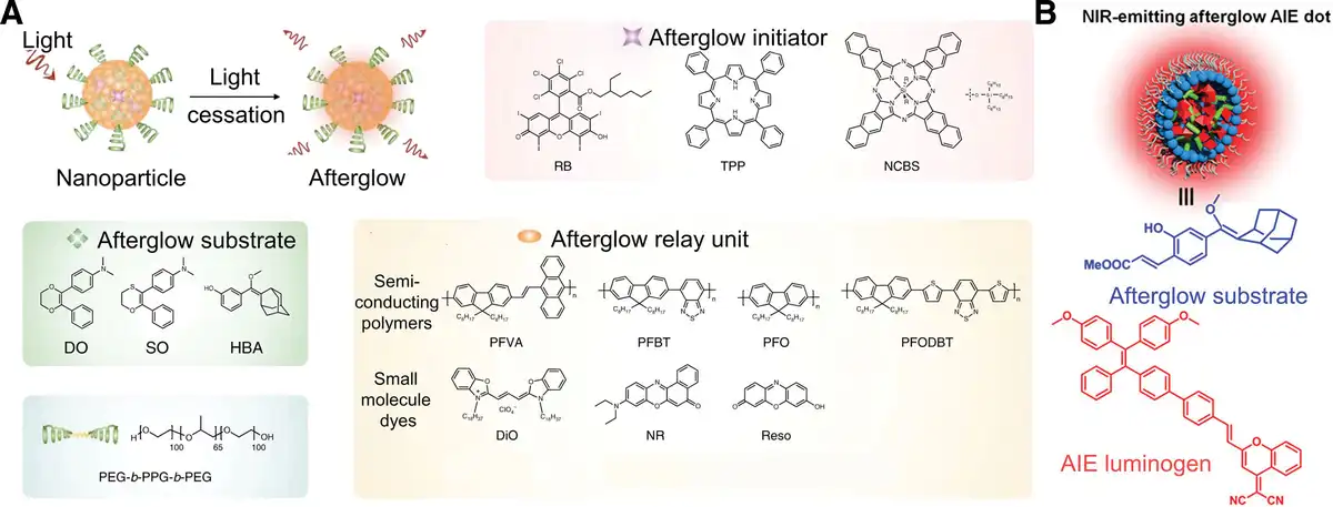
Figure 2.
Design strategy of coencapsulation of near-infrared OALPs. (A) Scheme illustrating the composition of afterglow luminescence nanoparticle (ALNP) and its chemical structure. Adapted with permission from Jiang et al.[27] (B) The chemical structural formulas of afterglow substrate and AIE luminogens. Adapted with permission from Ni et al.[28]
3.1.2 Responsive OALPs
The previously mentioned OALPs do not rely on biomarkers and their afterglow luminescence often remains in an undistinguished “always on” state, which heavily relies on the concentration difference between the lesion site and normal tissues during disease imaging, leading to poor specificity.[29,30] Responsive OALPs refer to afterglow luminescence regulated by biomarkers, aiming to further enhance signal specificity and diagnostic accuracy. Wu et al reported an OALP responsive to the biomarker hydrogen sulfide (H2S), which exhibits specific afterglow luminescence only under the influence of H2S (Fig. 3A).[31] First, the authors carefully designed F12+ as a chromophore responsive to H2S, ensuring that its spectrum extensively overlaps with that of afterglow nanoparticle (ANP) composed of the afterglow substrate MEH-PPV and the afterglow initiator NIR775 in the “always on” state. After incorporating F12+ into the nanoparticles, the fluorescence and singlet oxygen generation abilities of MEH-PPV and NIR775 are both quenched by F12+, attributed to the effective fluorescence resonance energy transfer (FRET) from MEH-PPV and NIR775 to F12+. In the presence of H2S, F12+ rapidly reverts to F2 with a significantly blue-shifted absorption, eliminating the FRET process and allowing F2-ANP to regain singlet oxygen production and fluorescence under light irradiation, thereby activating the afterglow luminescence. Additionally, Xu et al developed an activatable sonoafterglow nanoparticle (SNAP-M), which only activates its sonoafterglow luminescence in the presence of the specific disease biomarker (Fig. 3B).[32] The authors first prepared sonoafterglow nanoparticles, comprising a sonosensitizer NCBS, which acts as an afterglow initiator to generate 1O2 and an afterglow substrate Pro-DPAs with a peroxynitrite (ONOO−)-responsive cage moiety. In the presence of ONOO−, the responsive moiety on Pro-DPAs is decaged, activating the afterglow luminescence capability as the afterglow substrate. Due to the greater penetration of energy generated by ultrasound, it exhibits a brighter and deeper emission compared to previously reported light-induced afterglow luminescence, even reaching tissue depths of up to 4 cm. The innovation brought by sonoafterglow imaging technology not only lies in the brightness and depth of the signal but also provides a potential solution for a wider range of biomedical applications. Building upon the previous research on NIR-emitting afterglow AIE dots, Chen et al further designed and synthesized a dual-responsive OALPs sensitive to both ONOO− and pH.[33] In the presence of ONOO− at physiological pH, it exhibits activated near-infrared afterglow luminescence, with its intensity and lasting time significantly enhanced by introducing the AIE effect.
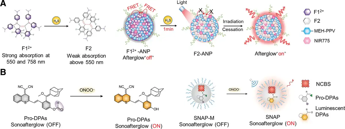
Figure 3.
Design strategy of coencapsulation of responsive OALPs. (A) The chemical structures of the F12+ before and after reacting to H2S to form F2, accompanied by a scheme illustrating the composition of F12+-ANP in the off state and F2-ANP in the on state. Adapted with permission from Wu et al.[31] (B) The chemical structures of the Pro-DPAs before and after reacting to ONOO− to form DPAs, accompanied by a scheme illustrating the composition of SNAP-M in the off state and SNAP in the on state. Adapted with permission from Xu et al.[32]
3.1.3 Ratiometric OALPs
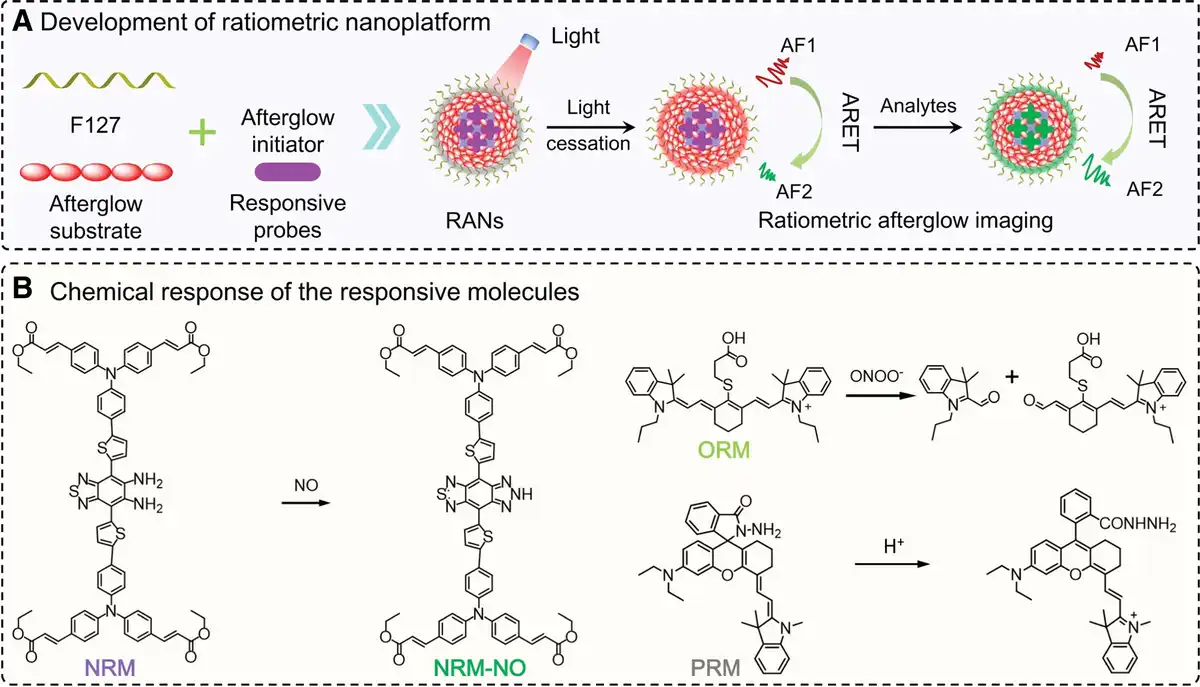
Although responsive OALPs can be selectively activated for specific biomarkers and quantitatively analyzed, the increasing decay of afterglow intensity over time adds complexity to quantitative analysis.[2] Additionally, the intensity of afterglow is influenced by factors such as laser power, irradiation time, and exposure time, further complicating the system.[34] Liu et al has reported a ratiometric afterglow nanoplatform (RAN) that can be used to tailor various responsive OALPs and precisely quantify specific biomarker analytes (Fig. 4).[35] The authors encapsulated responsive molecules (NRM, ORM, or PRM), afterglow substrates, and afterglow initiators within the same nanoparticle. Following light irradiation, the afterglow initiator generates singlet oxygen, prompting the release of shorter-wavelength afterglow luminescence (AF2) from the afterglow substrate, which is then gradually transferred to responsive molecules emitting longer-wavelength afterglow luminescence (AF1) via afterglow resonance energy transfer (ARET). Consequently, quantitative detection of biomarkers can be achieved by calculating the ratio of afterglow intensity (AF1/AF2) between the afterglow energy donor (afterglow substrate) and energy acceptor (responsive molecules). The ratios of afterglow intensities would be independent of interference factors other than analytes, which not only address the decay of afterglow intensity but also eliminate interference from various factors (such as laser power, irradiation time, and exposure time), thereby facilitating reliable quantitative analysis in biological systems.
3.2 Conjugation
In addition to the coencapsulation strategy, there is also an integrated conjugation strategy. Unlike the coencapsulation strategy that requires intermolecular resonance energy transfer, the conjugation strategy concentrates all functional units (including units for 1O2 generation units, 1O2 capturing units, and luminescent units) within a single molecule, utilizing intramolecular energy transfer for luminescence. Thus, this strategy of molecular OALPs eliminates the need for spectral overlap in coencapsulation and enhances system reliability.
3.2.1 Near-Infrared OALPs
The design strategies for near-infrared luminescent moieties primarily fall into 2 categories: (1) extending conjugated structures and (2) modulating the push-pull electronic capabilities of donor and acceptor groups. Therefore, conjugation can be utilized to extend afterglow substrates into the near-infrared region. However, not all afterglow substrates extended into the near-infrared region possess the capability to emit afterglow luminescence and also need to have the ability to generate 1O2. Yang et al utilized the characteristics of porphyrin derivatives, which possess both near-infrared emission wavelengths and excellent 1O2 generation capabilities (Fig. 5A).[36] They chemically conjugated the afterglow substrate (DO) with the NIR photosensitizer tetraphenylporphyrin (TPP), forming TPP-DO. When light irradiation, TPP-DO generates 1O2, oxidizing itself to an unstable TPP-DO-dioxetane intermediate. This intermediate spontaneously decomposes into the ester derivative TPP-DE, while simultaneously absorbing the energy released during the decomposition process to form the excited state TPP-DE*. TPP-DE* ultimately releases the absorbed energy in the form of NIR light as it returns to the ground state TPP-DE. This intramolecular energy transfer from the afterglow substrate to the energy release unit significantly enhances the afterglow brightness compared to intermolecular energy transfer. Semiconductor polymers comprise an extended π-conjugated structure. Recently, it has been discovered that poly(p-phenylenevinylene) (PPV)-based semiconductor polymers are also excellent afterglow substrates.[37] However, they can only emit visible light and have moderate 1O2 generation capability. Cui et al copolymerized the NIR photosensitizer TPP into the PPV to obtain PPV-TPP.[38] In comparison to PPV, PPV-TPP not only exhibits NIR afterglow luminescence but also possesses amplified afterglow intensity. Liao et al reported a novel NIR semiconductor polymer (named NIR-3) containing a donor–acceptor conjugated structure capable of producing intense afterglow luminescence in the NIR window (Fig. 5B).[39] NIR-3’s backbone structure contains abundant thiophene as the donor and introduces pyrido pyrazine as the acceptor. This donor–acceptor structure not only shifts the emission wavelength to the red but also enhances the intersystem crossing process, thus increasing the production of 1O2. Additionally, a large number of 1O2-reactive thiophene groups can effectively form unstable high-energy thiophene-dioxetane intermediates, followed by peroxide (O–O) bond cleavage to release energy, which is absorbed by NIR-3 itself to form excited state NIR-3* for afterglow luminescence. The integrated conjugation strategy eliminates the need for complex component modulation, allowing a single molecule to fulfill all the requirements for afterglow luminescence.
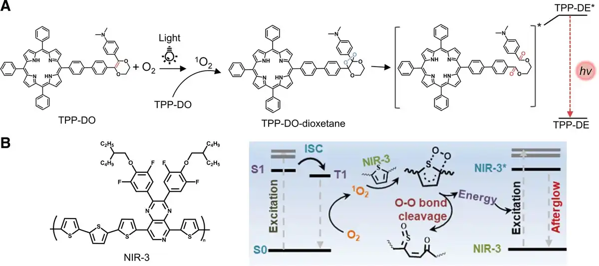
Figure 5.
Design strategy of conjugation of near-infrared OALPs. (A) The chemical structures of TPP-DO and proposed mechanism for the afterglow luminescence of TPP-DO. Adapted with permission from Yang et al.[36] (B) The chemical structures of NIR-3 and proposed mechanism for the afterglow luminescence of NIR-3. Adapted with permission from Liao et al.[39]
3.2.2 Responsive OALPs
The design strategies for responsive molecular OALPs primarily fall into 2 categories: (1) destruction and restoration of conjugated skeletons; (2) enhancement and inhibition of intramolecular charge transfer (ICT). Lei et al integrated responsive units, 1O2 generation units, 1O2 capturing units, and luminescent units into a single molecule (Fig. 6A).[40] When responsive units encounter H+, the probe undergoes a conformational change from a spirocyclic to an open structure, transitioning the molecular structure from nonconjugated to conjugated, ultimately leading to the activation of 1O2 and fluorescence. When light irradiates the open structure, the generated 1O2 oxidizes the 1O2 capturing unit to form unstable high-energy dioxetane intermediates. These intermediates gradually decompose and release excitation energy to the decomposed molecules themselves, resulting in afterglow luminescence. Additionally, the researchers have expanded the application scope of this afterglow molecular scaffold by introducing various biomarkers (ATP or metal ions) responsive units. The same research group also designed hemicyanine-based molecular scaffolds with responsive afterglow luminescence.[41] They similarly employed the “four-in-one” molecular design strategy, integrating responsive units, 1O2 generation units, 1O2 capturing units, and luminescent units into a single molecule, customizing it for the quantification and imaging of targets such as pH, superoxide anions, and aminopeptidase. The responsive units can modulate the activation of fluorescence and the generation of 1O2 by altering the ICT of the entire molecule, thus enabling activatable afterglow luminescence. The issue of tissue penetration depth during excitation can be addressed by ultrasound and similarly, X-rays can also overcome tissue penetration challenges.[42] However, organic molecules often contain light atoms (such as hydrogen, carbon, and oxygen) and exhibit weak spin-orbit coupling, resulting in weak X-ray absorption. Huang et al integrated responsive units, 1O2 generation units, 1O2 capturing units, and luminescent units into a single molecule, to prepare a molecular radioafterglow dynamic probe (MRAP) for radioafterglow luminescence (Fig. 6B).[43] They replaced hydrogen atoms in the benzene ring with heavy iodine atoms to form a new conjugated structure. The introduction of iodine atoms not only enhances the probe’s X-ray absorption but also allows a large number of electrons to exist as triplet excitons (Tn). In the absence of cathepsin B (CatB) response, the reduced electron-donating ability of the oxygen atom in the phenolic moiety inhibits the ICT, resulting in negligible radioafterglow luminescence and radiodynamic 1O2 generation. Upon encountering CatB, the responsive moiety is cleaved to liberate uncaged MRAP (IDPASU), restoring fluorescence and radiodynamic 1O2 generation. The specific process involves X-ray photons first exciting high-energy electrons, which then interact with surrounding atoms, initiating a secondary electron cascade with decreased energy. Upon absorption by IDPASU, a large number of electrons exist as triplet excitons, transitioning back to the ground state and exciting triplet oxygen (3O2) to 1O2. The in situ generated 1O2 reacts with IDPAs to form dioxetane intermediates. Eventually, these intermediates gradually decompose into the corresponding activated products (activated IDPAx), emitting radioafterglow luminescence.
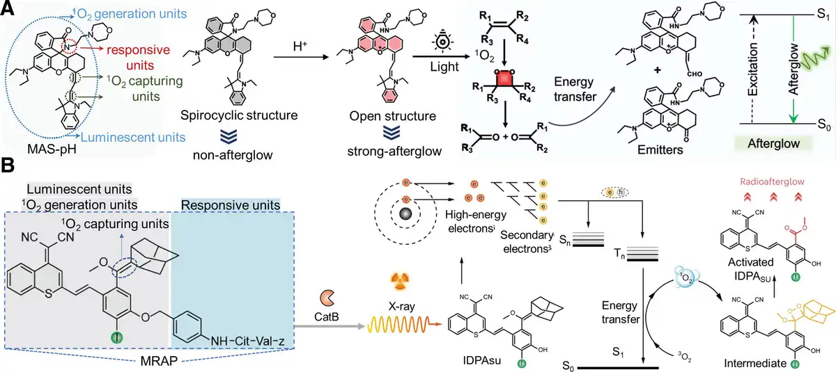
Figure 6.
Design strategy of conjugation of responsive OALPs. (A) The chemical structures of MAS-pH before and after responding to H+, accompanied by the proposed mechanism for the afterglow luminescence of the responsive molecules. Adapted with permission from Lei et al.[40] (B) The chemical structures of MRAP before and after responding to CatB to form IDPAsu, accompanied by the proposed mechanism for the radioafterglow luminescence of IDPAsu. Adapted with permission from Huang et al.[40]
The afterglow imaging offers higher SBR and sensitivity in vivo compared to fluorescence imaging. However, it requires several seconds to minutes for data acquisition, whereas fluorescence imaging only takes a few hundred microseconds. To overcome this limitation, further enhancement of the brightness of OALPs is necessary. Currently, the screening of high-brightness OALPs designed using a coencapsulation strategy still primarily relies on the random combination of different components, which is relatively complex. To address this challenge, we believe it is essential not only to consider the individual energy conversion efficiencies of the afterglow initiator, afterglow substrate, and afterglow relay unit but also the energy transfer efficiencies among these components. For example, to more effectively transfer the 1O2 generated by the afterglow initiator to the afterglow substrate, it is required that the afterglow substrate has a higher rate of producing 1,2-dioxetane intermediate when reacting with 1O2. The high oxidizability of the afterglow substrate is apparently beneficial to this process. Additionally, to enhance the transfer of emitted light energy from the afterglow substrate to the afterglow relay unit, improving the resonance energy transfer efficiency between them is crucial. This might require better spectral overlap between the emission spectrum of the afterglow substrate and the absorption spectrum of the afterglow relay unit. Therefore, optimizing both the individual properties and the interactions among the components is vital for advancing the brightness of OALPs.
For the OALPs designed using the conjugation strategy, although the intramolecular energy transfer from the afterglow substrate to the energy release unit significantly enhances afterglow brightness compared to intermolecular energy transfer, such probes still lack sufficient brightness. To address this challenge, we propose focusing on the relationship between the molecular structure of conjugated probes and their afterglow intensity, continuously improving their brightness.
For near-infrared OALPs, molecular imaging in the second near-infrared (NIR-II, 1000–1700 nm) window has already provided high-resolution imaging with subcentimeter tissue depth. In the future, utilizing OALPs emitting in the NIR-II window could further enhance penetration depth and imaging resolution. However, existing energy donors often emit at shorter wavelengths, requiring multiple energy transfers to successfully shift their emission to the NIR-II region, resulting in significant energy loss. To address this challenge, we propose chemically modifying the energy donors to directly extend their conjugation length, thus extending their emission wavelength to the NIR-II window.
Responsive OALPs enable quantitative analysis of target analytes. However, the presence of various interference factors in afterglow luminescence results in decreased signal reliability. To address this challenge, the design of ratiometric OALPs enables reliable quantification and molecular imaging of specific analytes. This approach mitigates interference from factors such as laser parameters, penetration depth, and probe concentration, significantly enhancing the reliability of afterglow signals.
The primary challenges in the design of ratiometric OALPs is the lack of responsive probes for constructing ratiometric OALPs. This is attributed to the inert nature of most probe structures, lacking reactive sites. Previously reported ratio afterglow probes have been limited to detecting reactive oxygen and nitrogen species or pH levels. However, many physiological and pathological processes involve the excessive production of these biomarkers, rendering precise and specific disease monitoring highly challenging. Therefore, there is still a need for the development of more specific responsive molecular probes, which remains to be explored.
4. Application in cancer theranostics
Cancer, as a malignant tumor, has emerged as a significant cause of human mortality, posing a grave threat to human health.[44] Consequently, considerable importance is placed on researching cancer theranostics, where targeted delivery to tumor sites using probes plays a crucial role.[45] The targeting of tumors can be classified into 2 categories. (1) Active targeting involves the use of specific targeting molecules or carriers to bind with tumor cells, enabling precise diagnosis and therapy.[46] (2) Passive targeting, on the other hand, capitalizes on physiological differences between tumor and normal tissues, such as the enhanced permeability and retention (EPR) effect, to facilitate the accumulation of drugs or diagnostic agents within tumor tissues.[47] The advantages of cancer theranostics lie in assisting in formulating personalized therapy plans, monitoring therapy efficacy, reducing delays and redundant examinations, and achieving personalized cancer therapy.[48,49]
Near-infrared emission wavelength is an ideal wavelength for biological imaging due to its low tissue absorption and scattering, as well as strong tissue penetration. Therefore, it is suitable for cancer theranostics, such as image-guided surgical therapy and monitoring the effects of photodynamic therapy, photothermal therapy, and radiodynamic therapy (Table 1). Responsive OALPs can respond to specific biomolecules or environmental conditions, such as granzyme B, peroxynitrite (ONOO−), and pH value. This responsiveness allows OALPs to provide specific diagnostic information in cancer treatment, help evaluate the effectiveness of chemotherapy and immunotherapy, and track the release of chemotherapy drugs. Ratiometric OALPs, based on responsive OALPs, address the issue of afterglow intensity attenuation over time and eliminate interference from various factors (such as laser power, irradiation time, and exposure time), making them more suitable for reliable quantitative analysis in biological systems.
Table 1
Properties and therapy pathways of designed OALPs.
| OALPs | Design strategy | λem* | Brightness† | Therapy pathways | Reference |
|---|---|---|---|---|---|
| AGL-AIE dots | Coencapsulation/near-infrared | 650 nm | 2.0 × 10⁶ | Surgical therapy | [28] |
| F12+-ANP-Gal | Coencapsulation/response/near-infrared | 780 nm | 3.0 × 10⁵ | Surgical therapy | [31] |
| SCAN | Coencapsulation/response | 500 nm | 3.0 × 10⁷ | Immunotherapy | [32] |
| RAN1 | Coencapsulation/ratiometer | AF1 = 600 nm AF2 = 830 nm | 4.0 × 10⁵ | Immunotherapy | [35] |
| Q-TPP-DO | Conjugation/near-infrared | 670 nm | 1.8 × 10⁸ | Surgical therapy | [36] |
| NIR-3 | Conjugation/near-infrared | 800–820 nm | 1.2 × 10⁶ | Photodynamic therapy | [39] |
| MRAP-cRGD | Conjugation/response/near-infrared | 770 nm | 1.3 × 10⁶ | Radiodynamic therapy | [43] |
| MAS-pH | Conjugation/response/near-infrared | 690–770 nm | 1.0 × 10⁶ | Chemotherapy | [40] |
| SPNCT | Coencapsulation | 600 nm | 1.4 × 10⁵ | Photothermal therapy | [50] |
| PA-AGL NPs | Coencapsulation/response | 625 nm | 1.2 × 10⁹ | Immunotherapy | [33] |
| PFODBT@CPPO | Coencapsulation/near-infrared | 690–770 nm | 1.0 × 10⁷ | Photodynamic therapy | [51] |
| TD-Grz-BHQ | Coencapsulation/response | 630 nm | 7.0 × 10⁷ | Immunotherapy | [52] |
| AIE/B-AGL-HCPT NPs | Coencapsulation/response/near-infrared | 660 nm | 2.0 × 10⁶ | Chemotherapy | [53] |
| APtN | Coencapsulation/response | 550 nm | 2.0 × 10⁵ | Chemotherapy | [54] |
SBR refers to the tumor to the paratumoral tissue unless specified.
*
λem is the highest afterglow emission wavelength.
†
The unit of afterglow brightness is p s-1 cm-2 sr-1.
4.1 Surgical therapy
The use of NIR fluorescence imaging for guiding cancer surgical therapy has emerged as a powerful strategy in clinical practice, aiding surgeons in faster and more accurate tumor resections.[55] Indocyanine green, approved by the Food and Drug Administration, has been explored for this application.[56] The significant advantage of NIR fluorescence, particularly its much lower tissue background noise, undoubtedly makes it a more ideal modality for guiding tumor resections during surgical therapy. Ni et al achieved precise surgical removal of abdominal metastatic tumors using the aforementioned NIR-emitting afterglow AIE dot (Fig. 7A).[28] Utilizing NIR-emitting afterglow AIE dots, it demonstrates effective passive tumor-targeting capability, illuminating nearly all tumor nodules, particularly submillimeter ones (Fig. 7B). Additionally, leveraging afterglow imaging enables minimal background interference. Subsequently, surgical therapy was performed to remove the tumors by surgeons who were unaware of the images and relied on their experience. A large number of tumors, most of which were >1 mm, were excised after unguided surgical therapy (Fig. 7C). This is because submillimeter tumors are in the early stages of cancer and are indeed difficult for surgeons to detect. In the second surgical therapy guided by afterglow imaging, the surgeon almost excised all tiny tumor nodules until there was no residual afterglow signal in the abdominal cavity. This demonstrates the excellent performance of NIR-emitting afterglow AIE dots in image-guided cancer surgical therapy. Wu et al reported a novel organic afterglow probe called F12+-ANP-Gal, which can be used for the localization of liver cancer during surgery.[31] In the study, F12+-ANP-Gal probe was used, and intense afterglow signals were generated through irradiation to accurately locate liver cancer tissue. This organic afterglow probe exhibits high sensitivity and noninvasiveness, allowing real-time imaging of liver cancer tissue and providing precise localization information for surgery. Additionally, the study demonstrated the ability of this organic afterglow probe to combine with tissue staining techniques, enabling precise delineation of the edges of liver cancer tissue. In conclusion, the organic afterglow probe F12+-ANP-Gal holds great potential as a valuable tool for guiding liver cancer surgery. Xie et al developed an amphiphilic poly(p-phenylenevinylene) derivatives that can self-assemble into the nanoagent (SPPVN) in biological solutions and emit near-infrared afterglow luminescence.[57] Compared to other reported near-infrared fluorescence imaging probes, SPPVN demonstrates superior sensitivity and is capable of detecting tumors with much smaller volumes. It can rapidly detect xenograft tumors as small as 1 mm3 and even tiny peritoneal metastatic tumors that are almost invisible to the naked eye.
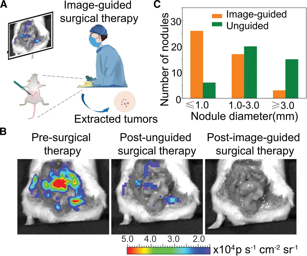
Figure 7.
OALPs for surgical therapy. (A) Scheme illustrating image-guided surgical therapy. (B) Afterglow imaging of the opened peritoneal cavity at various stages of surgical therapy in tumor-bearing mice, following intravenous injection of NIR-emitting afterglow AIE dots. (C) The size distribution of tumor nodules resected from the mouse in (B) after the first unguided surgical therapy and the second afterglow image-guided surgical therapy. Adapted with permission from Ni et al.[28]
4.2 Photodynamic therapy
When irradiated by light, the photosensitizer can generate reactive oxygen species (ROS) that possess cytotoxicity and can kill cancer cells, thereby achieving therapeutic effects through a process known as photodynamic therapy (PDT).[58] Due to its highly spatial and temporal control over the therapy process, PDT has been employed in various cancer therapy.[59] However, the unknown ROS dosage during cancer therapy may induce the uncertainty of anticancer efficacy.[60] Liao et al constructed afterglow nanoreporters using the semiconducting polymer NIR-3, for precise and noninvasive early monitoring of the effectiveness of PDT (Fig. 8A).[39] As the NIR-3’s afterglow luminescence is dependent on 1O2, the afterglow intensity correlates closely with the tumor inhibition rates. Experimental results demonstrate that with increasing injection dosage, mice exhibit enhanced afterglow luminescence in the tumor area (Fig. 8B). As the injection dosage of NIR-3 increases, the tumor inhibition rate also significantly increases and the afterglow intensity in the tumor region correlates with the tumor inhibition rate (Fig. 8C). The afterglow luminescence can be utilized as an early-stage therapy metric prior to tumor volume reduction, measuring the efficacy of 1O2-mediated anticancer activity. Wang et al developed a cyclic amplification of the afterglow luminescent nanoreporter called PFODBT@CPPO, which can be used to real-time visualize light-induced ROS and monitor ROS-mediated cancer treatment efficacy.[51] The afterglow intensity of the nanoreporter can also be utilized to adjust treatment parameters such as dosage and irradiation time, enabling personalized treatment plans and avoiding ineffective or excessive medical interventions.
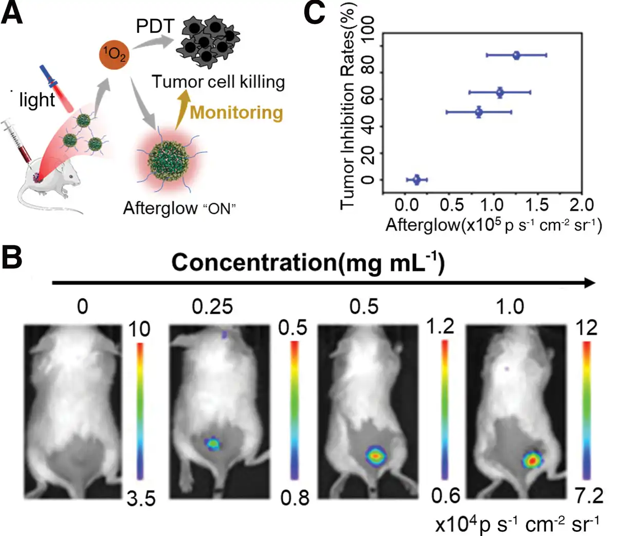
Figure 8.
OALPs for photodynamic therapy. (A) Scheme illustrating afterglow imaging monitoring photodynamic therapy. (B) Afterglow images of mice with subcutaneous tumors injected intratumorally with different concentrations of NIR-3. (C) Quantification of in vivo afterglow intensities of tumor as a function of tumor growth inhibition rates. Adapted with permission from Liao et al.[39]
4.3 Photothermal therapy
Photothermal therapy (PTT) effectively utilizes photothermal agents to convert light energy into heat, thereby killing cancer cells.[61] Zhen et al coencapsulated OALPs and photothermal agents to prepare nanococktail. Following intravenous administration, nanococktail passively targets and accumulates in tumors (Fig. 9A).[50] Subsequently, nanococktail can absorb light, converting light energy into heat to achieve PTT. During PTT, as the decomposition of 1,2-dioxetane is temperature-dependent, the afterglow intensity of nanococktail also exhibits temperature dependence, enabling monitoring of PTT via afterglow imaging (Fig. 9B). Furthermore, due to the high tissue penetration and imaging sensitivity of afterglow luminescence, it holds potential for monitoring deep-tissue temperatures.
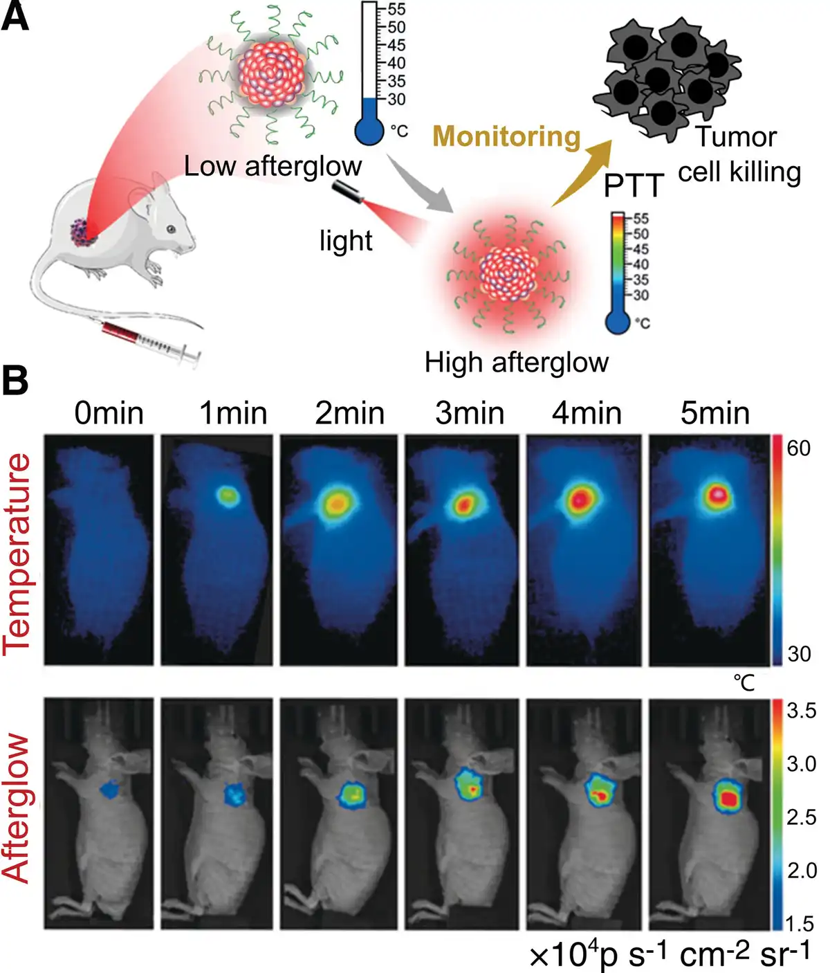
Figure 9.
OALPs for photothermal therapy. (A) Scheme illustrating afterglow imaging monitoring photothermal therapy. (B) Thermal and afterglow imaging of mice with subcutaneous tumors after intravenous injection of nanococktail, undergoing light irradiation for various durations. Adapted with permission from Zhen et al.[50]
4.4 Radiodynamic therapy
Compared to light, X-rays have greater tissue penetration capability, making them more suitable for deep-tissue diagnostic imaging and cancer therapy.[62] Inspired by PDT, X-rays can be utilized as an energy source to achieve radiodynamic therapy (RDT).[63] Due to the localized production of cytotoxic ROS by the therapeutic agent, RDT can minimize radiation damage to normal tissues. Huang et al utilized the previously mentioned MRAP and modified it with the tumor-targeting moiety cRGD to obtain MRAP-cRGD, which was successfully employed in brain tumor radiodynamic theranostics.[43] MRAP has both tumor-activatable afterglow luminescence and the ability to produce 1O2 (Fig. 10A). Under low-dose X-ray irradiation of 5 mGy, radioafterglow imaging (RAI) was performed, showing that the radioafterglow in orthotopic glioblastoma gradually increased, reaching its peak at 24 hours, with significantly higher afterglow intensity compared to other groups (Fig. 10B, C). Subsequently, tumor growth was continuously monitored via bioluminescence signal. The results revealed that following high-dose 0.8 Gy RDT, tumor growth was completely inhibited in the MRAP group, whereas other groups did not exhibit significant tumor regression or inhibition (Fig. 10D).
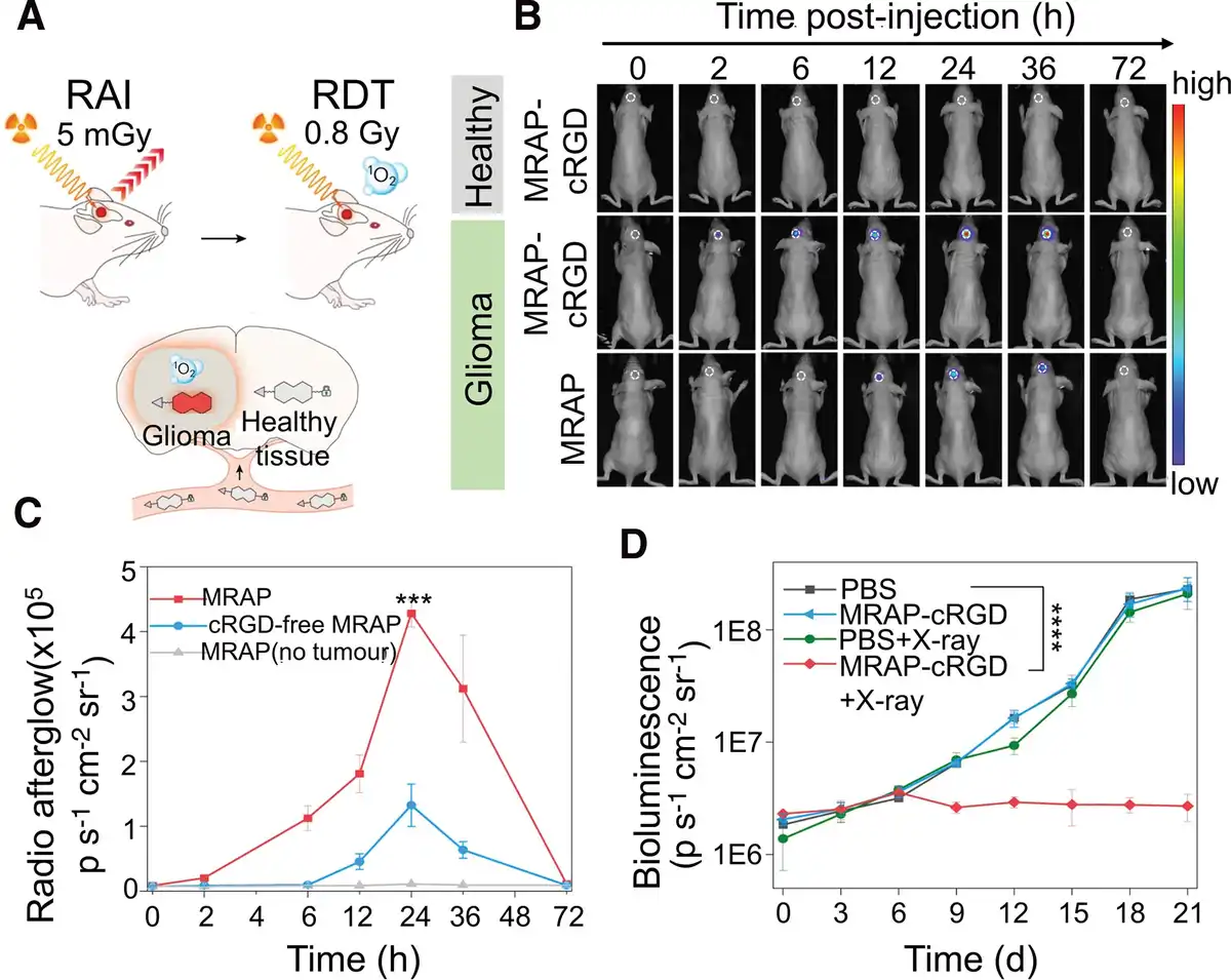
Figure 10.
OALPs for radiodynamic therapy. (A) Scheme illustrating RAI monitoring RDT. (B) Radioafterglow images of orthotopic glioblastoma (glioma) and healthy mice at different time points after an intravenous injection of MRAP-cRGD and MRAP. (C) The quantitative analysis of radioafterglow intensities in the mouse brain region is presented in (B). (D) Tumor growth of orthotopic glioblastoma mice after the indicated therapies. Adapted with permission from Huang et al.[43]
4.5 Chemotherapy
Chemotherapy is a method of cancer therapy by using specific drugs to kill or control the growth of cancer cells, thereby helping patients alleviate symptoms, control the disease, and extend survival.[64] However, chemotherapy drugs not only affect tumors but may also impact normal tissues. In order to mitigate damage to normal tissues, a series of prodrugs have been developed for cancer therapy, which are inactive until they reach the tumor microenvironment.[65] Upon response to biomarkers in the tumor microenvironment, they release active drugs, thus partially avoiding toxicity to normal tissues. He et al developed an afterglow protheranostic nanoassembly by coencapsulating an afterglow prodrug and an afterglow initiator.[54] The afterglow prodrug is comprised of the chemotherapy drug 5’-deoxy-5-fluorouridine (5DFUR) linked to an afterglow substrate via a boronic ester bond responsive to H2O2. Once the nanoassembly reaches the tumor microenvironment, it reacts with overexpressed H2O2, releasing 5DFUR and simultaneously activates afterglow luminescence (Fig. 11A). Hence, there is a good linear correlation (R2 = 0.9857) between the afterglow luminescence intensity and the release of chemotherapy drugs (Fig. 11B), enabling real-time tracking of drug activation and release in vivo, which holds promise for guiding physicians in formulating drug regimens. Gao et al also reported an activatable NIR afterglow protheranostic nanoassembly by coencapsulating afterglow prodrugs and NIR emissive AIE luminogens.[53] The afterglow prodrug consists of chemotherapy drug hydroxycamptothecin (HCPT) and an afterglow substrate linked through the carbonate group, which is caged by an ONOO−-responsive phenylborate group. The AIE luminogens serve as both the afterglow initiator and afterglow relay unit. The activatable NIR afterglow luminescence serves as the signal readout to monitor and quantify drug release status and theranostic response. Lei et al developed a pH-responsive afterglow probe called MAS-pH, which can be used to monitor the effects of the oxidative phosphorylation inhibitor tamoxifen on tumor glycolysis and chemotherapy outcomes in living mice.[40] The experimental results demonstrate that increasing the dose of tamoxifen leads to an increase in the afterglow signal, a decrease in tumor pH, an elevation in glycolytic levels, and an enhancement in chemotherapy outcomes. Therefore, MAS-pH shows great potential in evaluating tumor glycolysis, monitoring chemotherapy resistance, and screening effective approaches to regulate glycolysis.
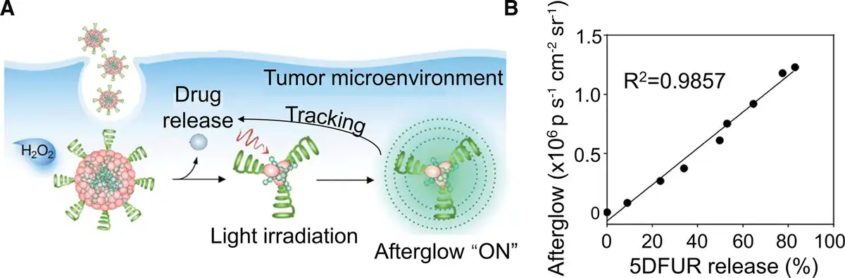
Figure 11.
OALPs for chemotherapy. (A) Scheme illustrating afterglow luminescence tracking drug release status. (B) Correlation between afterglow intensities and the percentage of 5DFUR release. Adapted with permission from He et al.[54]
4.6 Immunotherapy
While immunotherapy holds great promise in cancer therapy, patient responses vary widely across different types of cancer, resulting in significant individual differences.[66] Due to the proven correlation between response rates and therapy outcomes with the proinflammatory tumor microenvironment, there is a critical need for cancer immunotherapeutic agents capable of providing real-time feedback on the proinflammatory tumor microenvironment to guide immunotherapy.[67] Xu et al coencapsulated a M1-like macrophages (M1)-polarizing prodrug caged with a 1O2-cleavable moiety in the responsive sonoafterglow nanoparticles, creating a sonoafterglow cancer nanoimmunotheranostic probe (SCAN).[32] The sonodynamic effect (generation of 1O2) and sonoafterglow are activated only in the proinflammatory tumor microenvironment when high levels of ONOO– are present (Fig. 12A). The generated 1O2 by SCAN triggers the cleavage of the M1-polarizing prodrug, thereby activating immunotherapy in situ. Researchers defined the difference in sonoafterglow intensity between sequential doses as ΔS and investigated its relationship with the proinflammatory state levels of tumor M1 characteristics (Fig. 12B, C). Under the same ultrasound irradiation, SCAN induced a significantly higher number of M1 macrophages compared to the SCANc group (lacking the M1-polarizing prodrug) after the fourth dose (Fig. 12D). The increasing trend of M1 macrophage population was consistent with the sonoafterglow in each group. Tumor suppression experiments revealed that single sonodynamic therapy mediated by SCAN-C failed to eradicate the tumor, whereas the combination of immunotherapy with SCAN resulted in complete tumor eradication (Fig. 12E). These results confirm the efficacy of SCAN-mediated immunotherapy. Chen et al developed a novel organic afterglow probe called PA-AGL NPs, which can be used for fast screening of antitumor drugs capable of inducing immunogenic cell death (ICD).[33] Specifically, preirradiated PA-AGL nanoparticles can visualize the infiltration process of neutrophils by measuring the activated afterglow intensity of ONOO−. The activation of ICD requires the activation of inflammation-related immune cells, particularly neutrophils. ONOO−, as a ROS associated with inflammation, can be generated during the inflammatory process. Therefore, ICD drugs may promote the immunogenic conversion of tumors by activating the inflammatory response and immune cells. Liu et al developed an ultrasound-induced afterglow luminescent probes called RAN1, which enables reliable quantification of nitric oxide (NO) and can be used for afterglow imaging in macrophage-modulated immunotherapy.[35] The researchers first treated RAW264.7 macrophages with different macrophage polarization modulators and then performed afterglow imaging by administering RAN1 to these cells. It was observed that the afterglow intensity was correlated with the degree of macrophage polarization. Furthermore, the researchers observed a strong correlation between afterglow intensity, macrophage polarization, and the anticancer effect in 4T1 tumor-bearing mice. In conclusion, ratiometric afterglow imaging can serve as an effective method for evaluating macrophage polarization and screening modulators, enabling real-time monitoring of macrophage-modulated immunotherapy. The same research group has developed an ultrasound-induced afterglow luminescence nanoprobe called TD-Grz-BHQ, which can be used to monitor the granzyme B levels and the immune response in living mice.[52] The afterglow intensity of TD-Grz-BHQ was detected in CT-26 tumors following anti-PDL1 treatment and exhibited a higher level compared to the signal observed in 4T1 tumors. The higher afterglow intensity of TD-Grz-BHQ in CT-26 tumors following anti-PDL1 treatment can be attributed to the elevated levels of granzyme B, CD8+ T cells, and NK cells. In conclusion, ultrasound-induced afterglow luminescence nanoprobe can be used to monitor the immune response and evaluate the efficacy of immunotherapy.
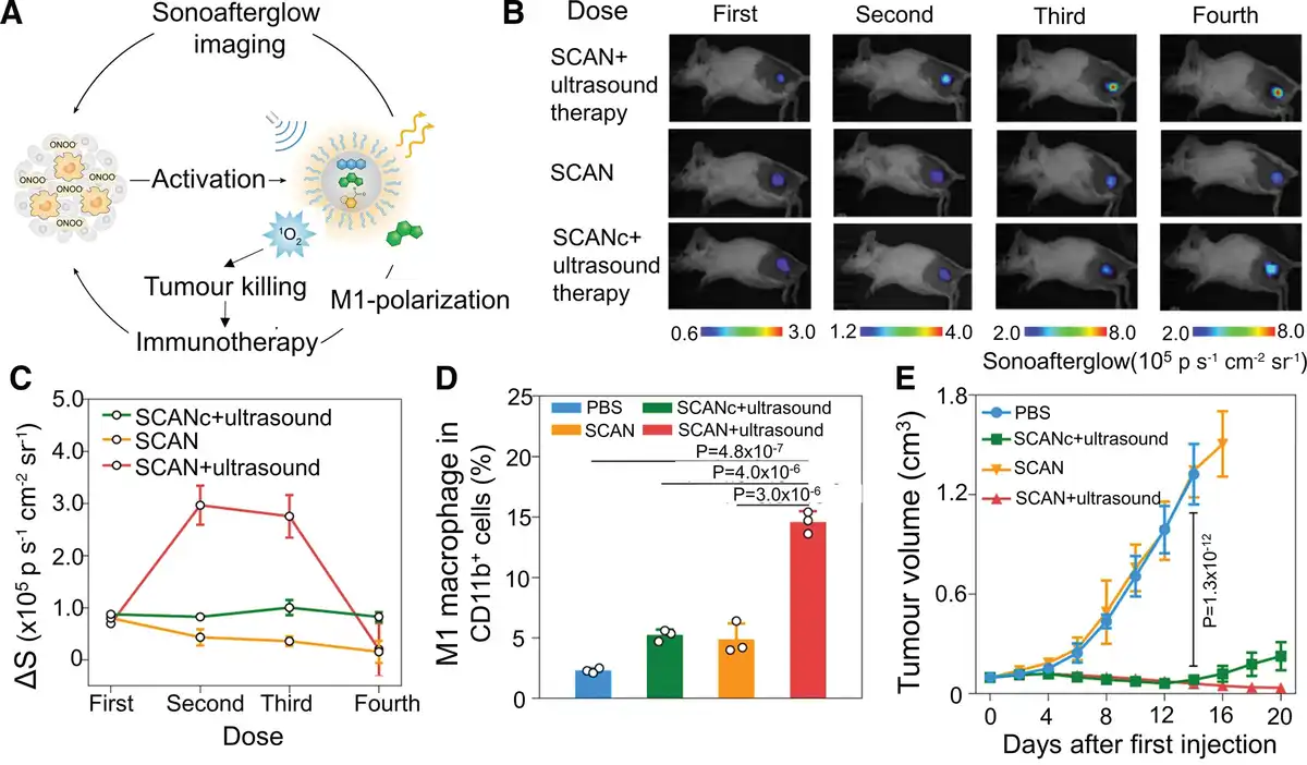
Figure 12.
OALPs for immunotherapy. (A) Scheme illustrating sonoafterglow-guided immunotherapy. (B) Sonoafterglow images of tumor-bearing mice administered in increasing doses after the indicated therapies. (C) Quantification of the difference in afterglow intensity (ΔS) between sequential doses is presented in (B). (D) Quantification of intratumoral M1 macrophages using flow cytometry. (E) Tumor growth of mice after the indicated therapies. Adapted with permission from Xu et al.[32]
5. The biosafety and metabolic pathways
Currently, OALPs used in cancer theranostics often need to be formulated into nanoparticles that integrate different components to function collectively and enhance tumor targeting and bioavailability. Therefore, the biocompatibility and metabolic pathways of these carriers are critical considerations. Organic nanoparticles are typically composed of natural or synthetic materials with high biocompatibility, such as proteins, polysaccharides, and lipids, making them more readily accepted by biological systems in vivo.[52] Moreover, to enhance the safety of carriers, their surfaces are commonly modified with polyethylene glycol, which effectively reduces protein adsorption in vivo, decreases immune system recognition and clearance, and reduces immunogenicity while prolonging circulation time in the bloodstream.[68] Additionally, organic nanoparticles can be tailored to target specific tissues or cells by adjusting their surface chemistry or attaching targeting molecules. This targeting helps to increase the local concentration of the drug, reducing adverse effects on healthy tissues. Regarding metabolic pathways, unlike small molecule probes or drugs, which are easily excreted through urine or metabolized by the liver, nanoparticles, due to the EPR effect, tend to accumulate at tumor sites.[69] However, larger nanoparticles are likely to remain in the reticuloendothelial system (such as the liver and spleen), which may lead to long-term biological accumulation and potential toxicity issues. Therefore, to enhance the metabolism and safety of nanoparticles, it is advisable to develop smaller and more uniformly distributed nanoparticles, as their reduced size facilitates renal excretion.[70]
6. Conclusions and outlook
Despite significant progress, there are considerable challenges that need to be addressed before clinical translation. We identify the following challenges in this field. (1) While X-ray and ultrasound-excited afterglows have resolved the issue of penetration depth, there is still a lack of OALPs emitting in the NIR-II window (1000–1700 nm), which can reduce light scattering in tissues. (2) In tumor hypoxic microenvironments, the efficiency of high oxygen-dependent type I photosensitizers in producing 1O2 is not optimal. Therefore, there is a need to develop OALPs mediated by other ROS, such as those based on type II photosensitizers with lower oxygen consumption. (3) Current afterglow imaging studies mainly focus on cancer research, neglecting other life-threatening or chronic diseases such as coronary heart disease, diabetes, neurological disorders, and autoimmune diseases. (4) There are few reports on ratiometric afterglow. More research efforts should be directed toward improving the quality and reliability of afterglow imaging using this strategy or developing new techniques such as afterglow lifetime imaging to further enhance imaging quality. In conclusion, OALPs offer significant advantages in disease diagnosis and therapy. We hope this review will inspire researchers interested in developing OALPs to collectively advance their application in the biomedical field.
Acknowledgments
This work was supported by the National Natural Science Foundation of China (52225310) and Tianjin Key Medical Discipline (Specialty) Construction Project.
Conflicts of interests
The authors declare that they have no conflicts of interest.
References
- [1] Kenry, Duan Y, Liu B. Recent advances of optical imaging in the second near-infrared window. Adv Mater. 2018;30(47):1802394.
- [2] Li J, Pu K. Development of organic semiconducting materials for deep-tissue optical imaging, phototherapy and photoactivation. Chem Soc Rev. 2019;48(1):38–71.
- [3] Cheng P, Pu K. Molecular imaging and disease theranostics with renal-clearable optical agents. Nat Rev Mater. 2021;6(12):1095–1113.
- [4] Culley S, Albrecht D, Jacobs C, et al. Quantitative mapping and minimization of super-resolution optical imaging artifacts. Nat Methods. 2018;15(4):263–266.
- [5] Cosco ED, Spearman AL, Ramakrishnan S, et al. Shortwave infrared polymethine fluorophores matched to excitation lasers enable non-invasive, multicolour in vivo imaging in real time. Nat Chem. 2020;12(12):1123–1130.
- [6] Yoon S, Kim M, Jang M, et al. Deep optical imaging within complex scattering media. Nat Rev Phys. 2020;2(3):141–158.
- [7] Jiang Y, Pu K. Molecular probes for autofluorescence-free optical imaging. Chem Rev. 2021;121(21):13086–13131.
- [8] Yeh H-W, Ai H-W. Development and applications of bioluminescent and chemiluminescent reporters and biosensors. Annu Rev Anal Chem (Palo Alto Calif). 2019;12:129–150.
- [9] Vacher M, Fdez Galván I, Ding B-W, et al. Chemi-and bioluminescence of cyclic peroxides. Chem Rev. 2018;118(15):6927–6974.
- [10] Yang M, Huang J, Fan J, et al. Chemiluminescence for bioimaging and therapeutics: recent advances and challenges. Chem Soc Rev. 2020;49(19):6800–6815.
- [11] Yan Y, Shi P, Song W, et al. Chemiluminescence and bioluminescence imaging for biosensing and therapy: in vitro and in vivo perspectives. Theranostics. 2019;9(14):4047–4065.
- [12] Syed AJ, Anderson JC. Applications of bioluminescence in biotechnology and beyond. Chem Soc Rev. 2021;50(9):5668–5705.
- [13] Shen H, Liao S, Li Z, et al. Organic afterglow nanoparticles in bioapplications. Chemistry. 2023;29(42):e202301209.
- [14] Wang X, Pu K. Molecular substrates for the construction of afterglow imaging probes in disease diagnosis and treatment. Chem Soc Rev. 2023;52(14):4549–4566.
- [15] Yang X, Waterhouse GI, Lu S, et al. Recent advances in the design of afterglow materials: mechanisms, structural regulation strategies and applications. Chem Soc Rev. 2023;52:8005–8058.
- [16] Wu Z, Midgley AC, Kong D, et al. Organic persistent luminescence imaging for biomedical applications. Mater Today Bio. 2022;17:100481.
- [17] Yang L, Gai S, Ding H, et al. Recent progress in inorganic afterglow materials: mechanisms, persistent luminescent properties, modulating methods, and bioimaging applications. Adv Opt Mater. 2023;11(11):2202382.
- [18] Xu M, Liu J, Su X, et al. Lanthanide-containing persistent luminescence materials with superbright red afterglow and excellent solution processability. Sci China Chem. 2021;64:2125–2133.
- [19] Li J, Chang X, Chen X, et al. Toxicity of inorganic nanomaterials in biomedical imaging. Biotechnol Adv. 2014;32(4):727–743.
- [20] Wang X, Zhong X, Li J, et al. Inorganic nanomaterials with rapid clearance for biomedical applications. Chem Soc Rev. 2021;50(15):8669–8742.
- [21] Yang M, Zeng Z, Lam JW, et al. State-of-the-art self-luminescence: a win–win situation. Chem Soc Rev. 2022;51:8815–8831.
- [22] Gao M, Yu F, Lv C, et al. Fluorescent chemical probes for accurate tumor diagnosis and targeting therapy. Chem Soc Rev. 2017;46(8):2237–2271.
- [23] Jeyamogan S, Khan NA, Siddiqui R. Application and importance of theranostics in the diagnosis and treatment of cancer. Arch Med Res. 2021;52(2):131–142.
- [24] Ko C-N, Li G, Leung C-H, et al. Dual function luminescent transition metal complexes for cancer theranostics: the combination of diagnosis and therapy. Coord Chem Rev. 2019;381:79–103.
- [25] Hong G, Antaris AL, Dai H. Near-infrared fluorophores for biomedical imaging. Nat Biomed Eng. 2017;1(1):0010.
- [26] Li C, Chen G, Zhang Y, et al. Advanced fluorescence imaging technology in the near-infrared-II window for biomedical applications. J Am Chem Soc. 2020;142(35):14789–14804.
- [27] Jiang Y, Huang J, Zhen X, et al. A generic approach towards afterglow luminescent nanoparticles for ultrasensitive in vivo imaging. Nat Commun. 2019;10(1):2064.
- [28] Ni X, Zhang X, Duan X, et al. Near-infrared afterglow luminescent aggregation-induced emission dots with ultrahigh tumor-to-liver signal ratio for promoted image-guided cancer surgery. Nano Lett. 2018;19(1):318–330.
- [29] Zeng Y, Dou T, Ma L, et al. Biomedical photoacoustic imaging for molecular detection and disease diagnosis: “Always-On” and “Turn-On” probes. Adv Sci. 2022;9(25):e2202384.
- [30] Li X, Yin C, Liew SS, et al. Organic semiconducting luminophores for near-infrared afterglow, chemiluminescence, and bioluminescence imaging. Adv Funct Mater. 2021;31(46):2106154.
- [31] Wu L, Ishigaki Y, Hu Y, et al. H2S-activatable near-infrared afterglow luminescent probes for sensitive molecular imaging in vivo. Nat Commun. 2020;11(1):446.
- [32] Xu C, Huang J, Jiang Y, et al. Nanoparticles with ultrasound-induced afterglow luminescence for tumour-specific theranostics. Nat Biomed Eng. 2023;7(3):298–312.
- [33] Chen C, Gao H, Ou H, et al. Amplification of activated near-infrared afterglow luminescence by introducing twisted molecular geometry for understanding neutrophil-involved diseases. J Am Chem Soc. 2022;144(8):3429–3441.
- [34] Duan X, Zhang GQ, Ji S, et al. Activatable persistent luminescence from porphyrin derivatives and supramolecular probes with imaging-modality transformable characteristics for improved biological applications. Angew Chem Int Ed. 2022;61(24):e202116174.
- [35] Liu Y, Teng L, Lyu Y, et al. Ratiometric afterglow luminescent nanoplatform enables reliable quantification and molecular imaging. Nat Commun. 2022;13(1):2216.
- [36] Yang L, Zhao M, Chen W, et al. A highly bright near-infrared afterglow luminophore for activatable ultrasensitive in vivo imaging. Angew Chem. 2024;136(4):e202313117.
- [37] Miao Q, Xie C, Zhen X, et al. Molecular afterglow imaging with bright, biodegradable polymer nanoparticles. Nat Biotechnol. 2017;35(11):1102–1110.
- [38] Cui D, Xie C, Li J, et al. Semiconducting photosensitizer-incorporated copolymers as near-infrared afterglow nanoagents for tumor imaging. Adv Healthcare Mater. 2018;7(18):1800329.
- [39] Liao S, Wang Y, Li Z, et al. A novel afterglow nanoreporter for monitoring cancer therapy. Theranostics. 2022;12(16):6883–6897.
- [40] Lei L, Yang F, Meng X, et al. Noninvasive imaging of tumor glycolysis and chemotherapeutic resistance via de novo design of molecular afterglow scaffold. J Am Chem Soc. 2023;145(44):24386–24400.
- [41] Liu Y, Teng L, Lou X-F, et al. “Four-In-One” design of a hemicyanine-based modular scaffold for high-contrast activatable molecular afterglow imaging. J Am Chem Soc. 2023;145(9):5134–5144.
- [42] Hong Z, Chen Z, Chen Q, et al. Advancing X-ray luminescence for imaging, biosensing, and theragnostics. Acc Chem Res. 2022;56(1):37–51.
- [43] Huang J, Su L, Xu C, et al. Molecular radio afterglow probes for cancer radiodynamic theranostics. Nat Mater. 2023;22(11):1421–1429.
- [44] Siegel RL, Miller KD, Wagle NS, et al. Cancer statistics, 2023. CA Cancer J Clin. 2023;73(1):17–48.
- [45] Rosenblum D, Joshi N, Tao W, et al. Progress and challenges towards targeted delivery of cancer therapeutics. Nat Commun. 2018;9(1):1410.
- [46] Attia MF, Anton N, Wallyn J, et al. An overview of active and passive targeting strategies to improve the nanocarriers efficiency to tumour sites. J Pharm Pharmacol. 2019;71(8):1185–1198.
- [47] Hui Y, Wibowo D, Liu Y, et al. Understanding the effects of nanocapsular mechanical property on passive and active tumor targeting. ACS Nano. 2018;12(3):2846–2857.
- [48] Zhang J, Ning L, Huang J, et al. Activatable molecular agents for cancer theranostics. Chem Sci. 2020;11(3):618–630.
- [49] Li J, Zhang Y, Qu J, et al. Phototheranostic nanoagents for imaging-guided treatment of oral cancer. Chin J Chem. 2023;41(13):1624–1636.
- [50] Zhen X, Xie C, Pu K. Temperature-correlated afterglow of a semiconducting polymer nanococktail for imaging-guided photothermal therapy. Angew Chem. 2018;130(15):4002–4006.
- [51] Wang Y, Song G, Liao S, et al. Cyclic amplification of the afterglow luminescent nanoreporter enables the prediction of anti-cancer efficiency. Angew Chem Int Ed Engl. 2021;60(36):19779–19789.
- [52] Wang Y, Yi Z, Guo J, et al. In vivo ultrasound-induced luminescence molecular imaging. Nat Photon. 2024;18:334–343.
- [53] Gao Z, Jia S, Ou H, et al. An activatable near-infrared afterglow theranostic prodrug with self-sustainable magnification effect of immunogenic cell death. Angew Chem Int Ed. 2022;61(40):e202209793.
- [54] He S, Xie C, Jiang Y, et al. An organic afterglow protheranostic nanoassembly. Adv Mater. 2019;31(32):1902672.
- [55] Li J, Zhang GQ, Zhang Y, et al. Building highly light-harvesting near-infrared AIEgens using triazole-based luminescent core for improved intravital afterglow imaging. Adv Funct Mater. 2023;33(19):2212380.
- [56] Egloff-Juras C, Bezdetnaya L, Dolivet G, et al. NIR fluorescence-guided tumor surgery: new strategies for the use of indocyanine green. Int J Nanomed. 2019;14:7823–7838.
- [57] Xie C, Zhen X, Miao Q, et al. Self-assembled semiconducting polymer nanoparticles for ultrasensitive near-infrared afterglow imaging of metastatic tumors. Adv Mater. 2018;30(21):1801331.
- [58] Gunaydin G, Gedik ME, Ayan S. Photodynamic therapy—current limitations and novel approaches. Front Chem. 2021;9:691697.
- [59] Lan M, Zhao S, Liu W, et al. Photosensitizers for photodynamic therapy. Adv Healthcare Mater. 2019;8(13):1900132.
- [60] Zhou L, Zhang P, Wang H, et al. Smart nanosized drug delivery systems inducing immunogenic cell death for combination with cancer immunotherapy. Acc Chem Res. 2020;53(9):1761–1772.
- [61] Zhi D, Yang T, O’hagan J, et al. Photothermal therapy. J Control Release. 2020;325:52–71.
- [62] Larue L, Mihoub AB, Youssef Z, et al. Using X-rays in photodynamic therapy: an overview. Photochem Photobiol Sci. 2018;17(11):1612–1650.
- [63] Liu J, Hu F, Wu M, et al. Bioorthogonal coordination polymer nanoparticles with aggregation-induced emission for deep tumor-penetrating radio-and radiodynamic therapy. Adv Mater. 2021;33(9):2007888.
- [64] Bukowski K, Kciuk M, Kontek R. Mechanisms of multidrug resistance in cancer chemotherapy. Int J Mol Sci. 2020;21(9):3233.
- [65] Liu L, Liu F, Liu D, et al. A smart theranostic prodrug system activated by reactive oxygen species for regional chemotherapy of metastatic cancer. Angew Chem. 2022;134(12):e202116807.
- [66] Conforti F, Pala L, Bagnardi V, et al. Cancer immunotherapy efficacy and patients’ sex: a systematic review and meta-analysis. Lancet Oncol. 2018;19(6):737–746.
- [67] Sadeghi Rad H, Monkman J, Warkiani ME, et al. Understanding the tumor microenvironment for effective immunotherapy. Med Res Rev. 2021;41(3):1474–1498.
- [68] Shi L, Zhang J, Zhao M, et al. Effects of polyethylene glycol on the surface of nanoparticles for targeted drug delivery. Nanoscale. 2021;13(24):10748–10764.
- [69] Llop J, Lammers T. Nanoparticles for cancer diagnosis, radionuclide therapy and theranostics. ACS Nano. 2021;15(11):16974–16981.
- [70] Du B, Yu M, Zheng J. Transport and interactions of nanoparticles in the kidneys. Nat Rev Mater. 2018;3(10):358–374.
This article is available under Open Access.
© 2024 The Authors