Degradable piezoelectric biomaterials for medical applications
Authors
Yuan Bai, Hongyu Meng, Zhou Li*, Zhong Lin Wang
- aBeijing Institute of Nanoenergy and Nanosystems, Chinese Academy of t Sciences, Beijing, China
- bCenter on Nanoenergy Research, School of Physical Science and Technology, Guangxi University, Nanning, China
- cSchool of p Nanoscience and Technology, University of Chinese Academy of Sciences, p Beijing, China
- dSchool of Material Science and Engineering, Georgia Institute of n Technology, Atlanta, Georgia, USA.
* Correspondence: Address: Zhou Li, Beijing Institute of Nanoenergy and 2 Nanosystems, Chinese Academy of Sciences, Beijing 101400, China. Email: zli@binn.cas.cn (Z. Li). 2
MedMat · 2024 · Vol. 1 · No. 1 · pp. 40-49
Abstract
The energy harvesting technology based on piezoelectricity promises to achieve a self-powered mode for portable medical electronic devices. Piezoelectric materials, as crucial components in electromechanical applications, have extensively been utilized in portable medical electronic devices. Especially, degradable piezoelectric biomaterials have received much attention in the medical field due to their excellent biocompatibility and biosafety. This mini-review mainly summarizes the types and structural characteristics of degradable piezoelectric biomaterials from degradable piezoelectric small-molecule crystals to piezoelectric polymers. Afterward, medical applications are briefly introduced, including energy harvester and sensor, actuator and transducer, and tissue engineering scaffold. Finally, from a material perspective, some challenges currently faced by degradable piezoelectric biomaterials are proposed.
Translations
Long abstracts in additional languages. The English article is the version of record.
中文zh-Hans
随着便携式医疗电子设备的快速发展,对自供电能源解决方案的需求日益迫切。基于压电效应的能量收集技术为这一领域带来了革命性的前景,有望实现无需外部电源的自主运行模式。然而,传统无机或不可降解聚合物在体内长期留存可能引发免疫反应、炎症甚至组织损伤,限制了其在植入式设备中的广泛应用。本文旨在系统综述可降解压电生物材料的研究进展,重点探讨其作为关键机电组件在解决上述安全性问题上的潜力与机制,为开发下一代安全高效的医疗电子器件提供理论依据和参考框架。
本综述深入分析了从可降解压电小分子晶体到各类功能性压电聚合物的结构特征与分类体系。研究涵盖了材料设计的核心策略,包括通过化学合成调控分子链的极化取向、优化结晶度以增强压电系数,以及构建纳米复合结构以提升机械柔韧性与生物相容性。文章详细梳理了不同类别材料的物理化学性质,特别是其在生理环境下的降解动力学行为与压电性能的耦合关系。通过对现有文献的系统整合,我们构建了从分子设计到宏观器件集成的完整技术路线图,为材料选择与应用匹配提供了详尽的评估标准。
综述揭示了可降解压电生物材料在多种医疗场景中的显著应用潜力。作为能量收集器,它们能有效利用人体运动产生的机械能转化为电能;作为传感器与执行器/换能器,可实现对生理信号的高灵敏度监测与治疗干预。特别是在组织工程支架领域,这类材料不仅提供结构支撑,还能通过自发电压刺激细胞增殖、分化及血管生成,展现出独特的生物活性调控功能。研究指出,材料的降解速率需与组织再生周期精确匹配,以确保在发挥功能的整个过程中维持稳定的机电输出,同时避免过早失效或残留毒性产物对周围组织的不良影响。
尽管前景广阔,可降解压电生物材料仍面临诸多挑战亟待解决。当前的主要局限在于部分材料的压电系数相对较低,难以满足高功率器件的需求;此外,长期体内环境下的性能稳定性、降解产物的代谢途径及其潜在细胞毒性仍需进一步验证。未来的工作应聚焦于开发新型高分子体系以平衡机械强度与柔韧性,优化表面改性技术以提升生物界面相容性,并建立标准化的体外/体内评价体系。通过跨学科合作攻克这些材料科学瓶颈,有望推动可植入式自供电医疗设备从实验室走向临床转化,最终实现更安全、更智能的个性化医疗解决方案。
Françaisfr
L'évolution rapide des dispositifs médicaux électroniques portables a engendré un besoin critique de solutions d'alimentation autonomes. La technologie de récupération d'énergie basée sur l'effet piézoélectrique promet une révolution dans ce domaine, offrant la possibilité d'un mode de fonctionnement auto-alimenté sans dépendance externe. Cependant, les matériaux piezoelectriques traditionnels, qu'ils soient inorganiques ou polymères non dégradables, posent des risques à long terme lorsqu'ils sont implantés, tels que des réactions immunitaires, une inflammation chronique et des dommages tissulaires potentiels. Cette revue miniature vise à synthétiser les avancées concernant les biomatériaux piézoélectriques biodégradables, en mettant l'accent sur leur biocompatibilité exceptionnelle et leur biosécurité pour résoudre ces problèmes de sécurité inhérents aux dispositifs implantables.
L'étude examine systématiquement la typologie et les caractéristiques structurelles des biomatériaux piézoélectriques biodégradables, s'étendant des cristaux moléculaires petits à base organique jusqu'aux polymères piézoélectriques complexes. L'approche méthodologique repose sur une analyse approfondie de la conception matérielle, incluant le contrôle de l'orientation de polarisation par synthèse chimique et l'optimisation du degré de cristallinité pour maximiser les coefficients piézoélectriques. Le texte détaille également les stratégies de construction de structures nanocomposites destinées à améliorer simultanément la flexibilité mécanique et la compatibilité biologique dans des environnements physiologiques hostiles, tout en décrivant rigoureusement la cinétique de dégradation couplée aux performances électromécaniques.
Les résultats principaux soulignent l'efficacité remarquable de ces matériaux dans diverses applications médicales avancées. En tant que récupérateurs d'énergie, ils convertissent efficacement l'énergie mécanique des mouvements corporels en électricité utilisable pour alimenter les dispositifs électroniques embarqués. Leurs rôles en tant que capteurs et actionneurs/transducteurs permettent une surveillance précise des signaux physiologiques et des interventions thérapeutiques ciblées. De plus, dans le domaine de l'ingénierie tissulaire, ces matériaux servent d'échafaudages intelligents qui non seulement soutiennent la structure mais stimulent également la prolifération cellulaire, la différenciation et l'angiogenèse grâce à leur auto-génération de tension électrique, démontrant ainsi une capacité unique à réguler les processus biologiques actifs.
Malgré ces promesses significatives, des défis majeurs subsistent du point de vue matériel. Les limitations actuelles incluent des coefficients piézoélectriques souvent insuffisants pour les applications nécessitant une forte puissance et des incertitudes concernant la stabilité à long terme dans l'environnement corporel ainsi que le métabolisme complet des produits de dégradation potentiels. L'avenir du domaine dépendra de la capacité à développer de nouveaux systèmes polymères équilibrant rigidité mécanique et flexibilité, d'améliorer les techniques de modification de surface pour une meilleure interface biologique, et d'établir des protocoles standardisés d'évaluation in vitro et in vivo. La résolution collaborative de ces obstacles scientifiques est essentielle pour transformer ces technologies prometteuses en solutions médicales personnalisées plus sûres et intelligentes destinées à la clinique.
Españoles
El rápido avance de los dispositivos médicos electrónicos portátiles ha generado una necesidad crítica de soluciones energéticas autónomas. La tecnología de recolección de energía basada en el efecto piezoeléctrico promete un futuro revolucionario, ofreciendo la posibilidad de operar sin fuentes externas de alimentación continua. Sin embargo, los materiales piezoeléctricos tradicionales, ya sean inorgánicos o polímeros no degradables, presentan riesgos significativos cuando se implantan a largo plazo, como reacciones inmunitarias, inflamación crónica y posibles daños tisulares que limitan su uso clínico generalizado. Esta mini-revisión tiene como objetivo sintetizar los avances en biomateriales piezoeléctricos biodegradables, destacando sus excepcionales propiedades de biocompatibilidad y biosseguridad para abordar estos problemas inherentes a la seguridad de los dispositivos implantables.
El estudio examina sistemáticamente las tipologías y características estructurales de los biomateriales piezoeléctricos biodegradables, abarcando desde cristales moleculares pequeños hasta polímeros piezoeléctricos complejos. El enfoque metodológico se centra en el análisis profundo del diseño material, incluyendo estrategias como la síntesis química para controlar la orientación de polarización y optimizar los grados de cristalinidad con el fin de maximizar los coeficientes piezoeléctricos. Asimismo, se detallan las técnicas de construcción de estructuras nanocompuestas destinadas a mejorar simultáneamente la flexibilidad mecánica y la compatibilidad biológica en entornos fisiológicos hostiles, describiendo rigurosamente la cinética de degradación acoplada al rendimiento electromecánico para establecer criterios de evaluación claros.
Los hallazgos principales subrayan el potencial notable de estos materiales en diversas aplicaciones médicas avanzadas. Como recolectores de energía, son capaces de convertir eficientemente la energía mecánica generada por los movimientos corporales en electricidad utilizable para alimentar dispositivos electrónicos integrados. Sus funciones como sensores y actuadores/transductores permiten una monitorización precisa de señales fisiológicas e intervenciones terapéuticas dirigidas. En el campo específico del andamiaje para ingeniería tisular, estos materiales no solo proporcionan soporte estructural sino que también estimulan la proliferación celular, diferenciación y angiogénesis mediante su voltaje auto-generado, demostrando así una capacidad única para regular procesos biológicos activos de manera efectiva.
A pesar de estas promesas significativas, persisten desafíos importantes desde la perspectiva del material. Las limitaciones actuales incluyen coeficientes piezoeléctricos a menudo insuficientes para aplicaciones que requieren alta potencia y preocupaciones sobre la estabilidad a largo plazo en el entorno corporal así como la metabolización completa de los productos de degradación potenciales. El futuro del campo dependerá de la capacidad para desarrollar nuevos sistemas poliméricos que equilibren rigidez mecánica y flexibilidad, mejorar las técnicas de modificación superficial para una mejor interfaz biológica y establecer protocolos estandarizados de evaluación in vitro e in vivo. La resolución colaborativa de estos obstáculos científicos es esencial para transformar estas tecnologías prometedoras en soluciones médicas personalizadas más seguras e inteligentes destinadas a la clínica.
日本語ja
携帯型医療電子デバイスの急速な発展に伴い、外部電源に依存しない自給電力型の解決策に対する需要が高まっています。圧電効果に基づくエネルギー収集技術は、この分野において革命的な展望をもたらす可能性があり、自立した動作モードの実現を約束しています。しかしながら、従来の無機材料や非分解性ポリマーは体内に長期残留するリスクがあり、免疫反応の誘発、炎症、さらには組織損傷を引き起こし得るため、埋め込み型デバイスへの応用が制限されています。本ミニレビューでは、これらの安全性課題を解決するための鍵となるコンポーネントとして、優れた生体適合性と生物学的安全性を持つ分解性圧電バイオマテリアルの研究動向を体系的に概説することを目的としています。
本研究は、分解性圧電小分子結晶から圧電ポリマーに至るまでの多様な材料のタイプと構造的特徴について包括的に分析しています。アプローチとしては、化学合成による鎖配列の極化方向制御や結晶度の最適化を通じた圧電係数の増強、ならびに機械的柔軟性と生体適合性を向上させるためのナノ複合構造設計といった材料設計のコア戦略を詳述します。生理環境下での分解動力学と圧電性能との相関関係を詳細に検討し、分子レベルの設計からマクロなデバイス統合に至るまでの包括的な技術ロードマップを構築しました。これにより、特定の医療応用に対する最適な材料選択およびマッチングのための評価基準を提供しています。
主要な知見として、分解性圧電バイオマテリアルがエネルギー収集器、センサー、アクチュエータ・トランスデューサー、ならびに組織工学スキャフォールドなど多様な医療分野で顕著な応用可能性を示すことが明らかになりました。特に、人体の運動に伴う機械的エネルギーを効率的に変換して電力を得る能力や、生理信号の高感度モニタリングと治療介入の実現においてその価値が確認されています。組織工学スキャフォールドにおいては、構造的支持だけでなく、自己発電電圧による細胞増殖・分化および血管新生の刺激という独自の生物活性制御機能を発揮し、再生医療における重要な役割を果たすことが示唆されました。
材料の観点から考察すると、現在直面している課題は依然として多く存在します。主な限界点としては、一部の材料において高電力デバイスに必要な圧電係数が相対的に低いこと、および体内環境下での長期性能安定性や分解産物の代謝経路・潜在的な細胞毒性に関する検証が不十分である点が挙げられます。今後の研究では、機械的強度と柔軟性のバランスを最適化する新規高分子系の開発、生体界面相容性を高める表面改質技術の向上、ならびに標準化された体外および体内評価体系の確立に向けた取り組みが必要です。これらの材料科学上のボトルネックを克服することで、より安全でスマートな個別化医療ソリューションの実現が期待されます。
العربيةar
مع التطور السريع للأجهزة الطبية الإلكترونية المحمولة، تزايدت الحاجة بشكل حرج إلى حلول طاقة ذاتية التشغيل. تعد تقنية جمع الطاقة القائمة على خاصية الكهرباء الانضغاطية واعدة بتحقيق وضع تشغيل مستقل دون الاعتماد على مصادر خارجية، مما يفتح آفاقاً جديدة في هذا المجال. ومع ذلك، فإن المواد الكهروضغطية التقليدية سواء كانت غير عضوية أو بوليمرات غير قابلة للتحلل قد تشكل مخاطر عند بقائها لفترات طويلة داخل الجسم، مثل استجابات المناعة والالتهابات المزمنة وحتى تلف الأنسجة، مما يحد من استخدامها الواسع في الأجهزة القابلة للزرع. تهدف هذه المراجعة القصيرة إلى تلخيص التقدم المحرز في المواد الحيوية الكهروضغطية القابلة للتحلل، مع التركيز على قدرتها الفائقة على التوافق الحيوي والسلامة البيولوجية لحل مشكلات السلامة الكامنة.
يستعرض هذا العمل بشكل منهجي أنواع وخصائص البنية للمواد الحيوية الكهروضغطية القابلة للتحلل، بدءاً من البلورات الجزيئية الصغيرة وحتى البوليمرات الكهروضغطية المعقدة. يعتمد النهج على تحليل عميق لاستراتيجيات تصميم المواد، بما في ذلك التحكم الكيميائي في اتجاه استقطاب السلاسل الجزيئية وتحسين درجة التبلور لتعزيز معاملات الكهرباء الانضغاطية، بالإضافة إلى بناء هياكل نانوية مركبة لتحسين المرونة الميكانيكية والتوافق الحيوي. يغطي النص بالتفصيل الخصائص الفيزيائية والكيميائية لفئات مختلفة من المواد، خاصة سلوكها الديناميكي للتحلل في البيئة الفسيولوجية وعلاقته بالأداء الكهروضغطي، مما يوفر إطاراً شاملاً لاختيار المواد المناسبة وتطبيقاتها.
تُظهر النتائج الرئيسية إمكانات تطبيق كبيرة لهذه المواد في سيناريوهات طبية متنوعة. كجهاز لتجميع الطاقة، يمكنها تحويل الميكانيكية الناتجة عن حركة الجسم بكفاءة إلى طاقة كهربائية؛ وكأجهزة استشعار ومشغلات/محوّلات، تتيح مراقبة دقيقة للإشارات الفسيولوجية والتدخل العلاجي المستهدف. وفي مجال هندسة الأنسجة على وجه الخصوص، لا توفر هذه المواد دعماً هيكلياً فحسب بل تحفز أيضاً تكاثر الخلايا وتمايزها وتكوين الأوعية الدموية من خلال الجهد الكهربائي الذاتي الذي تولده، مما يبرز وظيفتها الفريدة في تنظيم النشاط البيولوجي بشكل فعال.
على الرغم من الآفاق الواعدة، لا تزال هناك تحديات كبيرة تواجه المواد الحيوية الكهروضغطية القابلة للتحلل. تشمل القيود الحالية انخفاض معاملات الكهرباء الانضغاطية في بعض المواد مقارنة بالمتطلبات العالية للأجهزة ذات الطاقة الكبيرة، بالإضافة إلى الحاجة لمزيد من التحقق من الاستقرار طويل الأمد والأداء في البيئة الداخلية ومسارات استقلاب نواتج التحلل المحتملة. يجب أن يركز العمل المستقبلي على تطوير أنظمة بوليمرية جديدة توازن بين القوة الميكانيكية والمرونة، وتحسين تقنيات تعديل السطح لتعزيز التوافق الحيوي، وإنشاء معايير تقييم موحدة خارج الجسم وداخله. إن التغلب على هذه العقبات العلمية من خلال التعاون متعدد التخصصات سيؤدي إلى دفع الأجهزة الطبية القابلة للزرع ذاتية التشغيل نحو التطبيق السريري الفعلي.
Keywords
Full Text
1. Introduction
The rapid advancement in materials science and electronic technology has led to portable medical electronic devices transitioning toward miniaturization, lightweight, and intelligence.[1–2–3–4] These devices are increasingly being designed for integration with accessories or implanted in vivo for health monitoring and disease therapy. However, most of these devices still rely on the traditional battery power supply mode, contributing to environmental pollution and escalating user costs.
The human body offers a rich energy source,[5] offering an ideal alternative for powering portable medical electronic devices by harnessing energy from bodily activities. Energy harvesting technologies based on triboelectricity and piezoelectricity have demonstrated the capability to harvest mechanical energy from human activities such as movement,[6–7–8] breathing,[9,10] and heartbeats.[11,12] These technologies pave the way for a self-powered model for portable medical electronic devices, eliminating the dependency on conventional power supplies.[13–14–15–16]
Piezoelectric materials, recognized for their unique ability to convert mechanical energy into electrical energy and vice versa, play a pivotal role in self-powered portable medical electronic devices.[17–18–19–20] These materials exhibit an asymmetric crystal structure that, when subjected to external forces, allows for the displacement of positive and negative ions within their unit cells. This displacement leads to the generation of electrical charge across the material.[21] The Curie brothers discovered this phenomenon in quartz and Rochelle salt crystals in 1880, which further explored the piezoelectric properties of materials with similar asymmetric centers.[22] Over time, various piezoelectric materials have developed rapidly, ranging from rigid crystals to flexible polymers.[23–24–25] In recent years, piezoelectric materials have exhibited significant potential in implant-related medical applications, such as self-powered electrical stimulation therapy, energy harvesting, and physiological signal sensing.[26–27–28–29–30–31] These applications impose stringent requirements on the biosafety, biocompatibility, biodegradability, and even mechanical flexibility of piezoelectric materials.
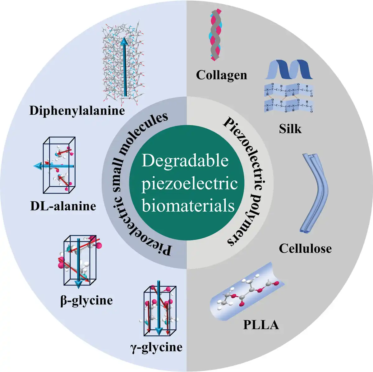
Piezoelectric biomaterials have garnered significant attention in biomedicine due to excellent biocompatibility, biosafety, and biodegradability.[32–33–34–35] Specially, degradable piezoelectric biomaterials are ideal candidates for developing implantable medical devices. These materials can be degraded and absorbed in vivo after completing their function, avoiding the need for additional surgical removal and reducing the pain and expense to patients. Composed of small-molecule crystals (such as amino acids and diphenylalanine [FF]) and polymers (such as proteins, polysaccharides, and poly(l-lactic acid) (PLLA)) (Figure 1),[36,37] these degradable piezoelectric materials demonstrate promising potential for applications in biomechanical energy harvesting, physiological signal monitoring, and tissue repair, heralding a new era in medical device innovation.
Figure 1.
The schematic of degradable piezoelectric biomaterials includes small molecules such as piezoelectric amino acids (glycine, dl-alanine, etc) and peptides (diphenylalanine, etc), as well as piezoelectric polymers such as proteins (collagen, silk, etc), polysaccharides (cellulose, chitin, chitosan, etc), and synthetic polymers (poly-l-lactic acid, etc).
2. Degradable piezoelectric biomaterials
2.1. Degradable piezoelectric small-molecule crystals
Many natural biological small-molecule crystals exhibit piezoelectricity due to the intrinsic lack of symmetry within their molecular structures. Amino acids are crucial to various physiological functions as the building blocks of proteins. Among the 20 natural amino acids, 17 display piezoelectric properties at ambient temperature.[38] This piezoelectric effect in amino acids is attributed to stress-induced polarization, where dipole moments orient from the carboxyl group toward the amino group. Specifically, glycine and dl-alanine exhibit piezoelectric characteristics akin to those observed in quartz crystals.[39]
Glycine primarily exists in 3 polycrystalline forms: α, β, and γ, each with distinct piezoelectric properties (Figure 2A).[40] The α-glycine molecules are organized in an antiparallel conformation, neutralizing dipole moments and resulting in the absence of piezoelectricity. This form is classified within the P21/c space group. In contrast, β-glycine and γ-glycine exhibit piezoelectric characteristics and are categorized under the P21 and P32 space groups, respectively.[37] Notably, β-glycine possessed an exceptionally high shear piezoelectric coefficient d16 with a predicted value of 195 pm V−1 and a measured value of 178 pm V−1.[41] This pronounced effect was attributed to the supramolecular stacking of glycine molecules enhancing the alignment of electric dipoles along specific directions.[38] Nonetheless, it is important to note that β-glycine is metastable, tending to spontaneously transition to the stable α and γ phases upon exposure to air.[42] While shear piezoelectricity is a notable feature in piezoelectric biomaterials, the longitudinal piezoelectric coefficient generally has greater significance and practical applicability. γ-Glycine, with its helically oriented dipole along the axis, exhibited superior longitudinal piezoelectric coefficients d33 of 9.93 pm V−1, underscoring its potential utility.[41] In addition, dl-alanine crystallizes in an orthorhombic symmetry to form a racemic mixture exhibiting significant piezoelectricity, with a d33 (9.1 pm V−1) comparable to γ-glycine (Figure 2B).[43]
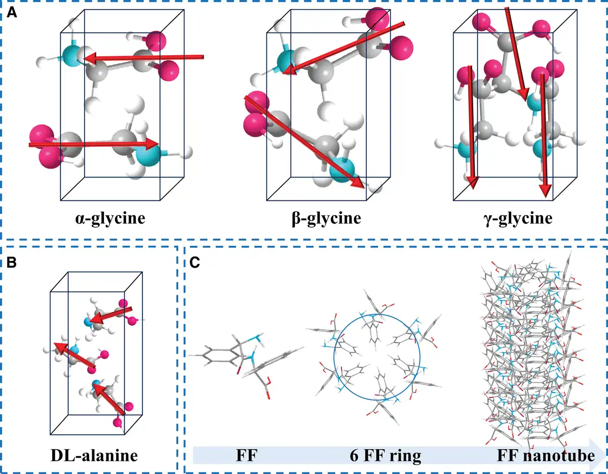
Figure 2.
Schematic diagram of some representative piezoelectric small-molecule crystals. (A) Three crystal structures of the glycine molecule. (B) The crystal structures of dl-alanine. (C) The process of forming diphenylalanine from phenylalanine monomers.
Peptides comprised of at least 2 amino acids connected via peptide bonds have captured significant interest due to their potent piezoelectricity and the variety of their self-assembly structures.[44,45] FF peptide, in particular, is the most representative piezoelectric peptide (Figure 2C). FF monomers self-assemble into various micro-nanostructures, driven by intermolecular forces, including hydrogen bonds, electrostatic interactions, solvent-mediated forces, and π–π stacking.[46,47] FF peptide nanotubes exhibited substantial shear piezoelectricity d15 (60 ± 10 pm V−1), but the d33 of FF nanostructures ranged between 9.9 and 17.9 pm V−1.[34,36,48] The coassembly of macromolecular groups and FF peptides is an effective strategy to enhance piezoelectric properties. The designed tert-butyloxycarbonyl (Boc)-β,β-diphenyl-Ala-OH (Dip)-Dip showed exceptional d33,eff (73.1 ± 13.1 pm V−1).[49]
Despite biological small-molecule crystals possessing high piezoelectricity and biocompatibility, their intrinsic rigidity and brittleness limit compatibility with the flexible modulus of tissues. Integrating piezoelectric small-molecule crystals with flexible substrates has emerged as an effective strategy to confer flexibility. In this approach, flexible polymers serve as substrates and significantly influence crystal growth through their functional groups, playing a pivotal role in enhancing piezoelectricity and modulating the flexibility of small-molecule crystals.[50,51]
2.2. Degradable piezoelectric polymers
In addition to degradable biological small-molecule crystals mentioned above, a variety of degradable polymers not only demonstrate excellent piezoelectricity but also possess inherent flexibility. These polymers are typically semicrystalline and exhibit inherent piezoelectric properties due to well-organized structures with low symmetry. Chemical compositions do not solely determine their piezoelectric attributes; the 3-dimensional structural configurations are also essential for piezoelectricity.
Due to the inherent piezoelectricity of amino acids, numerous proteins have demonstrated piezoelectric responses, such as collagen, silk proteins, viruses, and poly(γ-benzyl-l-glutamate) (PBLG).[52] Collagen has been the subject of extensive study for its piezoelectric properties since the discovery of the piezoelectric effect in bone (Figure 3A).[53,54] The prevailing theory suggests that the piezoelectric effect in collagen results from the accumulation of dipole moments along the peptide chain and the hydrogen bonding interactions within the supramolecular structure.[55] The highest measured d14 in collagen was 12 pm V−1.[56] Compared to collagen, silk exhibits a higher d33 (Figure 3B). An electrospun-induced, highly oriented, electrically polarized silk film achieved a d33 of 38 pm V−1.[57] The M13 phage has also attracted considerable attention due to its considerable d33 (13.2 pm V−1) and scalability.[58] However, concerns regarding its biosafety have curtailed its further application in vivo.
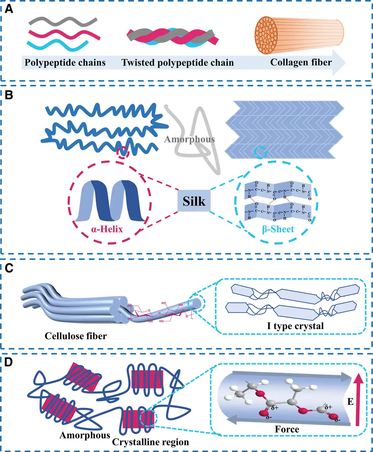
Figure 3.
Schematic diagram of some representative degradable piezoelectric polymers. (A) The collagen fiber is formed from 3 polypeptide chains. (B) Two crystal structures of silk. (C) The cellulose fiber is composed of I-type crystals. (D) Synthetic polymer poly-l-lactic acid.
Polysaccharides are large biomolecules found extensively in animals and plants. They consist of numerous monosaccharides linked by glycosidic bonds. Cellulose and chitin, as representatives of polysaccharides, possess hierarchical fiber structures with low symmetry, demonstrating notable piezoelectric properties.[34] Cellulose possesses alternating highly ordered crystalline and amorphous regions, stabilized by hydrogen bonds and van der Waals forces (Figure 3C). The source of cellulose’s piezoelectricity lies in the orderly dipoles formed by the strong hydrogen bonds between the oxygen and hydrogen atoms within each unit cell. Theoretical calculations indicated that the piezoelectric coefficient of cellulose ranged from 4.3 to 36.4 pm V−1.[35] The arrangement of cellulose crystals would be effectively achieved through mechanical stretching and electric field polarization to enhance its piezoelectric properties. Ultrathin-oriented cellulose nanocrystal (CNC) films prepared using assisted shear force and electric field exhibited a high d25 of 210 pm V−1.[59] Direct current-assisted confinement cell technology further stabilized the vertical alignment of CNC rods, ensuring that all dipole moments are oriented perpendicular to the film surface, thereby yielding a more practical d33 of 19.3 ± 2.9 pm V−1.[60] Similarly, chitin and its deacetylated derivative chitosan, with structures akin to cellulose, demonstrated reported maximum d33 of 9.49 and 18.4 pm V−1, respectively.[61,62]
Laboratory-synthesized polymers significantly expand the diversity and versatility of piezoelectric materials. Adjusting the polymer chain orientations to create dipoles can induce piezoelectricity in synthetic polymers. Modifying groups susceptible to hydrolysis or oxidative degradation can also regulate their degradability.[35] PLLA is a commonly used degradable synthetic piezoelectric polymer. Due to its helical chain structure, PLLA has shear piezoelectricity parallel to the z-axis. Its d14 ranged from 9 to 19 pm V−1.[35,63] Its piezoelectricity depends on the crystallinity and dipole arrangement of the C=O bond of PLLA (Figure 3D).[64] The thermal annealing process can control the crystallinity of PLLA, and external electric field and mechanical stretching can align the dipoles parallel to each other and perpendicular to the molecular chain direction, thus increasing its shear piezoelectric properties.[32,65]
2.3. Degradability of piezoelectric biomaterials
Since amino acids and peptides are water soluble and degrade into alkaline molecules in an aqueous environment, they can be reabsorbed as nutrients in vivo.[66,67] Polymers are made up of the copolymerization of many small chemical units. Their relatively complex degradation process necessitates breaking long chains into smaller molecules. This process is usually sluggish under natural conditions, but it can be accelerated with the assistance of enzymes, fungi, and other catalysts. For instance, silk and collagen necessitate enzyme catalysis under physiological conditions to degrade into amino acids the human body can absorb.[67] Cellulose cannot be directly degraded and absorbed in the human body and can only be broken down into carbon dioxide and water by microorganisms and fungal enzymes.[68,69] Chitin can be hydrolyzed by lysozyme in the human body.[70] Similarly, PLLA degrades slowly in aqueous solutions, but microorganisms can speed up its degradation into carbon dioxide and water.[71] Since these piezoelectric polymers are semicrystalline, their degradation in aqueous solutions occurs in 2 stages. First, water penetrates the amorphous region, breaking the polymer chains with the aid of a catalyst. Then, degradation spreads from the amorphous region to the crystalline region until complete decomposition into small molecular compounds occurs.
3. Medical applications
3.1. Energy harvester and sensor
Piezoelectric materials are the core component of piezoelectric nanogenerators (PENGs). Specifically, degradable piezoelectric materials provide new opportunities for self-powered transient implantable bioelectronics and sensors (Figure 4). The degradable PENGs not only facilitate converting energy from human movements into power for wearable and implantable electronics but also ensure their degradation and absorption postutilization, thereby circumventing the need for secondary processing. Su et al notably enhanced the crystallinity and piezoelectric constant of self-assembled FF films by applying a 7-kV voltage. Under an exerted force of 90 N, fabricated PENG demonstrated a open-circuit voltage (Voc) of 3.4 V, an short-circuit current (Isc) of 235 nA, and a power density of 9.98 W m−3, surpassing several nanogenerators predicated on degradable biomaterials.[72] The output power of PENG based on degradable piezoelectric materials is typically in the nanowatt range, which is lower than that of other energy harvesting devices, but its unique degradable properties remain highly appealing in bioelectronic devices.
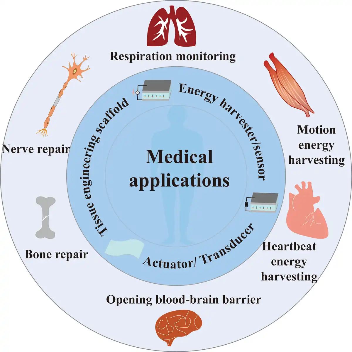
Figure 4.
The applications of piezoelectric materials for medical devices: fabricated piezoelectric nanogenerators based on biodegradable piezoelectric biomaterials for energy harvesting and sensing; as actuator and transducer for cleaning thrombi; as a tissue engineering scaffold for tissue repair such as bone regeneration, nerve repair, and wound healing.
In addition to energy harvesting, the sensing prioritizes signal correlation over the magnitude of energy output. Force sensors employing degradable piezoelectric materials manifest superior suitability for in vivo detection of physiological signals. Curry et al constructed an implantable pressure sensor based on PLLA. This sensor comprised a PLLA piezoelectric material, a molybdenum electrode, and a polylactic acid (PLA) packaging layer. This PLLA film pressure sensor, upon implantation within the abdominal cavity of mice, was capable of monitoring diaphragm contraction pressure.[73] Furthermore, Yang et al developed a glycine-polyvinyl alcohol (PVA) heterostructure film characterized by a sandwich structure wherein the crystalline glycine layer self-assembles and aligns autonomously between 2 PVA films. This configuration significantly amplified its macroscopic piezoelectric properties, achieving a piezoelectric coefficient (d33) of 5.3 pm V−1. The PENG with glycine-PVA film was implanted under the skin of the thigh and chest areas, producing Vpp signals of >150 mV and peak-peak voltage (Vpp) of >20 mV, respectively.[50] However, the existing literature mainly focuses on degradable pressure sensors designed for movement and respiration monitoring, with limited studies addressing the monitoring of small physiological signals such as heartbeat, pulse, and blood flow.[73–74–75]
3.2. Actuator and transducer
Utilizing the inverse piezoelectric effect, intelligent medical tweezers crafted from PLLA fibers could generate vibrations by applying a voltage at a specific frequency, effectively cleaning thrombi in blood vessels.[76] In addition, Nguyen et al adopted an electrospinning process to create highly oriented glycine-polycaprolactone (PCL) nanofibers. This composite exhibited an exceptionally effective piezoelectric coefficient (d33) of 19 pm V−1. The fabricated ultrasound transducer generated significant levels of ultrasound to open the blood–brain barrier, facilitating the auxiliary delivery of paclitaxel for treating mice with an orthotopic glioblastoma tumor model. The survival rate of animals receiving glycine-PCL device-based ultrasound-mediated paclitaxel treatment was nearly double that of animals receiving ultrasound treatment from a current state-of-the-art implantable ultrasound transducer.[77] These intelligent actuators and transducers fabricated from degradable piezoelectric materials have demonstrated significant practical value in the biomedical field.
3.3. Tissue engineering scaffold
Electricity plays a crucial role in the functionality of biological tissues, with many human body components exhibiting piezoelectricity.[53,54] Early studies on the piezoelectricity of bone revealed that it primarily stems from collagen, contributing to bone regeneration.[78] This discovery has spurred further research into using piezoelectric materials in tissue engineering.[79,80] Degradable piezoelectric materials, particularly, have garnered attention for their excellent biocompatibility and degradability, making them an attractive option for tissue engineering applications. These materials have been used in creating electroactive scaffolds for bone regeneration,[81,82] nerve repair,[83–84–85–86] and electroactive dressings for wound healing.[87,88] The electricity generated by the deformation of implanted piezoelectric material regulates the physiological electrical environment of cells, causing a series of changes in cells, such as migration, proliferation, differentiation, activation of ion channels, and activation of signaling pathways, and ultimately promotes tissue repair.
4. Challenges
Although degradable piezoelectric biomaterials hold promising application prospects in implantable transient medical electronics, some challenges persist (Figure 5).
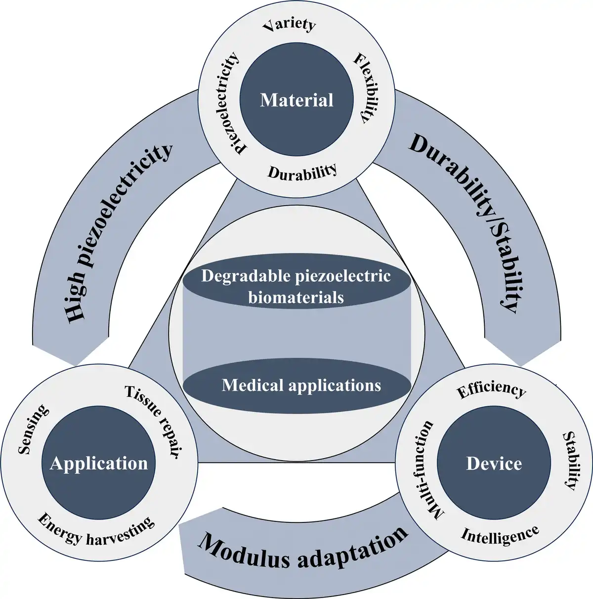
Figure 5.
Challenges of degradable piezoelectric biomaterials for medical applications: material and tissue modulus adaptation issues; the low and impractical piezoelectric coefficient issues; the contradiction between degradability and durability and stability.
(1) A primary issue resides in the mismatch between the modulus of current degradable piezoelectric biomaterials and biological tissues. Specifically, degradable piezoelectric small-molecule crystals demonstrate inherent rigidity, characterized by a modulus exceeding gigapascals, while degradable piezoelectric polymers possess moduli within the megapascal range, surpassing that of human skin tissue. Concurrently, achieving high piezoelectricity necessitates materials featuring extensive crystalline regions, whereas the attainment of flexibility demands materials with a preponderance of amorphous regions. Thus, striking an equilibrium among piezoelectricity, flexibility, and application requisites emerges as a paramount concern in developing future self-powered implantable medical and wearable electronic devices.
(2) Furthermore, the piezoelectric coefficient of degradable piezoelectric biomaterials significantly underperforms compared to inorganic piezoelectric materials. More efficient methods are needed to manipulate dipoles/polarizations within biomaterials to realize enhanced piezoelectric properties. Moreover, many degradable piezoelectric biomaterials exhibit exceptionally high shear piezoelectric coefficients, starkly contrasting the often low or negligible longitudinal/transverse piezoelectric coefficients (Table 1). Because out-of-plane stress represents the most prevalent mechanical stimulus, device designs require specific configurations to transduce such stress into shear stress, thereby amplifying device complexity and diminishing practical utility.
(3) The stability and durability of biodegradable piezoelectric materials have attracted significant attention. Numerous polymorphs with strong piezoelectricity are metastable, and electromechanical coupling devices are subjected to numerous strain cycles in practical scenarios, potentially impairing the piezoelectric properties of materials exhibiting relatively low stability. Addressing this challenge mandates innovative approaches in material and device structural design.
(4) Although extensive research has been dedicated to applying degradable piezoelectric materials in bone regeneration, wound repair, and nerve repair, their involvement in tumor diagnosis, treatment, and drug delivery remains scarce. The comparative maturity of research on other piezoelectric materials in these fields suggests a potential avenue for broadening the applicability of degradable piezoelectric materials, thereby enhancing their utility and impact in these critical fields.
Table 1
Summary of piezoelectric biomaterials.
| Type | Materials | Highest piezoelectric coefficient | Medical applications |
|---|---|---|---|
| Amino acids | β-glycine | d16 = 178 pm V−1[41] | Pressure sensor[89] Face masks for filtration[90] Bone regeneration[91] Motions and physiological signals monitoring[50,74,92–93–94] Ultrasonic transducer[77] Energy harvesting[43,95,96] |
| d22 = 4.7 pm V−1[41]] | |||
| d33 = 19 pm V−1[77] | |||
| γ-glycine | d11 = 1.7 pm V−1[41] | ||
| d16 = 6 pm V−1[41] | |||
| d22 = −1.1 pm V−1[41] | |||
| d33 = 10 pm V−1[97] | |||
| dl-alanine | d33 = 9.1 pm V−1[43] | ||
| Acetylated tryptophan | dik = 47 pm V−1[95] | ||
| Valine | d16 = 7.72 pm V−1[98] | ||
| d22 = 12.37 pm V−1[98] | |||
| d34 = 5.32 pm V−1[98] | |||
| Peptides | Self-assembly and coassembly FF | d14 = −10 pm V−1[99] | Energy harvesting[100–101–102–103] |
| d15 = 80 pm V−1[99] | |||
| d16 = 13.8 pm V−1[104] | |||
| d33 = 73.1 pm V−1[49] | |||
| def = 35.5 pm V−1[102] | |||
| Hyp–Phe–Phe | d16 = 27.3 pm V−1[103] | ||
| d35 = 27.3 pm V−1[103] | |||
| d36 = 17.1 pm V−1[103] | |||
| Ala–Hyp–Gly | d25 = 25 pm V−1[105] | ||
| Oligopeptide | d33 = 9.8 pm V−1[45] | ||
| Proteins | Collagen | d14 = −12.00 pm V−1[56] | Ultrasonic transducer[106] Energy harvesting[57,107] Motions and physiological signals monitoring[108,109] Biting force monitoring[110] |
| d15 = 6.21 pm V−1[56] | |||
| d31 = -4.84 pm V−1[56] | |||
| d33 = 2.6 pm V−1[111] | |||
| Silk | d14 = 1.5 pm V−1[112] | ||
| d33 = 56.7 pm V−1[113] | |||
| Viruses | d33 = 13.2 pm V−1[107] | ||
| PBLG | d33 = 27 pm V−1[114] | ||
| Polysaccharides | Cellulose | d25 = 210 pm V−1[59] | Physiological signal monitoring[115] Energy harvesting[116] Scaffolds for collagen formation of osteoblasts[117] Cardiorespiratory monitoring[118] Motion sensing[119] |
| d31 = 27 pm V−1[120] | |||
| d33 = 19.3 ± 2.9 pm V−1[60] | |||
| Chitosan | d33 = 18.4 pm V−1[61] | ||
| Chitin | d33 = 3.98 pm V−1[121] | ||
| Synthesized polymers | PLLA | d14 = 19 pm V−1[63] | Bone regeneration[81,122–123–124] Wound healing[87,125] Ultrasonic transducer[63,126] Enhancing cell adhesion[127] Face masks for filtration[75,128] Motion sensing[31] Nerve repair[83,86,129] |
Ala, alanine; FF, diphenylalanine; Gly, glycine; Hyp, hydroxyproline; PBLG, poly(γ-benzyl-L-glutamate); Phe, phenylalanine; PLLA, poly(L-lactic acid).
Conflicts of interests
The authors declare that they have no conflicts of interest.
Funding
This work was supported by the National Key Research and Development Program of China (2022YFB3804700, 2021YFB3200303), the National Natural Science Foundation of China (T2125003, 52203325), Beijing Natural Science Foundation (L212010), and the Fundamental Research Funds for the Central Universities.
Author contributions
Zhou Li and Yuan Bai designed the research title. Yuan Bai and Hongyu Meng wrote the paper. Zhou Li revised the manuscript. Zhou Li is the primary one responsible for the final content. All authors read and approved the final manuscript.
References
- [1] Libanori A, Chen G, Zhao X, et al. Smart textiles for personalized healthcare. Nat Electron. 2022;5:142.
- [2] Chen G, Xiao X, Zhao X, et al. Electronic textiles for wearable point-of-care systems. Chem Rev. 2022;122:3259.
- [3] Chen G, Li Y, Bick M, et al. Smart textiles for electricity generation. Chem Rev. 2020;120:3668.
- [4] Yin J, Wang S, Di Carlo A, et al. Smart textiles for self-powered biomonitoring. Med-X. 2023;1:3.
- [5] Zou Y, Bo L, Li Z. Recent progress in human body energy harvesting for smart bioelectronic system. Fundam Res. 2021;1:364.
- [6] Gai Y, Bai Y, Cao Y, et al. A gyroscope nanogenerator with frequency up-conversion effect for fitness and energy harvesting. Small. 2022;18:e2108091.
- [7] Lai Z, Xu J, Bowen CR, et al. Self-powered and self-sensing devices based on human motion. Joule. 2022;6:1501.
- [8] Gai Y, Wang E, Liu M, et al. A self-powered wearable sensor for continuous wireless sweat monitoring. Small Methods. 2022;6:e2200653.
- [9] Li C, Luo R, Bai Y, et al. Molecular doped biodegradable triboelectric nanogenerator with optimal output performance. Adv Funct Mater. 2024;n/a:2400277.
- [10] Liu M, Tan P, Xue J, et al. A portable self-powered turbine spirometer for rehabilitation monitoring on COVID-19. Adv Mater Technol. 2023;8:2300328.
- [11] Liu Z, Hu Y, Qu X, et al. A self-powered intracardiac pacemaker in swine model. Nat Commun. 2024;15:507.
- [12] Ouyang H, Liu Z, Li N, et al. Symbiotic cardiac pacemaker. Nat Commun. 2019;10:1821.
- [13] Sun M, Li Z, Yang C, et al. Nanogenerator-based devices for biomedical applications. Nano Energy. 2021;89:106461.
- [14] Ouyang H, Li Z, Gu M, et al. A bioresorbable dynamic pressure sensor for cardiovascular postoperative care. Adv Mater. 2021;33:e2102302.
- [15] Jiang D, Shi B, Ouyang H, et al. Emerging implantable energy harvesters and self-powered implantable medical electronics. ACS Nano. 2020;14:6436.
- [16] Gai Y, Jiang Y, Li Z. Advances in health rehabilitation devices based on triboelectric nanogenerators. Nano Energy. 2023;116:108787.
- [17] Xi Y, Cheng S, Chao S, et al. Piezoelectric wearable atrial fibrillation prediction wristband enabled by machine learning and hydrogel affinity. Nano Res. 2023;16:11674.
- [18] Yang Y, Luo R, Chao S, et al. Improved pharmacodynamics of epidermal growth factor via microneedles-based self-powered transcutaneous electrical stimulation. Nat Commun. 2022;13:6908.
- [19] Wan X, Wang Z, Zhao X, et al. Flexible and highly piezoelectric nanofibers with organic–inorganic coaxial structure for self-powered physiological multimodal sensing. Chem Eng J. 2023;451:139077.
- [20] Hu B, Xue J, Jiang D, et al. Wearable exoskeleton system for energy harvesting and angle sensing based on a piezoelectric cantilever generator array. ACS Appl Mater Interfaces. 2022;14:36622.
- [21] Sekhar MC, Veena E, Kumar NS, et al. A review on piezoelectric materials and their applications. Cryst Res Technol. 2023;58:2200130.
- [22] Khorsand Zak A, Yazdi ST, Abrishami ME, et al. A review on piezoelectric ceramics and nanostructures: fundamentals and fabrications. J Aust Ceram Soc. 2024.
- [23] Costa CM, Cardoso VF, Martins P, et al. Smart and multifunctional materials based on electroactive poly(vinylidene fluoride): recent advances and opportunities in sensors, actuators, energy, environmental, and biomedical applications. Chem Rev. 2023;123.
- [24] You Y-M, Liao W-Q, Zhao D, et al. An organic-inorganic perovskite ferroelectric with large piezoelectric response. Science. 2017;357.
- [25] Xiang Z, Xu L, Shan Y, et al. Tumor microenvironment-responsive self-assembly of barium titanate nanoparticles with enhanced piezoelectric catalysis capabilities for efficient tumor therapy. Bioact Mater. 2024;33:251.
- [26] Wang C, Hu Y, Liu Y, et al. Tissue-adhesive piezoelectric soft sensor for in vivo blood pressure monitoring during surgical operation. Adv Funct Mater. 2023;33:2303696.
- [27] Ji J, Yang C, Shan Y, et al. Research trends of piezoelectric nanomaterials in biomedical engineering. Adv NanoBiomed Res. 2023;3:2200088.
- [28] Liu Z, Xu L, Zheng Q, et al. Human motion driven self-powered photodynamic system for long-term autonomous cancer therapy. ACS Nano. 2020;14:8074.
- [29] Deng W, Zhou Y, Libanori A, et al. Piezoelectric nanogenerators for personalized healthcare. Chem Soc Rev. 2022;51:3380.
- [30] Cui X, Xu L, Shan Y, et al. Piezocatalytically-induced controllable mineralization scaffold with bone-like microenvironment to achieve endogenous bone regeneration. Sci Bull. 2024.
- [31] Shan Y, Wang E, Cui X, et al. A biodegradable piezoelectric sensor for real-time evaluation of the motor function recovery after nerve injury. Adv Funct Mater. 2024;n/a:2400295.
- [32] Chorsi MT, Curry EJ, Chorsi HT, et al. Piezoelectric biomaterials for sensors and actuators. Adv Mater. 2019;31:1802084.
- [33] Lay R, Deijs GS, Malmstrom J. The intrinsic piezoelectric properties of materials - a review with a focus on biological materials. RSC Adv. 2021;11:30657.
- [34] Li J, Long Y, Yang F, et al. Degradable piezoelectric biomaterials for wearable and implantable bioelectronics. Curr Opin Solid State Mater Sci. 2020;24:100806.
- [35] Liu T, Wang Y, Hong M, et al. Advances in biodegradable piezoelectrics for medical implants. Nano Today. 2023;52:101945.
- [36] Ali M, Bathaei MJ, Istif E, et al. Biodegradable piezoelectric polymers: recent advancements in materials and applications. Adv Healthc Mater. 2023;12:2300318.
- [37] Kim D, Han SA, Kim JH, et al. Biomolecular piezoelectric materials: from amino acids to living tissues. Adv Mater. 2020;32:1906989.
- [38] Wang R, Sui J, Wang X. Natural piezoelectric biomaterials: a biocompatible and sustainable building block for biomedical devices. ACS Nano. 2022;16:17708.
- [39] Jeon B, Han D, Yoon G. Piezoelectric characteristics of PVA/dl-alanine polycrystals in d(33) mode. iScience. 2023;26:105768.
- [40] Boldyreva E. Glycine: the gift that keeps on giving. Israel J Chem. 2021;61:828.
- [41] Guerin S, Stapleton A, Chovan D, et al. Control of piezoelectricity in amino acids by supramolecular packing. Nat Mater. 2018;17:180.
- [42] Isakov D, Petukhova D, Vasilev S, et al. In situ observation of the humidity controlled polymorphic phase transformation in glycine microcrystals. Cryst Growth Des. 2014;14:4138.
- [43] Guerin S, O’Donnell J, Haq EU, et al. Racemic amino acid piezoelectric transducer. Phys Rev Lett. 2019;122:047701.
- [44] Cai C, Lin J, Lu Y, et al. Polypeptide self-assemblies: nanostructures and bioapplications. Chem Soc Rev. 2016;45:5985.
- [45] Petroff CA, Cassone G, Šponer J, et al. Intrinsically polar piezoelectric self-assembled oligopeptide monolayers. Adv Mater. 2021;33:2007486.
- [46] Anderson J, Lake PT, McCullagh M. Initial aggregation and ordering mechanism of diphenylalanine from microsecond all-atom molecular dynamics simulations. J Phys Chem B. 2018;122:12331.
- [47] Zhao J, Liu Q, Tong X, et al. Rational design of bio-inspired peptide electronic materials toward bionanotechnology: strategies and applications. Adv Funct Mater. 2024:2401466.
- [48] Kholkin A, Amdursky N, Bdikin I, et al. Strong piezoelectricity in bioinspired peptide nanotubes. ACS Nano. 2010;4:610.
- [49] Basavalingappa V, Bera S, Xue B, et al. Diphenylalanine-derivative peptide assemblies with increased aromaticity exhibit metal-like rigidity and high piezoelectricity. ACS Nano. 2020;14:7025.
- [50] Yang F, Li J, Long Y, et al. Wafer-scale heterostructured piezoelectric bio-organic thin films. Science. 2021;373:337.
- [51] Yu Q, Bai Y, Li Z, et al. Interface-induced high piezoelectric γ-glycine-based flexible biodegradable films. Nano Energy. 2024;121:109196.
- [52] Sun Y, Zeng K, Li T. Piezo-/ferroelectric phenomena in biomaterials: a brief review of recent progress and perspectives. Sci China-Phys Mech Astron. 2020;63:278701.
- [53] Kapat K, Shubhra QTH, Zhou M, et al. Piezoelectric nano-biomaterials for biomedicine and tissue regeneration. Adv Funct Mater. 2020;30:1909045.
- [54] Nain A, Chakraborty S, Barman SR, et al. Progress in the development of piezoelectric biomaterials for tissue remodeling. Biomaterials. 2024;307:122528.
- [55] Zhang Y, Herling M, Chenoweth DM. General solution for stabilizing triple helical collagen. J Am Chem Soc. 2016;138:9751.
- [56] Denning D, Kilpatrick JI, Fukada E, et al. Piezoelectric tensor of collagen fibrils determined at the nanoscale. ACS Biomater Sci Eng. 2017;3:929.
- [57] Sencadas V, Garvey C, Mudie S, et al. Electroactive properties of electrospun silk fibroin for energy harvesting applications. Nano Energy. 2019;66:104106.
- [58] Lee BY, Zhang J, Zueger C, et al. Virus-based piezoelectric energy generation. Nat Nanotechnol. 2012;7:351.
- [59] Csoka L, Hoeger IC, Rojas OJ, et al. Piezoelectric effect of cellulose nanocrystals thin films. ACS Macro Lett. 2012;1:867.
- [60] Wang J, Carlos C, Zhang Z, et al. Piezoelectric nanocellulose thin film with large-scale vertical crystal alignment. ACS Appl Mater Interfaces. 2020;12:26399.
- [61] Praveen E, Murugan S, Jayakumar K. Investigations on the existence of piezoelectric property of a bio-polymer - chitosan and its application in vibration sensors. RSC Adv. 2017;7:35490.
- [62] Hoque NA, Thakur P, Biswas P, et al. Biowaste crab shell-extracted chitin nanofiber-based superior piezoelectric nanogenerator. J Mater Chem A. 2018;6:13848.
- [63] Curry EJ, Le TT, Das R, et al. Biodegradable nanofiber-based piezoelectric transducer. Proc Natl Acad Sci USA. 2020;117:214.
- [64] Zhao G, Huang B, Zhang J, et al. Electrospun poly(l-lactic acid) nanofibers for nanogenerator and diagnostic sensor applications. Macromol Mater Eng. 2017;302:1600476.
- [65] Sultana A, Ghosh SK, Sencadas V, et al. Human skin interactive self-powered wearable piezoelectric bio-e-skin by electrospun poly-l-lactic acid nanofibers for non-invasive physiological signal monitoring. J Mater Chem B. 2017;5:7352.
- [66] Wang W, Wu Z, Dai Z, et al. Glycine metabolism in animals and humans: implications for nutrition and health. Amino Acids. 2013;45:463.
- [67] Reitzer L. Catabolism of amino acids and related compounds. EcoSal Plus. 2005;1.
- [68] Zhao L, Zhang J, Zhao D, et al. Biological degradation of lignin: a critical review on progress and perspectives. Ind Crop Prod. 2022;188:115715.
- [69] Datta R. Enzymatic degradation of cellulose in soil: a review. Heliyon. 2024;10:e24022.
- [70] Tomihata K, Ikada Y. In vitro and in vivo degradation of films of chitin and its deacetylated derivatives. Biomaterials. 1997;18:567.
- [71] Walton M, Cotton NJ. Long-term in vivo degradation of poly-l-lactide (PLLA) in bone. J Biomater Appl. 2007;21:395.
- [72] Su Y, Liu J, Yang D, et al. Electric field-assisted self-assembly of diphenylalanine peptides for high-performance energy conversion. ACS Mater Lett. 2023;5:2317.
- [73] Curry EJ, Ke K, Chorsi MT, et al. Biodegradable piezoelectric force sensor. Proc Natl Acad Sci USA. 2018;115:909.
- [74] Cheng Y, Xu J, Li L, et al. Boosting the piezoelectric sensitivity of amino acid crystals by mechanical annealing for the engineering of fully degradable force sensors. Adv Sci. 2023;10:e2207269.
- [75] Le TT, Curry EJ, Vinikoor T, et al. Piezoelectric nanofiber membrane for reusable, stable, and highly functional face mask filter with long-term biodegradability. Adv Funct Mater. 2022;32:2113040.
- [76] Tajitsu Y, Kawai S, Kanesaki M, et al. Microactuators with piezoelectric polylactic acid fibers-toward the realization of tweezers for biological cells. Ferroelectrics. 2004;304:195.
- [77] Chorsi MT, Le TT, Lin F, et al. Highly piezoelectric, biodegradable, and flexible amino acid nanofibers for medical applications. Sci Adv. 2023;9:eadg6075.
- [78] Fukada E, Yasuda I. On the piezoelectric effect of bone. J Phys Soc Jpn. 1957;12:1158.
- [79] Luo R, Shi B, Luo D, et al. Self-powered electrical stimulation assisted skin wound therapy. Sci Bull. 2023;68:1740.
- [80] Konuk Tokak E, Çetin Altindal D, Akdere E, et al. In-vitro effectiveness of poly-β-alanine reinforced poly(3-hydroxybutyrate) fibrous scaffolds for skeletal muscle regeneration. Mater Sci Eng C Mater Biol Appl. 2021;131:112528.
- [81] Das R, Curry EJ, Le TT, et al. Biodegradable nanofiber bone-tissue scaffold as remotely-controlled and self-powering electrical stimulator. Nano Energy. 2020;76:105028.
- [82] Yang C, Ji J, Lv Y, et al. Application of piezoelectric material and devices in bone regeneration. Nanomaterials. 2022;12:4386.
- [83] Wu P, Chen P, Xu C, et al. Ultrasound-driven in vivo electrical stimulation based on biodegradable piezoelectric nanogenerators for enhancing and monitoring the nerve tissue repair. Nano Energy. 2022;102:107707.
- [84] Si JZ, Yang YH, Xing XL, et al. Controlled degradable chitosan/collagen composite scaffolds for application in nerve tissue regeneration. Polym Degrad Stab. 2019;166:73.
- [85] Jiang F, Shan Y, Tian J, et al. Poly(l-lactic acid) nanofiber-based multilayer film for the electrical stimulation of nerve cells. Adv Mater Interfaces. 2023;10:2202474.
- [86] Yang Y, Yin X, Wang H, et al. Engineering a wirelessly self-powered and electroconductive scaffold to promote peripheral nerve regeneration. Nano Energy. 2023;107:108145.
- [87] Das R, Le TT, Schiff B, et al. Biodegradable piezoelectric skin-wound scaffold. Biomaterials. 2023;301:122270.
- [88] Chen YX, Ye MQ, Song LW, et al. Piezoelectric and photothermal dual functional film for enhanced dermal wound regeneration via upregulation of Hsp90 and HIF-1α. Appl Mater Today. 2020;20:100756.
- [89] J AS, S EF, B SS, et al. Bio-compatible piezoelectric material based wearable pressure sensor for smart textiles. Smart Mater Struct. 2022;31:125015.
- [90] Wang Z, Zhang Z, Peng Z, et al. Self-charging and long-term face masks leveraging low-cost, biodegradable and sustainable piezoelectric nanofiber membrane. Nano Mater Sci. 2024.
- [91] Alazzawi M, Kadim Abid Alsahib N, Turkoglu Sasmazel H. Core/shell glycine-polyvinyl alcohol/polycaprolactone nanofibrous membrane intended for guided bone regeneration: development and characterization. Coatings. 2021;11:1130.
- [92] Ali M, Hoseyni SM, Das R, et al. A flexible and biodegradable piezoelectric based wearable sensor for non invasive monitoring of dynamic human motions and physiological signals. Adv Mater Technol. 2023;8:2300347.
- [93] Zheng W, Li T, Jin F, et al. Interfacial polarization locked flexible β-phase glycine/Nb(2) CT(x) piezoelectric nanofibers. Small. 2024:e2308715.
- [94] Li J, Carlos C, Zhou H, et al. Stretchable piezoelectric biocrystal thin films. Nat Commun. 2023;14:6562.
- [95] Wang Y, Liu S, Li L, et al. Manipulating the piezoelectric response of amino acid-based assemblies by supramolecular engineering. J Am Chem Soc. 2023;145:15331.
- [96] Zhang Z, Li X, Peng Z, et al. Active self-assembly of piezoelectric biomolecular films via synergistic nanoconfinement and in-situ poling. Nat Commun. 2023;14:4094.
- [97] Heredia A, Meunier V, Bdikin IK, et al. Nanoscale ferroelectricity in crystalline γ-glycine. Adv Funct Mater. 2012;22:2996.
- [98] Yuan H, Chen Y, Lin R, et al. Modified Stranski–Krastanov growth of amino acid arrays toward piezoelectric energy harvesting. ACS Appl Mater Interfaces. 2022;14:46304.
- [99] Vasilev S, Zelenovskiy P, Vasileva D, et al. Piezoelectric properties of diphenylalanine microtubes prepared from the solution. J Phys Chem Solids. 2016;93:68.
- [100] Tao K, Xue B, Li Q, et al. Stable and optoelectronic dipeptide assemblies for power harvesting. Mater Today (Kidlington). 2019;30.
- [101] Lee J-H, Heo K, Schulz-Schönhagen K, et al. Diphenylalanine peptide nanotube energy harvesters. ACS Nano. 2018;12:8138.
- [102] Yuan H, Han P, Tao Z, et al. Peptide coassembly to enhance piezoelectricity for energy harvesting. ACS Appl Mater Interfaces. 2022;14:6538.
- [103] Bera S, Guerin S, Yuan H, et al. Molecular engineering of piezoelectricity in collagen-mimicking peptide assemblies. Nat Commun. 2021;12:2634.
- [104] Tao K, Hu W, Xue B, et al. Bioinspired stable and photoluminescent assemblies for power generation. Adv Mater. 2019;31:1807481.
- [105] Guerin S, Syed TAM, Thompson D. Deconstructing collagen piezoelectricity using alanine-hydroxyproline-glycine building blocks. Nanoscale. 2018;10:9653.
- [106] Joseph J, Singh SG, Vanjari SRK. Piezoelectric micromachined ultrasonic transducer using silk piezoelectric thin film. IEEE Electron Device Lett. 2018;39:749.
- [107] Lee J-H, Lee JH, Xiao J, et al. Vertical self-assembly of polarized phage nanostructure for energy harvesting. Nano Lett. 2019;19.
- [108] Niu Q, Chen J, Fan S, et al. Silk nanoribbon films with enriched silk II structure and enhanced piezoelectricity for self-powered implantable and wearable devices. Nano Today. 2024;56:102228.
- [109] Sohn C, Kim H, Han J, et al. Generating electricity from molecular bonding-correlated piezoresponse of biodegradable silk nanofibers. Nano Energy. 2022;103:107844.
- [110] Liu J-H, Li W-D, Jia J, et al. Structure-regenerated silk fibroin with boosted piezoelectricity for disposable and biodegradable oral healthcare device. Nano Energy. 2022;103:107787.
- [111] Zhou Z, Qian D, Minary-Jolandan M. Molecular mechanism of polarization and piezoelectric effect in super-twisted collagen. ACS Biomater Sci Eng. 2016;2:929.
- [112] Yucel T, Cebe P, Kaplan DL. Structural origins of silk piezoelectricity. Adv Funct Mater. 2011;21:779.
- [113] Joseph J, Saraswathi S, Agarwal A, Singh SG, Vanjari SRK. Silk piezoelectric thin films: Materials to devices. Paper presented at: 2016 IEEE SENSORS; October 30–November 3, 2016.
- [114] Nguyen D-N, Yu SM, Moon W. Electrospinning of poly(γ-benzyl–α,l-glutamate) microfibers for piezoelectric polymer applications. J Appl Polym Sci. 2018;135:46440.
- [115] Ghosh SK, Mandal D. Bio-assembled, piezoelectric prawn shell made self-powered wearable sensor for non-invasive physiological signal monitoring. Appl Phys Lett. 2017;110:123701.
- [116] Patil SR, Chougale MY, Kim J, et al. Nature-driven edible black soldier fly (BSF) insect larvae derived chitin biofilm for sustainable multifunctional energy harvesting. Adv Sustain Syst. 2024;8:2300312.
- [117] Szewczyk PK, Berniak K, Knapczyk-Korczak J, et al. Mimicking natural electrical environment with cellulose acetate scaffolds enhances collagen formation of osteoblasts. Nanoscale. 2023;15:6890.
- [118] Maity K, Mondal A, Saha MC. Cellulose nanocrystal-based all-3D-printed pyro-piezoelectric nanogenerator for hybrid energy harvesting and self-powered cardiorespiratory monitoring toward the human-machine interface. ACS Appl Mater Interfaces. 2023;15:13956.
- [119] Shao D, Wang C, Li W, et al. Natural ginkgo tree leaves as piezo-energy harvesters. J Mater Chem C. 2022;10:15016.
- [120] Yang C, Kim J-H, Kim J-H, et al. Piezoelectricity of wet drawn cellulose electro-active paper. Sens Actuator A Phys. 2009;154:117.
- [121] Kim K, Ha M, Choi B, et al. Biodegradable, electro-active chitin nanofiber films for flexible piezoelectric transducers. Nano Energy. 2018;48:275.
- [122] Vinikoor T, Dzidotor GK, Le TT, et al. Injectable and biodegradable piezoelectric hydrogel for osteoarthritis treatment. Nat Commun. 2023;14:6257.
- [123] Liu Y, Dzidotor G, Le TT, et al. Exercise-induced piezoelectric stimulation for cartilage regeneration in rabbits. Sci Transl Med. 2022;14:eabi7282.
- [124] Li Y, Chen J, Liu S, et al. Biodegradable piezoelectric polymer for cartilage remodeling. Matter. 2024;7:1631.
- [125] Zhang Z, Wang L, Zhang Q, et al. Effective electrical stimulation by a poly(l-lactic acid)/vitamin B2-based piezoelectric generator promotes wound healing. Eur Polym J. 2023;189:111962.
- [126] Yousry YM, Wong V-K, Ji R, et al. Shear mode ultrasonic transducers from flexible piezoelectric PLLA fibers for structural health monitoring. Adv Funct Mater. 2023;33:2213582.
- [127] Polak M, Berniak K, Szewczyk PK, et al. PLLA scaffolds with controlled surface potential and piezoelectricity for enhancing cell adhesion in tissue engineering. Appl Surf Sci. 2023;621:156835.
- [128] Yu Y-H, Huang Y, Zhang J-F, et al. Self-powered, biodegradable, and antibacterial air filters based on piezoelectric poly(l-lactic acid) nanofibrous membranes. ACS Appl Polym. Mater. 2023;5:10426.
- [129] Xiong F, Wei S, Wu S, et al. Aligned electroactive electrospun fibrous scaffolds for peripheral nerve regeneration. ACS Appl Mater Interfaces. 2023;15:41385.
This article is available under Open Access.
© 2024 The Authors