Nanomedicine-enabled strategies for navigating the blood–brain barrier in Parkinson disease
Authors
Xiao Xiao, Keyang Xu, Kexin Ni, Yongzhen Su, Yadan Shi, Mengyao Wu, Xiuqi Hu, Jianxian Ge, Jianfeng Zeng*
- aDepartment of Radiology, The First Affiliated Hospital of Soochow University, State Key Laboratory of Radiation Medicine and Protection, School for Radiological and Interdisciplinary Sciences (RAD-X), Collaborative Innovation Center of Radiological Medicine of Jiangsu Higher Education Institutions, Soochow University, Suzhou, China
- bBiomedical Basic Research Center (BBRC) of Jiangsu Province, Suzhou, China.
* Correspondence: Address: Jianfeng Zeng, Department of Radiology, The First Affiliated Hospital of Soochow University, State Key Laboratory of Radiation Medicine and Protection, School for Radiological and Interdisciplinary Sciences (RAD-X), Collaborative Innovation Center of Radiological Medicine of Jiangsu Higher Education Institutions, Soochow University, Biomedical Basic Research Center (BBRC) of Jiangsu Province, 199 Ren’ai Road, Suzhou Industrial Park, Suzhou, Jiangsu 215000, China. E-mail address: jfzeng@suda.edu.cn (J. Zeng).
MedMat · 2026 · Vol. 3 · No. 3 · pp. 296-316
Abstract
Parkinson disease (PD) is still mainly treated with symptomatic therapies, while effective disease-modifying interventions remain limited. Although emerging therapeutics such as biologics, nucleic acid drugs, neurotrophic factors, and gene-based approaches offer new opportunities, their clinical translation is often restricted by inadequate brain delivery. Nanomedicine has, therefore, attracted increasing interest because it can improve drug stability, protect fragile cargos, enable controlled release, and support multifunctional therapeutic design. Nevertheless, the blood–brain barrier (BBB) remains the principal obstacle to effective brain-targeted therapy in PD. This review discusses nanomedicine-based delivery strategies for PD from the perspective of BBB navigation and classifies them into 2 major categories: strategies that cross the BBB and strategies that bypass it. BBB-crossing approaches include carrier-mediated transport, receptor-mediated transport, adsorptive and biomimetic transcytosis, and physically assisted methods such as focused ultrasound and photothermal modulation. BBB-bypassing approaches include intranasal delivery, meningeal lymphatic-associated pathways, skull–meninges-related microchannel routes, and local intracranial administration such as convection-enhanced delivery. We summarize their mechanisms, representative applications, advantages, and limitations, and compare them in terms of cargo suitability, delivery precision, repeatability, safety, and translational feasibility. Overall, nanomedicine provides important opportunities to expand the therapeutic potential of both conventional drugs and emerging biologics for PD. Future progress will depend on developing cargo-matched and clinically realistic delivery systems that balance brain access, therapeutic efficacy, safety, and manufacturability.
Translations
Long abstracts in additional languages. The English article is the version of record.
Chinesezh-Hans
帕金森病(PD)目前主要依赖对症治疗,而有效的疾病修饰干预手段仍然有限。尽管生物制剂、核酸药物、神经营养因子及基因疗法等新兴治疗策略为临床带来了新机遇,但其转化应用常受限于脑部递送效率不足的问题。纳米医学因此受到广泛关注,因其能够提高药物稳定性、保护脆弱载荷、实现可控释放并支持多功能治疗设计。然而,血脑屏障(BBB)仍是阻碍PD有效靶向治疗的根本障碍。本综述旨在从穿越和绕过血脑屏障的视角出发,系统梳理基于纳米技术的帕金森病递送策略,分析其机制与应用前景。
本文对现有研究进行了系统性归纳与框架性分类,将相关策略划分为两大类:穿越BBB的策略与绕过BBB的策略。穿越类方法包括载体介导转运、受体介导转运、吸附及仿生跨胞吞作用,以及聚焦超声和光热调节等物理辅助手段;绕过类方法则涵盖鼻内递送、脑膜淋巴通路、颅骨-脑膜相关微通道途径,以及局部颅内给药如对流增强输送。文章详细阐述了各类方法的科学机制,列举了代表性应用案例,并深入探讨了不同策略在载荷适用性、递送精度、可重复性、安全性及转化可行性等方面的具体表现与差异。
综述分析表明,纳米医学为扩展传统药物和新兴生物制剂的治疗潜力提供了重要机遇。通过优化载体设计,这些技术能够有效克服血脑屏障的物理化学限制,实现治疗成分在帕金森病模型中的精准递送。不同策略展现出独特的优势:物理辅助方法如聚焦超声可暂时开放BBB,而仿生跨胞吞作用则能利用内源性转运机制提高入脑效率;鼻内和淋巴通路则为非侵入性给药提供了新路径。然而,各类技术均存在局限性,例如部分物理方法的重复安全性尚需验证,局部给药的创伤性及长期生物相容性问题仍需进一步评估,且不同载荷对特定递送系统的匹配度要求各异。
本综述的显著意义在于为帕金森病的脑靶向治疗提供了全面的策略对比与选择依据,强调了未来研发必须平衡脑部渗透性、治疗效果、安全性及可制造性等多重因素。尽管纳米医学前景广阔,但目前的挑战仍集中在开发能够精准匹配载荷特性且符合临床现实需求的递送系统上。未来的工作需聚焦于解决转化可行性问题,特别是在大规模生产标准化、长期体内安全性评价以及复杂病理环境下的稳定性方面取得突破。只有通过构建更加成熟和可靠的纳米递送平台,才能真正实现从实验室研究到临床应用的有效跨越,为帕金森病患者带来实质性的治疗获益。
Françaisfr
La maladie de Parkinson (MP) est actuellement traitée principalement par des thérapies symptomatiques, tandis que les interventions modifiant la progression de la maladie restent limitées. Bien que des approches thérapeutiques émergentes telles que les biologicues, les médicaments à base d'acides nucléiques, les facteurs neurotrophiques et les stratégies génétiques offrent de nouvelles opportunités, leur traduction clinique est souvent entravée par une délivrance cérébrale inadéquate. La nanomédecine suscite donc un intérêt croissant car elle peut améliorer la stabilité des médicaments, protéger les charges fragiles, permettre une libération contrôlée et soutenir la conception thérapeutique multifonctionnelle. Néanmoins, la barrière hémato-encéphalique (BHE) demeure l'obstacle principal à une thérapie ciblée efficace dans la MP.
Cette revue examine les stratégies de délivrance basées sur la nanomédecine pour la MP sous l'angle de la navigation au niveau de la BHE, en les classant en deux catégories majeures : celles qui traversent la BHE et celles qui la contournent. Les approches de franchissement incluent le transport médié par des vecteurs, le transport récepteur-médié, la transcytose adsorptive et biomimétique, ainsi que les méthodes assistées physiquement telles que l'ultrason focalisé et la modulation photothermique. Les approches de contournement comprennent la délivrance intranasale, les voies associées au système lymphatique méningé, les routes microcanalaires liées aux os du crâne et aux méninges, ainsi que l'administration intracrânienne locale comme le transport par convection améliorée.
L'analyse synthétise leurs mécanismes d'action, des applications représentatives, ainsi que leurs avantages et limites respectifs. Les résultats indiquent que la nanomédecine offre des opportunités importantes pour étendre le potentiel thérapeutique tant des médicaments conventionnels que des biologicues émergents dans le traitement de la MP. La comparaison met en lumière les différences entre ces stratégies concernant l'adéquation du chargement, la précision de délivrance, la répétabilité, la sécurité et la faisabilité translationnelle. Chaque méthode présente un profil distinct : certaines permettent une libération contrôlée précise, tandis que d'autres offrent des voies non invasives mais avec des défis spécifiques en termes de biodistribution.
En conclusion, les progrès futurs dépendront du développement de systèmes de délivrance réalistes cliniquement et adaptés aux charges thérapeutiques, équilibrant l'accès cérébral, l'efficacité thérapeutique, la sécurité et la fabricabilité. Bien que prometteuse, cette approche nécessite une attention particulière à la traduction clinique pour surmonter les obstacles restants liés à la complexité de la BHE et aux exigences de production industrielle. La revue souligne qu'une sélection rigoureuse des stratégies en fonction du type de cargaison est essentielle pour maximiser l'efficacité tout en minimisant les risques potentiels, ouvrant ainsi la voie vers une thérapie plus efficace contre la maladie de Parkinson à long terme.
Españoles
La enfermedad de Parkinson (EP) se trata principalmente con terapias sintomáticas, mientras que las intervenciones modificadoras de la enfermedad siguen siendo limitadas. Aunque nuevas estrategias terapéuticas como los biológicos, fármacos basados en ácidos nucleicos, factores neurotróficos y enfoques genéticos ofrecen oportunidades prometedoras, su traducción clínica a menudo se ve restringida por una entrega cerebral inadecuada. La nanomedicina ha atraído un interés creciente debido a que puede mejorar la estabilidad de los medicamentos, proteger cargas frágiles, permitir la liberación controlada y apoyar el diseño terapéutico multifuncional. Sin embargo, la barrera hematoencefálica (BHE) sigue siendo el obstáculo principal para una terapia dirigida efectiva en la EP.
Esta revisión discute las estrategias de administración basadas en nanomedicina para la EP desde la perspectiva de la navegación a través de la BHE y las clasifica en dos categorías principales: estrategias que cruzan la BHE y aquellas que la evitan. Los enfoques de cruce incluyen el transporte mediado por portadores, el transporte mediado por receptores, la transcitosis adsorptiva y biomimética, así como métodos asistidos físicamente como los ultrasonidos focalizados y la modulación fototérmica. Las estrategias que evitan la BHE comprenden la administración intranasal, las vías asociadas al sistema linfático meníngeo, las rutas de microcanales relacionadas con el cráneo y las meninges, y la administración local intracraneal como la entrega por mejora de convección.
El análisis resume los mecanismos subyacentes, aplicaciones representativas, ventajas y limitaciones de cada enfoque. Los hallazgos indican que la nanomedicina proporciona oportunidades importantes para expandir el potencial terapéutico tanto de fármacos convencionales como de biológicos emergentes en el tratamiento de la EP. Se comparan las estrategias en términos de adecuación de la carga, precisión de entrega, repetibilidad, seguridad y viabilidad traslacional. Cada método presenta ventajas distintivas: los métodos físicos pueden abrir temporalmente la BHE, mientras que la transcitosis biomimética aprovecha mecanismos endógenos para mejorar la eficiencia cerebral; sin embargo, persisten desafíos como la necesidad de validar la seguridad a largo plazo en aplicaciones repetitivas y evaluar la biocompatibilidad de las administraciones locales.
La importancia de esta revisión radica en proporcionar una base comparativa completa para el desarrollo futuro de terapias dirigidas al cerebro. Los avances futuros dependerán del desarrollo de sistemas de administración que sean realistas clínicamente y coincidan con las cargas terapéuticas, equilibrando el acceso cerebral, la eficacia, la seguridad y la fabricabilidad. Aunque prometedor, este campo enfrenta retos significativos en cuanto a la estandarización para producción masiva y la evaluación rigurosa de la seguridad in vivo antes de su implementación clínica generalizada. La selección cuidadosa de estrategias según las características específicas del fármaco es esencial para maximizar los beneficios terapéuticos mientras se minimizan los riesgos, abriendo así el camino hacia una terapia más efectiva contra la enfermedad de Parkinson a largo plazo.
Japaneseja
パーキンソン病(PD)は現在、対症療法が中心であり、疾患修飾的な介入手段はまだ限られています。生物製剤、核酸医薬品、神経栄養因子、遺伝子ベースのアプローチなどの新興治療法は新たな機会を提供していますが、その臨床応用は脳内への十分な薬物送達という課題によって制限されることが多いです。ナノメディシンはこの点で注目されており、薬剤の安定性向上や脆弱な荷物の保護、制御放出の実現、多機能治療設計のサポートが可能であるためです。しかしながら、血脳関門(BBB)はパーキンソン病における有効な脳標的療法の主要な障壁であり続けます。本総説では、ナノメディシンに基づくPDの治療戦略をBBBの通過と回避という観点から分類し、そのメカニズムと応用について詳述します。
本稿は、関連するアプローチを「BBBを横断する戦略」と「BBBを迂回する戦略」の2つの主要カテゴリに体系的に分類して議論しています。BBB横断法には、キャリア介在輸送、受容体介在輸送、吸着および生体模倣トランスサイトーシスが含まれ、さらに集束超音波や光熱調節といった物理的補助手法も含まれます。一方、迂回法としては、鼻内投与、硬膜リンパ系関連経路、頭蓋骨-硬膜関連マイクロチャネル経路、および対流強化送達(convection-enhanced delivery)に代表される局所脳内投与などが挙げられます。各手法のメカニズム、代表的な応用例、利点と限界が詳細に説明され、荷物の適合性や送達の精度、反復可能性、安全性、そして臨床転用における実現性の観点から比較検討されています。
総説の結果として、ナノメディシンは従来の薬剤および新興の生物製剤の治療的潜在性を拡大する上で重要な機会を提供することが示されました。これらの戦略は、BBBという物理化学的な障壁を克服し、パーキンソン病モデルにおいて治療成分を脳内に効率的に到達させることを可能にします。各アプローチには固有の利点があり、例えば集束超音波による一時的なBBB開放や受容体を利用した生体内輸送経路の利用などです。しかしながら、物理的手法の反復安全性の確認が必要であったり、局所投与における侵襲性や長期生物適合性の評価が課題となったりするなど、各手法には明確な限界が存在し、荷物の特性に合わせた最適なシステムの選択が不可欠であることが強調されます。
本総説は、パーキンソン病の脳標的治療に対する包括的な戦略比較と選択指針を提供する点で重要な意義を持ちます。今後の進展は、脳へのアクセス、治療効果、安全性、製造可能性をバランスよく考慮した、荷物をマッチさせた臨床的に現実的なデリバリーシステムの開発に依存しています。ナノメディシンには大きな展望がありますが、大規模生産の標準化や長期体内での安全性評価など、転用における課題はまだ多く残されています。今後はこれらの技術的・実用的な障壁を克服し、より成熟した信頼性の高いナノデリバリープラットフォームを構築することで、基礎研究から臨床応用への確かな架け橋を作り出し、パーキンソン病患者に対する本質的な治療成果をもたらすことが期待されます。
Arabicar
تُعالج مرض باركنسون (PD) حاليًا بشكل رئيسي بالعلاجات العرضية، بينما تظل التدخلات المعدلة للمرض محدودة. وعلى الرغم من أن العلاجات الناشئة مثل البيولوجيات والأدوية القائمة على الأحماض النويا والعوامل العصبية والتوجهات الجينية تقدم فرصًا جديدة، إلا أن ترجمتها السريرية غالبًا ما تكون مقيدة بعدم كفاية توصيل الدواء إلى الدماغ. وقد جذبت تقنية النانو الطبية اهتمامًا متزايدًا لأنها يمكنها تحسين استقرار الأدوية وحماية الشحنات الهشة وتمكين الإطلاق المتحكم فيه ودعم التصميم العلاجي متعدد الوظائف. ومع ذلك، تظل الحاجز الدموي الدماغي (BBB) العقبة الرئيسية أمام العلاج الموجه الفعال للدماغ في مرض باركنسون.
تناقش هذه المراجعة استراتيجيات التوصيل القائمة على النانو الطبي لمرض باركنسون من منظور التنقل عبر الحاجز الدموي الدماغي وتصنفها إلى فئتين رئيسيتين: الاستراتيجيات التي تعبر الحاجز وتلك التي تتجاوزه. تشمل طرق عبور BBB النقل الوسيط بالناقلات، والنقل الوسيط بالمستقبلات، والانتقال الخلوي الماص والتقليدي الحيوي، بالإضافة إلى الطرق المساعدة فيزيائيًا مثل الموجات فوق الصوتية المركزة وتعديل الحرارة الضوئية. وتشمل طرق التجاوز التوصيل عبر الأنف، والطرق المرتبطة بالجهاز اللمفاوي السحائي، وطرق القنوات الدقيقة المتعلقة بالجمجمة والسحايا، والتوصيل الموضعي داخل الجمجمة مثل التوصيل المعزز بالتدفق.
نلخص آلياتها وتطبيقاتها النموذجية ومزاياها وقيودها، ونقارنها من حيث ملاءمة الشحنة ودقة التوصيل وقابلية التكرار والسلامة والقابلية للانتقال السريري. تشير النتائج إلى أن تقنية النانو الطبي توفر فرصًا مهمة لتوسيع الإمكانات العلاجية لكل من الأدوية التقليدية والبيولوجيات الناشئة لمرض باركنسون. وتبرز المراجعة كيف يمكن لهذه الاستراتيجيات التغلب على القيود الفيزيائية والكيميائية للحاجز الدموي الدماغي، مع الإشارة إلى أن كل طريقة لها نقاط قوة وضعف خاصة بها؛ فبعضها يسمح بإطلاق محكوم دقيق بينما يوفر البعض الآخر طرقًا غير جراحية لكنها تواجه تحديات في التوزيع الحيوي.
تتمثل الأهمية الرئيسية لهذه المراجعة في توفير إطار عمل شامل لمقارنة الاستراتيجيات واختيار الأنسب منها، مع التأكيد على أن التقدم المستقبلي سيعتمد على تطوير أنظمة توصيل واقعية سريريًا ومتوافقة مع الشحنة العلاجية التي توازن بين الوصول للدماغ والفعالية والسلامة وقابلية التصنيع. وعلى الرغم من الآفاق الواعدة لتقنية النانو الطبي، إلا أن التحديات لا تزال قائمة في مجالات مثل التوحيد القياسي للإنتاج واسع النطاق وتقييم السلامة طويلة الأمد داخل الجسم الحي قبل التطبيق السريري الشامل. إن الاختيار الدقيق للاستراتيجيات بناءً على خصائص الدواء ضروري لتحقيق أقصى استفادة علاجية مع تقليل المخاطر المحتملة.
Keywords
Full Text
Publisher’s version of record. Read the original PDF · View publisher record. Text and figures are presented under CC BY 4.0; individual figure credits are retained.
Author notes
Xiao Xiao, Keyang Xu, and Kexin Ni contributed equally to this work.
Graphical abstract
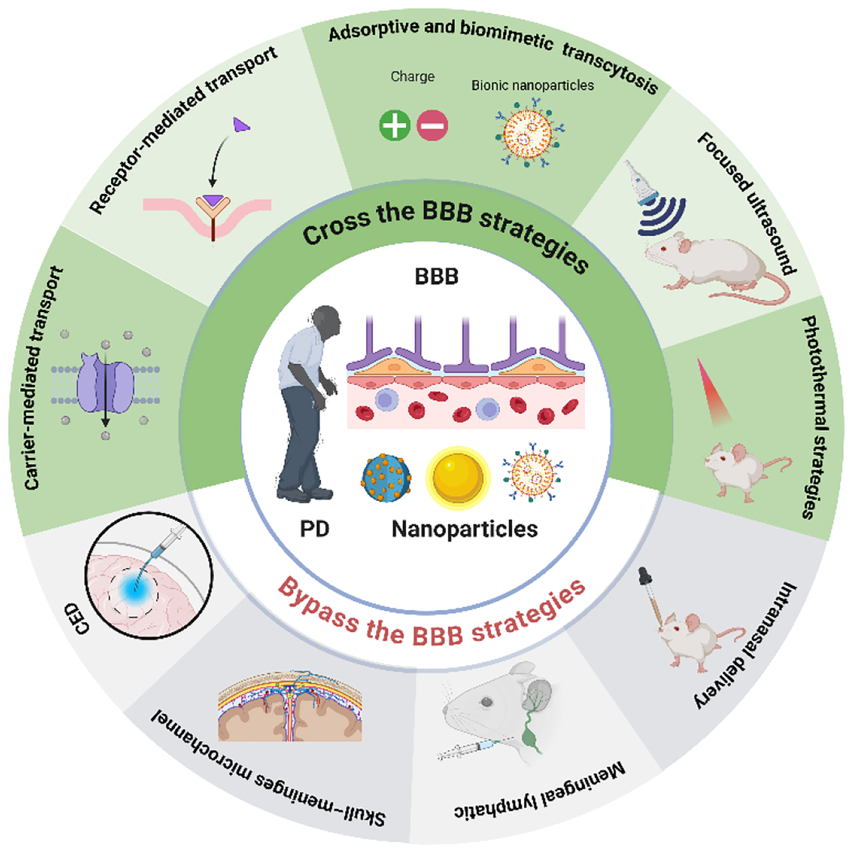
1. Introduction
Parkinson disease (PD) is the second most common neurodegenerative disorder worldwide, clinically characterized by progressive loss of dopaminergic neurons in the substantia nigra pars compacta, striatal dopamine depletion, and a broad spectrum of motor and nonmotor symptoms. Current pharmacotherapies offer symptomatic relief but remain largely palliative; long-term use is frequently constrained by diminishing efficacy, motor fluctuations, dyskinesia, and nonmotor side effects, without halting or reversing the underlying neurodegenerative process[1,2]. This therapeutic shortfall is exacerbated by the rapidly escalating global burden of the disease. PD is the fastest-growing neurological disorder in terms of prevalence and disability; the World Health Organization projects that neurodegenerative diseases, including PD and Alzheimer disease, will become the second leading cause of death globally by 2040, surpassing cancer-related mortality. The total number of PD patients worldwide is expected to reach approximately 25.2 million by 2050, imposing immense socioeconomic costs on healthcare systems and caregivers[3]. These converging factors collectively underscore the urgent and critical need for disease-modifying therapies that can effectively and safely deliver therapeutic agents to the brain.
As the understanding of PD pathogenesis has expanded from dopaminergic deficiency to include α-synuclein aggregation, oxidative stress, mitochondrial dysfunction, neuroinflammation, lysosomal impairment, and disordered neuroimmune interactions (Figure 1), the therapeutic landscape has also broadened from traditional small-molecule replacement strategies to biologics, nucleic acid therapeutics, neurotrophic factors, and gene- or cell-based interventions[4–8]. However, the development of more effective therapies for PD is constrained not only by the complexity of the disease itself but also by the difficulty of delivering therapeutically meaningful amounts of drugs to the brain[9]. Many promising agents show strong neuroprotective or disease-modifying potential in principle, yet fail to achieve sufficient intracerebral exposure after systemic administration[10]. This limitation is especially pronounced for proteins, antibodies, nucleic acids, and other macromolecular therapeutics, whose pharmacological promise is often offset by poor stability, rapid peripheral clearance, and restricted access to the central nervous system (CNS)[11]. Therefore, in PD, the key challenge is often not simply whether a therapeutic molecule can be identified, but whether it can be delivered efficiently, safely, and reproducibly to disease-relevant brain regions[12].
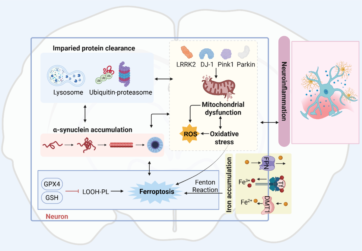
Major pathogenic mechanisms involved in Parkinson disease. This schematic illustrates the interconnected pathogenic processes underlying Parkinson disease. Impaired protein clearance, mitochondrial dysfunction, oxidative stress, iron accumulation, ferroptosis, and neuroinflammation interact to promote α-synuclein accumulation and dopaminergic neuronal injury. PD-related genes, including LRRK2, DJ-1, PINK1, and Parkin, contribute to mitochondrial and proteostatic dysfunction, further aggravating cellular stress.
In this context, nanomedicine has emerged as a highly attractive platform for PD therapy. By enabling drug protection, controlled release, multifunctional integration, and flexible surface engineering, nanoplatforms offer opportunities to improve the therapeutic performance of both conventional drugs and emerging biologics without necessarily redesigning the active molecule itself[13]. At the same time, nanomedicine does not automatically solve the brain delivery problem. The blood–brain barrier (BBB), as a highly selective neurovascular interface, continues to represent the central biological obstacle to effective CNS therapy. Even when nanocarriers improve drug stability or circulation, they still face the fundamental challenge of crossing or circumventing this barrier in a manner that is efficient, regionally relevant, and clinically feasible[14].
Accordingly, current nanomedicine-based approaches for PD can be understood from a delivery perspective as 2 broad and complementary categories: strategies designed to cross the BBB and strategies intended to bypass it[15,16]. The former include carrier-mediated, receptor-mediated, adsorptive, biomimetic, and physically assisted approaches that attempt to negotiate directly with the barrier[17]. The latter seek alternative routes to the brain, such as intranasal delivery, meningeal-associated pathways, skull–meninges-related channels, or local intracranial administration[18–20]. Each category offers distinct opportunities, but each is also shaped by different constraints in transport efficiency, regional distribution, repeatability, safety, and translational practicality.
In this review, we first summarize the current pharmacotherapy of PD and its limitations, and then discuss why nanomedicine provides a meaningful therapeutic and delivery framework for this disease. We next examine the BBB as the central obstacle to brain delivery, followed by a systematic discussion of nanomedicine strategies for crossing or bypassing the BBB. Finally, we compare these 2 strategy groups from a translational perspective and outline the key challenges and future directions for clinically realistic nanomedicine design in PD. Through this framework, we aim to highlight that the future of PD brain delivery lies not in identifying a single universally optimal route, but in developing cargo-matched, target-aware, and clinically translatable delivery strategies.
2. Current treatment strategies for PD and their limitations
Current pharmacotherapy for PD remains predominantly symptomatic[21]. Levodopa, as the gold standard, effectively ameliorates motor symptoms. However, its long-term use is inevitably associated with motor complications such as wearing-off phenomena and dyskinesia, and its efficacy progressively declines with disease progression[22]. Dopamine agonists, monoamine oxidase-B inhibitors, and catechol-O-methyltransferase inhibitors are frequently employed as adjunctive agents to prolong dopaminergic stimulation, yet they are equally incapable of halting the neurodegenerative process[23]. Nondopaminergic drugs, such as anticholinergics and amantadine, provide only supportive symptomatic benefit[24–26]. From a drug delivery perspective, current therapies carry inherent shortcomings: even for levodopa, the cornerstone of clinical management, brain uptake is highly variable due to extensive peripheral metabolism and competition with large neutral amino acids, making precise control of central exposure difficult to achieve[22].
For patients with advanced or medically refractory disease, deep brain stimulation can markedly improve symptoms by modulating pathological activity in the basal ganglia circuits[27]. Nevertheless, the procedure is invasive, resource-intensive, and associated with surgical risks, hardware-related complications, and neuropsychiatric adverse effects[28]. With respect to regenerative and restorative strategies, cell replacement therapies using fetal tissue, induced pluripotent stem cells, or embryonic stem cell-derived dopaminergic neurons have raised considerable hope[29], but limited graft survival, insufficient functional integration, immunological concerns, and safety issues have prevented their translation into widely applicable clinical solutions. Taken together, neither existing pharmacological nor surgical interventions can arrest disease progression[30].
With increasing insight into the molecular pathogenesis of PD, disease-modifying therapies have become a central research priority. Representative strategies include reducing the α-synuclein burden, interfering with its aberrant aggregation and cell-to-cell propagation, and suppressing the expression of disease-causing genes using antisense oligonucleotides or small interfering RNAs[31,32]. However, the most promising disease-modifying candidates, such as anti-α-synuclein antibodies, RNA-based therapeutics, and neurotrophic factors, are biologics or nucleic acid-based agents. Following systemic administration in their free form, these molecules confront a 3-fold delivery barrier: negligible penetration across the BBB, rapid enzymatic degradation in plasma, and the consequent failure to reach therapeutically effective concentrations within the brain parenchyma[33]. In essence, the core challenge is not the lack of pharmacologically active molecules, but the inability of these molecules to access their intracranial targets in sufficient quantities.
This delivery predicament is not unique to disease-modifying approaches; existing small-molecule drugs such as levodopa likewise suffer from highly variable brain uptake and poorly controllable BBB penetration. The key delivery bottleneck facing pharmacotherapy for PD can therefore be formulated as follows: how to deliver centrally active therapeutic molecules, whether small molecules or biologics, across the BBB at sufficient concentrations and with spatiotemporal control to reach the diseased regions[34]. Nanomedicine offers a systematic engineering framework to overcome this bottleneck. In contrast to conventional formulations, nanocarriers can simultaneously fulfill multiple functions: protecting fragile payloads from enzymatic degradation, prolonging systemic circulation half-life, actively traversing the BBB via mechanisms such as receptor-mediated transcytosis and adsorptive-mediated transcytosis (AMT), or bypassing the barrier through routes such as intranasal delivery and the meningeal lymphatic pathway. It is precisely this versatile and engineerable nature that positions nanomedicine as one of the most promising platforms for solving the brain delivery challenge in PD.
3. Nanomedicine provides distinct therapeutic and delivery advantages for PD
The progression of PD is driven collectively by α-synuclein aggregation, oxidative stress, mitochondrial dysfunction, ferroptosis, neuroinflammation, and other factors[35]. However, conventional drugs are largely ineffective at blocking these pathological processes due to low BBB penetration efficiency, short half-life, poor targeting, and significant systemic toxic side effects. The limitations of current pharmacotherapy have prompted increasing interest in therapeutic platforms that can improve drug performance without necessarily redesigning the active pharmaceutical ingredient itself. In this context, nanomedicine has emerged as a promising strategy for PD[14].
One important advantage of nanomedicine lies in its ability to improve the delivery profile of existing small-molecule drugs. Many conventional agents suffer from suboptimal aqueous solubility, rapid clearance, poor metabolic stability, or unfavorable systemic distribution[36]. Nanocarriers can mitigate some of these limitations by enhancing drug solubilization, prolonging circulation time, modulating biodistribution, and reducing premature degradation[37]. Such formulation-level optimization is particularly attractive in PD, where long-term treatment is often required and where maintaining more stable therapeutic exposure may help improve efficacy and tolerability. In this sense, nanomedicine does not simply replace established drugs, but may expand the therapeutic value of already available pharmacological agents[38].
Nanomedicine is also especially relevant for the delivery of biologics and other vulnerable therapeutic cargos that are difficult to use in conventional formulations. Proteins, peptides, nucleic acids, and other macromolecular therapeutics are often limited by poor stability, enzymatic degradation, rapid systemic elimination, or loss of bioactivity before reaching the desired site of action[39,40]. By providing a protective microenvironment, nanoplatforms can help preserve the integrity of these cargos and improve their in vivo persistence[41]. This feature is of particular importance for emerging PD therapies aimed at reducing α-synuclein burden, modulating pathogenic gene expression, or restoring cellular function, many of which rely on fragile biological molecules rather than traditional small drugs[42].
Beyond simple drug protection, nanomedicine offers opportunities for controlled and multifunctional therapy. Through rational design of material composition and nanostructure, nanocarriers can be tailored to achieve sustained release, stimulusresponsive release, or codelivery of multiple therapeutic agents[43,44]. These properties are potentially valuable in PD, a multifactorial neurodegenerative disorder involving dopaminergic neuron loss, α-synuclein aggregation, oxidative stress, mitochondrial dysfunction, and neuroinflammation[45]. Because a single intervention may be insufficient to address such a complex pathological network, nanoplatforms provide an attractive basis for combination therapy and multimodal intervention. In addition, some nanomaterials may contribute auxiliary functions such as reactive oxygen species (ROS) scavenging, imaging contrast, or energy responsiveness, further broadening their therapeutic potential[46,47].
Another major strength of nanomedicine is its high degree of engineerability. Nanoparticles can be tuned in terms of size, shape, surface charge, material composition, ligand decoration, and biomimetic coating, creating opportunities to optimize their biological behavior in a more versatile manner than is usually possible with free drugs alone[48,49]. This structural tunability is particularly important in brain-targeted therapy, where therapeutic success depends not only on the intrinsic activity of the cargo but also on whether it can be delivered to the relevant brain regions in sufficient amounts and with acceptable safety[50,51]. Thus, the value of nanomedicine in PD lies not merely in carrying drugs, but in enabling programmable interactions between therapeutic agents and biological barriers.
Leveraging these engineering advantages, nanomedicines can transcend the limitations of conventional therapies, enabling precision design against the core pathologies of PD and advancing toward disease-modifying treatment. For instance, α-syn aggregation represents a hallmark pathological event that can be targeted through nanomaterial–protein interactions. Nanomaterials can prevent fibrillization through specific surface interactions with hydrophobic regions of α-syn; for example, carboxyl groups on the surface of quantum dots can induce the disassembly of preformed α-syn fibrils into nontoxic monomers[52,53]. Regarding the progressive loss of dopaminergic neurons in the nigrostriatal pathway, which is the pathological alteration directly responsible for clinical symptoms, nanocarriers are required to achieve specific delivery to and long-term protection of this pathway. Encapsulating levodopa in PEGylated liposomes or poly(lactic-co-glycolic acid) nanoparticles enables sustained intracranial drug release, significantly reducing the “on–off” phenomenon and motor complications associated with oral administration[54]. For large-molecule proteins susceptible to enzymatic degradation, such as glial cell line-derived neurotrophic factor (GDNF), nanocarriers not only provide effective protection but can also be surface modified with ligands to achieve precise targeting of dopaminergic neurons[55].
Neuroinflammation acts as a disease amplifier throughout the entire course of PD, with its core pathological feature being the excessive activation of microglia into a pro-inflammatory M1 phenotype and the concomitant release of abundant inflammatory cytokines, which necessitates the precise targeting of activated microglia by nanocarriers. Certain nanomaterials possess intrinsic immunomodulatory activities; cerium oxide nanoparticles can simultaneously scavenge ROS and inhibit microglial activation[56]. Oxidative stress and mitochondrial dysfunction, as the final common pathways leading to neuronal death in PD, have driven the development of mitochondria-targeted antioxidant nanosystems. By modifying nanocarriers with the lipophilic cation triphenylphosphine, therapeutic agents can be accumulated in the mitochondrial matrix, driven by the mitochondrial membrane potential, thereby significantly improving mitochondrial function and reducing ROS production[57]. The characteristic microenvironment of the PD brain, including elevated ROS levels and increased matrix metalloproteinase (MMP) activity, provides natural triggers for the design of stimuli-responsive smart nanocarriers. For example, block copolymer nanoparticles containing ROS-sensitive chemical bonds such as thioether or boronate ester linkages undergo rapid drug release under the high-ROS conditions in the PD brain[58]; and nanoparticles modified with MMP-9-sensitive peptides can release drugs specifically at inflammatory sites, further enhancing therapeutic safety and efficacy[59].
Despite remarkable progress in nanomedicine design based on PD pathological features, numerous challenges remain: nanoplatforms are not inherently capable of efficient brain delivery, nor do they guarantee clinical benefit simply by virtue of nanoscale design. Their real promise lies in the possibility of rational engineering: by improving drug stability, supporting multifunctional therapy, and offering flexible interfaces for biological interaction, nanomedicine creates new opportunities to enhance PD treatment. At the same time, these opportunities can only be realized if the central obstacle to brain delivery, that is, the BBB, is effectively addressed[60]. Importantly, this barrier is not unique to nanomedicine; it represents a fundamental challenge for virtually all therapeutic agents aimed at the CNS. Thus, the key question is not whether nanomedicine faces the BBB, but how its engineerability can be leveraged to address a barrier that equally limits conventional therapies. This leads to the next question: why brain delivery remains so difficult and how the BBB continues to constrain nanomedicine-based interventions in PD.
4. The BBB as the central obstacle to brain delivery in PD
The BBB is a highly specialized and selectively permeable interface that regulates molecular exchange between the systemic circulation and the CNS. By maintaining the biochemical stability of the brain microenvironment and limiting the entry of harmful substances, the BBB plays an essential role in preserving neuronal function and CNS homeostasis[61]. However, the same properties that protect the brain also create a major obstacle for therapeutic intervention in general, regardless of whether the drug is a conventional small molecule, a biologic, or a nanoparticle-based formulation. Efficient and sustained delivery to specific brain regions remains exceptionally difficult for the vast majority of therapeutic agents[62].
Importantly, the BBB is not a single anatomical wall, but a dynamic and integrated neurovascular system. Its structural basis is formed primarily by brain microvascular endothelial cells (BMECs), which are tightly connected and supported by pericytes, astrocytic end-feet, and the basement membrane (Figure 2)[63]. Among these components, BMECs constitute the principal physical barrier by severely restricting paracellular diffusion and tightly regulating transcellular transport[64]. Pericytes contribute to vascular stability, barrier integrity, and local homeostatic regulation, while astrocytes help maintain BBB properties through close neurovascular signaling and modulation of endothelial function. Together, these cellular and extracellular elements establish a highly selective interface that is fundamentally different from the vasculature of most peripheral tissues[65].
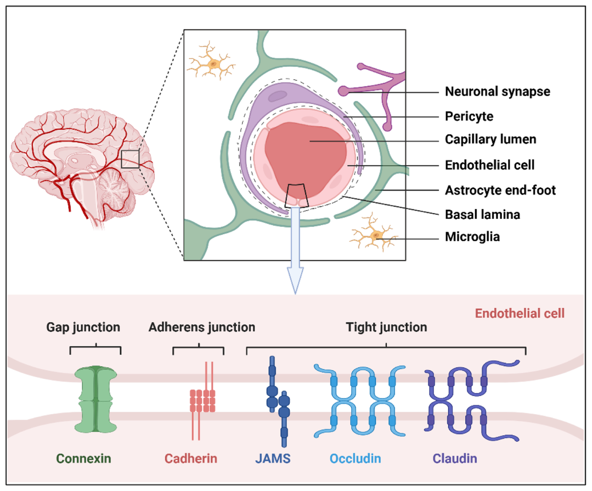
Structural organization of the BBB and the neurovascular unit. This schematic illustrates the cellular architecture of the BBB and the neurovascular unit. Brain microvascular endothelial cells form the vascular wall and are closely associated with the basement membrane, pericytes, astrocytic end-feet, neuronal elements, and microglia, which together contribute to BBB formation and maintenance. The lower panel highlights the major intercellular junctional complexes between adjacent endothelial cells, including tight junctions, adherens junctions, and gap junctions, with representative components such as claudin, occludin, junctional adhesion molecules, cadherins, and connexins.
From a drug delivery perspective, the restrictive nature of the BBB arises from several interrelated functional features. First, the intercellular junctions between BMECs greatly limit passive paracellular movement of solutes[66]. Second, transcellular trafficking across the BBB is tightly controlled, allowing only selected endogenous molecules to enter the brain through specialized transport systems[67]. Third, endothelial cells express metabolic enzymes and active efflux transporters, which can degrade therapeutic molecules or pump them back into the circulation, thereby further reducing intracerebral exposure[68]. As a result, although some small molecules can partially access the CNS, most macromolecules, biologics, and nanoparticle-based systems cannot simply diffuse into the brain in therapeutically meaningful amounts[69].
These BBB properties are particularly relevant in the context of nanomedicine. While nanocarriers can improve drug stability, prolong circulation, and support multifunctional therapeutic design, they do not inherently possess efficient brain-penetrating capacity[70]. On the contrary, their size, surface properties, and interactions with biological interfaces often make them even more dependent on active transport mechanisms, transient barrier modulation, or alternative delivery routes. In other words, the challenge in PD is not merely to develop nanomedicine formulations, but to design nanomedicine systems that can interact with the BBB in a controlled, effective, and safe manner[71].
Another important consideration is that the BBB in PD should not be viewed as a completely static barrier. Aging, chronic neuroinflammation, vascular dysfunction, and disease progression may alter BBB integrity and transport behavior in a region- and stage-dependent manner[36]. Astrocyte activation, endothelial dysfunction, and neurovascular remodeling have all been implicated in BBB dysregulation in neurodegenerative conditions, including PD[72–74]. Although these changes may in some cases create transient opportunities for drug entry, they also introduce additional heterogeneity and uncertainty into delivery design and therapeutic evaluation[75]. Therefore, successful nanomedicine-based intervention in PD requires not only overcoming the BBB as a general barrier, but also accounting for its dynamic pathological context[76].
Taken together, the BBB remains the central biological constraint on brain delivery in PD. Its structural selectivity, tightly regulated transport behavior, and disease-related variability explain why many therapeutics fail to achieve adequate exposure in the target brain regions, even when they show promising pharmacological activity in principle[77,78]. This challenge has driven the development of 2 broad categories of nanomedicine-based delivery strategies: those designed to cross the BBB and those intended to bypass it. The following sections discuss these 2 approaches in detail.
5. Nanomedicine strategies for crossing the BBB
Given that the BBB remains the principal obstacle to brain delivery in PD, considerable effort has been devoted to the development of nanomedicine strategies capable of crossing this barrier. Unlike bypass approaches, these strategies seek to negotiate directly with the BBB by harnessing endogenous transport mechanisms, promoting vesicular translocation, or temporarily increasing barrier permeability. Their appeal lies in the potential to achieve brain delivery while preserving the anatomical integrity of the delivery route and avoiding more invasive procedures[17]. Based on their underlying principles, current approaches can be grouped into transport-mediated systems, adsorptive or biomimetic transcytosis strategies, and physically assisted BBB-opening technologies (Figure 3).
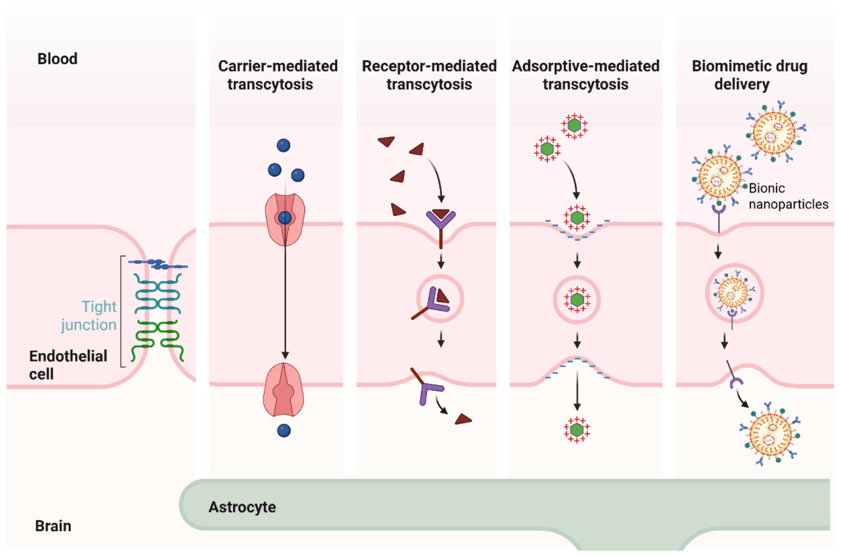
Major biological transport mechanisms for crossing the BBB. This schematic summarizes several representative biological pathways by which therapeutic agents or nanomedicines may traverse the BBB, including carrier-mediated transcytosis, receptor-mediated transcytosis, adsorptive-mediated transcytosis, and biomimetic drug delivery. Carrier-mediated transcytosis exploits endogenous solute transport systems to facilitate BBB passage. Receptor-mediated and biomimetic strategies rely on ligand-receptor interactions on the luminal surface of endothelial cells to trigger endocytosis and vesicular transcellular transport. Adsorptive-mediated transcytosis is driven by electrostatic interactions between positively charged nanomedicines and negatively charged components of the endothelial surface. Together, these mechanisms provide distinct routes for enhancing brain delivery across the BBB.
5.1 Carrier-mediated transport-based strategies
Carrier-mediated transport (CMT) is one of the major endogenous transport mechanisms that support nutrient exchange across the BBB. To meet the metabolic demands of the brain, brain endothelial cells express a variety of saturable and bidirectional transporters responsible for the uptake of essential small polar molecules, including glucose, amino acids, vitamins, fatty acids, and other nutrient-related substrates. These carriers recognize specific ligands and undergo conformational transitions that enable substrate translocation across the endothelial barrier. Based on this principle, nanomedicine platforms can be functionalized with transporter-recognizable substrates or substrate-mimicking moieties in an attempt to exploit these physiological transport systems and enhance brain delivery[79].
Compared with passive diffusion, CMT-based strategies are attractive because they rely on intrinsic BBB transport routes rather than nonspecific permeabilization. In principle, such approaches may improve brain entry while preserving barrier integrity and reducing the need for invasive intervention[9]. For PD therapy, this concept is especially relevant for cargos that cannot achieve sufficient intracerebral exposure in free form but may benefit from transporter-assisted entry when incorporated into a nanoplatform. In addition, because some endogenous transport systems are highly expressed at the BBB, they provide a biologically rational interface for the design of brain-targeted nanocarriers[65].
Several representative studies have explored this strategy. For example, a polymer containing choline- and acetylcholine-like moieties was reported to cross the BBB under the mediation of choline- and acetylcholine-related transport systems, thereby enabling the delivery of protein therapeutics into the CNS[80]. In another study, a gold nanoparticle-based composite nanovehicle loaded with plasmid DNA was modified with docosahexaenoic acid (DHA) and nerve growth factor (NGF). In this design, DHA was intended to facilitate BBB transport through interaction with fatty acid-related transport pathways, while NGF was further used to promote neuronal targeting after brain entry[81]. These examples illustrate how CMT-inspired nanodesign can be combined with secondary targeting elements to improve not only BBB interaction but also subsequent distribution toward disease-relevant cells.
Although CMT is conceptually attractive, its practical application still faces several key challenges. First, the number and transport capacity of endothelial carriers are inherently limited, and saturation and competition effects exist. This necessitates nanocarrier designs that balance high affinity with low endogenous interference. To this end, 1 can select carriers that are highly expressed on brain endothelial cells and possess relatively large transport cavities as targets, and employ nonnatural amino acid/sugar mimetics or allosteric modulators to avoid competition with natural substrates for the active site, thereby achieving selective delivery without sacrificing overall transport flux. Second, most carriers are naturally adapted to small-molecule substrates and exhibit limited compatibility with complex nanoplatforms; however, this can be addressed by multivalent ligand modification or biomimetic mineralization strategies to enhance apparent affinity, thereby improving the transport efficiency of larger-sized carriers. Furthermore, the heterogeneity of transporter expression across species, brain regions, and pathological conditions suggests that clinical translation should incorporate individualized assessment or imaging guidance[82–84]. Importantly, these challenges are not inherent flaws of CMT-based strategies, but rather drivers for innovation in precision nanoengineering. Compared with nonspecific permeabilization or invasive approaches, CMT offers a physiologically compatible and regulatable route for brain delivery, and with rational design, it still holds the potential to achieve efficient and low-toxicity brain-targeted delivery.
5.2 Receptor-mediated transport-based strategies
Receptor-mediated transport (RMT) remains one of the most intensively investigated routes for brain-targeted nanomedicine, largely because it harnesses endogenous endocytic and transcytotic machinery expressed on the luminal membrane of the BBB[85]. Unlike carrier-mediated systems, which are evolutionarily tuned for small nutrients, RMT exploits ligand-receptor recognition to trigger vesicular trafficking of macromolecular cargos, an attractive feature for delivering proteins, antibodies, nucleic acids, and multifunctional nanocarriers that otherwise show negligible free penetration into the brain[55]. In PD, this strategy gains additional relevance from the need to not only cross the BBB but also reach vulnerable dopaminergic neurons, a dual requirement that has spurred considerable innovation in ligand design and nanoplatform engineering.
A broad palette of ligands, including transferrin, lactoferrin, Angiopep-2, dopamine-related moieties, and RVG29-derived peptides, has been conjugated to nanoparticles to promote selective engagement with endothelial receptors[86,87]. Although each ligand-receptor pair has its own binding kinetics and tissue distribution profile, a common rationale underpins these designs: to enhance brain accumulation while, in many cases, conferring secondary neuronal tropism after extravasation[9]. This dual-targeting potential is particularly coveted in PD, where therapeutic efficacy depends on precise delivery to disease-relevant cell populations. Among the most widely explored are lactoferrin-modified systems, which benefit from the fact that lactoferrin receptors are present on both brain endothelium and neurons. For instance, a lactoferrin-functionalized platform that releases nitric oxide and paeoniflorin under near-infrared (NIR) stimulation successfully combined gas therapy with pharmacological intervention in a PD model[88]; Similarly, lactoferrin-decorated black phosphorus (BP) nanosheets loaded with paeoniflorin demonstrated favorable biocompatibility and brain-targeting performance[89]. Transferrin receptor-targeted carriers, by contrast, have been particularly valuable for macromolecular biologics, as exemplified by transferrin-modified liposomes delivering the anti-α-synuclein antibody SynO4, which achieved markedly higher antibody accumulation in brain cells than free antibody[90]. These cases illustrate that while the same RMT principle applies, the choice of ligand substantially influences the type of cargo that can be efficiently shuttled and the subsequent intracellular fate.
Beyond simple ligand decoration, recent work has pushed RMT toward multifunctional, stimuli-responsive architectures that integrate BBB crossing with disease-microenvironment sensing. A ROS-responsive, carrier-free nanomedicine was designed to exploit dopamine receptor-mediated uptake, enabling BBB passage and neuronal internalization before releasing rasagiline mesylate and dopamine specifically in the oxidative PD milieu[91]. Angiopep-2-modified nanoparticles have been used for codelivery of rasagiline and the P-glycoprotein inhibitor tariquidar, achieving significantly enhanced brain targeting relative to free drug[92]. RVG29-based systems further expand the toolkit by recognizing nicotinic acetylcholine receptor-related targets on both endothelium and neurons. They have been employed for codelivery of miR-133b and curcumin to simultaneously mitigate α-synuclein pathology, attenuate neuroinflammation, and promote repair[93]. Notably, RVG29-functionalized red blood cell membrane-coated curcumin nanoparticles have shown prolonged circulation and reduced reticuloendothelial clearance, underscoring how RMT can be effectively merged with biomimetic coating strategies to enhance overall pharmacokinetic performance[94]. Collectively, these examples demonstrate that RMT is not a monolithic tactic but a flexible backbone onto which multiple layers of functionality, such as stimulus responsiveness, combination therapy, and immune evasion, can be grafted. Beyond therapeutic delivery, RMT has also been extended to diagnostic applications; for instance, a HClO-activatable NIR-IIb fluorescent probe (SQ6-RENps) was recently reported to cross the BBB via VHPKQHR peptide-mediated transport, enabling real-time monitoring of intracerebral hypochlorous acid levels to reflect PD severity and evaluate therapeutic responses[95].
Despite these advances, the clinical translation of RMT-based nanomedicines faces persistent hurdles that go far beyond the initial receptor-binding step. The most fundamental bottleneck is that binding does not equate to productive transcytosis: after endocytosis, the vast majority of nanoparticles are either sequestered in lysosomes for degradation or recycled back to the vascular lumen, with only a minute fraction successfully reaching the brain parenchyma. This low trafficking efficiency is compounded by competitive inhibition from endogenous ligands (eg, transferrin in serum) and receptor saturation at high nanomedicine doses, which collectively cause in vitro targeting performance to deteriorate sharply in vivo[96–98]. Moreover, the rapid adsorption of plasma proteins onto nanoparticle surfaces, forming a protein corona, can physically mask targeting ligands, further undermining receptor recognition and contributing to the frequent discrepancy between cell-based assays and animal results[99]. Target specificity poses another layer of difficulty: receptors such as the transferrin receptor are widely expressed in peripheral organs (liver, spleen), leading to off-target accumulation that not only reduces brain bioavailability but also raises systemic toxicity risks[100]. Receptor density, trafficking kinetics, and accessibility also vary across species, disease stages, and brain regions, making it perilous to extrapolate preclinical findings directly to human PD pathology[15,101]. Even multivalent ligand modification, while boosting apparent affinity, may alter nanoparticle size, surface charge, and pharmacokinetics in ways that impair extravasation and tissue penetration. Safety considerations further complicate the picture; antibody- or peptide-based ligands can elicit immunogenicity, and chronic administration may disrupt receptor homeostasis and compromise BBB integrity[102].
Addressing these challenges will require a coordinated, multipronged shift in design philosophy. Future research should move beyond the pursuit of high-affinity binding toward low-affinity, high-selectivity ligands that minimize competition and off-target effects, coupled with anti-protein-corona surface modifications to preserve targeting function in biological fluids. Enhancing lysosomal escape and intracellular trafficking, for instance via pH-responsive or fusogenic moieties, could significantly increase the fraction of internalized nanoparticles that undergo transcytosis. Equally important is the adaptation of RMT systems to pathological features, such as by designing ligands that recognize disease-upregulated receptors or by incorporating microenvironment-triggered release to ensure cargo deployment only at sites of active neurodegeneration[103–105]. Ultimately, the success of receptor-mediated strategies will depend not on a single breakthrough but on the synergistic optimization of ligand design, nanoparticle engineering, and a deeper understanding of BBB dynamics under disease conditions, a goal that demands iterative feedback between preclinical models and clinically relevant benchmarks.
5.3 Adsorptive and biomimetic transcytosis strategies
In addition to carrier- and receptor-mediated pathways, nanomedicine platforms may also cross the BBB through adsorptive or biomimetic interactions with the brain endothelium. These approaches are attractive because they do not rely exclusively on a single endogenous transporter or receptor. Instead, they seek to enhance BBB translocation by either increasing nonspecific but exploitable interactions with the endothelial surface or by endowing nanocarriers with biological interfaces that more closely resemble native cells or extracellular vesicles[106,107]. In the context of PD, such strategies are of particular interest because they can be integrated with multifunctional nanoplatform design and may offer broader flexibility for delivering diverse therapeutic cargos.
AMT is primarily driven by electrostatic interactions between positively charged carriers and negatively charged domains on the luminal surface of BBB endothelial cells[106]. The endothelial membrane contains anionic components, such as glycosaminoglycan-rich structures, that can promote the adsorption of cationic nanomaterials or macromolecular therapeutics. After this initial surface interaction, the adsorbed carriers may undergo endocytic uptake, intracellular vesicular trafficking, and subsequent exocytotic release on the abluminal side, thereby completing transcytosis across the barrier[108]. From a design perspective, AMT can be promoted by using cationic polymers, surface charge engineering, cell-penetrating peptides, or other positively charged surface modifications[79,109]. Compared with highly specific receptor-targeted systems, AMT may provide a more generally accessible route for BBB interaction, particularly for nanosystems that are difficult to functionalize with precise ligand-receptor pairs.
Representative studies suggest that AMT-based concepts can be combined with more complex vesicular formulations. For example, a delivery system based on small apoptotic bodies was used to encapsulate antisense oligonucleotide-containing nanocomplexes, taking advantage of membrane-associated interactions with BBB endothelial glycocalyx components to facilitate endothelial uptake and transcytosis[33]. This example highlights an important feature of AMT-inspired strategies: electrostatic adsorption alone is often not the whole story, and successful BBB transfer may depend on the coordinated contribution of surface charge, membrane composition, and vesicular trafficking behavior. Thus, AMT should be understood less as a single mechanism and more as a design principle for enhancing endothelial interaction and transcellular transport.
Biomimetic transcytosis strategies take this concept further by equipping nanocarriers with cell-derived or cell-like interfaces to improve circulation, immune evasion, BBB interaction, and downstream targeting. Over the past decade, biomimetic delivery systems have attracted growing attention because cell membranes, extracellular vesicles, and related biological coatings can endow nanoparticles with functions that are difficult to reproduce using purely synthetic materials[110]. Depending on the source of the membrane or vesicle, these functions may include prolonged systemic circulation, reduced reticuloendothelial clearance, inflammatory tropism, neuronal affinity, or enhanced compatibility with the BBB[111,112]. For PD therapy, such features are particularly appealing because the disease requires not only access to the CNS, but often selective action within neuroinflammatory or neurodegenerative microenvironments.
Several membrane-based biomimetic strategies have been explored in PD models. Neural stem cell membrane-coated nanoparticles have been reported to improve the targeted delivery of antisense oligonucleotides while also supporting endogenous neural stem cell recruitment for neuronal repair[113]. Red blood cell membrane-based systems have been used to prolong circulation and reduce immune clearance, and when combined with stimulus-responsive cores, they can further support controlled therapeutic release after brain delivery[114]. Neuronal membrane-coated nanoparticles have also been developed to target activated microglia and modulate neuroinflammation, whereas macrophage membrane-based nanosystems may exploit inflammation-associated homing features while carrying mitochondria-protective or antiinflammatory payloads[115]. In addition, engineered exosome-like systems have shown considerable promise because their intrinsic intercellular communication properties and nanoscale dimensions make them attractive vehicles for RNA drugs, small molecules, and combination formulations. For instance, exosome-based hybrid systems have been designed for the codelivery of SNCA-targeting siRNA and curcumin, aiming to simultaneously reduce α-synuclein pathology and regulate immune activation in PD-related settings[116]. A recent study further developed a biomimetic nanosystem (Cur-Exo@Pt-PB) consisting of curcumin-derived exosomes encapsulating platinum-Prussian blue nanozymes, which effectively traverses the BBB by modulating protein corona composition to evade immune clearance while synergistically exerting antioxidant effects and inducing microglial M2-polarized antiinflammatory responses, thereby achieving effective therapy for PD[117].
Despite their conceptual appeal, adsorptive-mediated and biomimetic transcytosis strategies face distinct limitations. In AMT-based systems, increased positive charge can enhance BBB interaction but may also increase nonspecific binding, serum protein adsorption, cytotoxicity, and off-target uptake. The resulting gain in endothelial association does not always translate into efficient or selective delivery to the brain parenchyma[118]. Biomimetic systems, while often offering improved biological performance, come at the cost of greater complexity. First, manufacturing scalability and batch-to-batch consistency remain fundamental obstacles, as most biomimetic nanoplatforms are synthesized at the milligram scale using nonstandardized laboratory procedures that are often incompatible with large-scale production[119]. Second, inadequate storage and transport stability represent another critical issue, since membrane proteins and lipids are susceptible to denaturation, oxidation, aggregation during storage, imposing stringent requirements on cold-chain management[120]. Third, immunogenicity risks cannot be overlooked; although biomimetic strategies aim to evade immune clearance through “self” recognition, the introduction of exogenous biological materials may still trigger unintended immune activation, and long-term biosafety data remain largely lacking[121]. Moreover, although exosomes and membrane-coated nanoparticles are often described as naturally brain-targeting, their in vivo trafficking and BBB transport mechanisms are not always fully understood or quantitatively validated[122]. From a clinical translation perspective, no biomimetic nanoplatform has yet been approved for PD treatment, largely due to the absence of harmonized regulatory guidelines specifically tailored to such complex nanomedicines, insufficient systematic evaluation of biodistribution and pharmacokinetics, and interspecies differences that make it difficult to replicate preclinical efficacy in humans[43]. Consequently, while adsorptive and biomimetic strategies expand the design space of BBB-crossing nanomedicine for PD, their successful translation will depend on achieving a workable balance between biological sophistication, mechanistic clarity, safety, and manufacturability. Future research should focus on developing scalable and standardized manufacturing processes, establishing robust quality control systems, and conducting thorough assessments of long-term biosafety and immunogenicity.
5.4 Focused ultrasound-assisted BBB opening
Focused ultrasound (FUS) represents a distinct class of BBB-crossing strategies because it does not primarily rely on ligand-mediated interaction with endothelial transport systems. Instead, it uses externally applied acoustic energy to locally and transiently modulate BBB permeability, thereby creating a physical window for the entry of therapeutic agents into the brain[123]. In the field of PD, FUS has attracted growing interest not only because of its established use in image-guided neuromodulatory or ablative procedures, but also because its combination with intravenously administered microbubbles has opened a new avenue for noninvasive, region-specific brain delivery of macromolecules, gene vectors, and nanosystems[124].
The therapeutic application of FUS in PD initially centered on thermal ablation. By focusing ultrasound energy onto defined deep brain targets, such as the subthalamic nucleus or related motor circuits, FUS can induce localized tissue heating and thereby alleviate symptoms such as tremor[125–127]. However, from a drug delivery perspective, the more transformative development has been the emergence of microbubble-assisted focused ultrasound (MB+FUS). In this approach, systemically administered microbubbles oscillate in the ultrasound field within the targeted cerebral vasculature, generating mechanical effects at the neurovascular interface. These effects can temporarily loosen endothelial tight junctions and enhance vesicular transport activity, leading to a reversible increase in BBB permeability[128]. Compared with systemic chemical modulation or invasive local administration, MB+FUS offers several important advantages, including regional precision, temporal controllability, and the possibility of repeated noninvasive intervention[129].
However, the physical window opened by ultrasound alone does not guarantee efficient delivery of free therapeutic agents. Even in the region of barrier opening, free drugs often fail to cross effectively or are rapidly cleared from the circulation. The introduction of nanocarriers significantly improves this process. Nanocarriers can encapsulate macromolecules, nucleic acids, or unstable drugs into particles of appropriate size, enabling them to pass through the transient channels induced by ultrasound, while also prolonging circulation time and reducing peripheral organ exposure[130,131]. Surface modification with targeting ligands enhances local accumulation, and environment-responsive designs allow triggered release at the lesion site. Together, these features reduce systemic toxicity and protect the cargo from enzymatic degradation[132]. Furthermore, nanoplatforms can carry gene-editing tools or enable combination therapy, greatly expanding the repertoire of deliverable drugs[133]. Thus, the value of nanocarriers lies in transforming the local opening effect of ultrasound into meaningful therapeutic benefit in the brain, rather than serving merely as an auxiliary tool for barrier traversal.
These features make MB+FUS particularly attractive for PD therapies that are pharmacologically promising but delivery-limited. Neurotrophic factors such as GDNF and brain-derived neurotrophic factor have long been recognized for their ability to support dopaminergic neuron survival and repair, yet their clinical application has been severely restricted by insufficient brain delivery. To address this problem, FUS-based systems have been developed to combine circulating carriers with ultrasound-triggered BBB opening. For example, a microbubble–liposome complex loaded with neurotrophic factors was reported to achieve targeted intracerebral delivery after FUS irradiation, resulting in significant behavioral and pathological improvement in PD model mice (Figure 4A). This type of strategy illustrates the value of FUS as a delivery amplifier: rather than replacing the nanocarrier, it enables a locally gated entry process that can enhance the effectiveness of otherwise poorly brain-penetrant therapeutics[134].
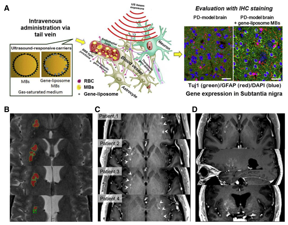
Focused ultrasound-assisted BBB opening for brain delivery. (A) Schematic illustration of intravenous administration of gene-liposome microbubbles followed by focused ultrasound exposure to induce localized BBB opening and facilitate intracerebral gene delivery. Immunohistochemical staining of PD-model brains shows gene expression in the substantia nigra after treatment with gene-liposome microbubbles[134]. Copyright 2020, Elsevier B.V. (B) Representative T2-weighted MRI acquired during an MR-guided focused ultrasound (MRgFUS) procedure, with the colormap indicating the relative ultrasound dose delivered to the putamen. (C) Contrast-enhanced T1-weighted MRI from individual patients showing gadolinium extravasation in the sonicated putamen (arrows), indicating localized BBB disruption. (D) Representative contrast-enhanced T1-weighted MRI in orthogonal planes illustrating the three-dimensional configuration of the BBB opening [135]. Copyright 2022, The Authors. Movement Disorders published by Wiley Periodicals LLC on behalf of the International Parkinson and Movement Disorder Society.
Magnetic resonance-guided focused ultrasound (MRgFUS) has further improved the translational potential of this approach by integrating real-time imaging with precisely controlled acoustic targeting. In a phase I clinical study involving PD patients with GBA1 mutations, MRgFUS combined with microbubbles was used to transiently open the BBB in the putamen in order to improve the intracerebral delivery of glucocerebrosidase (GCase) enzyme therapy. Preliminary findings suggested that the procedure was feasible and safe, with BBB integrity largely restored within 24 h and no evidence of long-term disruption (Figure 4B–D). Although still early, such studies provide important clinical proof-of-principle that FUS-assisted BBB opening may become a realistic platform for enabling brain delivery of biologics in PD[135].
The main challenge for FUS-assisted BBB opening is not proof of concept, but parameter control, reproducibility, and long-term neurovascular safety. Its delivery outcome depends heavily on ultrasound parameters such as acoustic pressure, pulse duration, frequency, exposure time, and cavitation behavior, all of which must be optimized to balance delivery efficiency against tissue safety[136]. In addition, microbubble formulation, circulation stability, and acoustic responsiveness can substantially influence treatment reproducibility. Repeated BBB opening also raises important questions regarding cumulative neurovascular effects, inflammatory responses, and long-term safety, particularly in elderly or neurodegenerative populations[137,138]. Therefore, FUS should be viewed not simply as a BBB-opening technique, but as a highly promising yet parameter-sensitive enabling technology whose clinical value in PD will depend on careful standardization, imaging guidance, and integration with appropriately designed therapeutic cargos.
5.5 Photothermal strategies for BBB modulation and brain delivery
Photothermal strategies represent another physically assisted approach for enhancing brain delivery across the BBB. Unlike transporter- or receptor-dependent nanomedicine systems, these approaches rely on photothermal agents that absorb NIR light and convert optical energy into localized heat, thereby transiently increasing BBB permeability and facilitating drug entry into the brain[139,140]. In the context of PD, photothermal-assisted delivery has attracted interest because it combines relatively low invasiveness with spatial controllability and can be integrated with multifunctional nanoplatform design. Notably, the photothermal agent itself is typically a nanomaterial (eg, BP nanosheets and polydopamine nanoparticles). Beyond serving as the energy converter, the nanocarrier encapsulates and protects the therapeutic cargo, enables brain targeting through surface modification, and can achieve stimulus-responsive drug release. This dual or triple functionality is difficult to replicate with small-molecule photothermal agents or with free drugs alone[89]. Rather than serving solely as a carrier property, the photothermal effect itself becomes an active component of the delivery process[141].
The effectiveness of this strategy depends largely on the properties of the photothermal agent. Ideally, photothermal materials used for BBB modulation should exhibit strong NIR absorption, high photothermal conversion efficiency, good photostability, biodegradability, and acceptable biocompatibility[142,143]. Upon NIR irradiation, localized heating at the vascular interface may disrupt endothelial integrity or transiently increase barrier permeability, thereby enabling the delivery of therapeutic cargos that would otherwise show poor brain penetration. At the same time, this mode of delivery also offers the possibility of coupling BBB modulation with stimulus-responsive drug release, which is particularly attractive for multifunctional nanosystems in PD[114,142].
BP-based nanomaterials are among the most widely investigated photothermal platforms in this context because of their high photothermal conversion efficiency and biodegradability[144]. For example, polyethylene glycol (PEG)-modified BP nanosheets loaded with matrine were developed as a multifunctional nanomedicine for PD (Figure 5A). In this system, NIR-triggered photothermal effects promoted BBB penetration, while matrine contributed antioxidant activity through regulation of oxidative stress-related pathways (Figure 5B). As a result, the combined platform was reported to exert synergistic antiparkinsonian effects through ROS scavenging and reduction of α-synuclein aggregation[145]. This example illustrates how photothermal assistance can be integrated with a therapeutic cargo that is pharmacologically relevant but otherwise limited by insufficient brain exposure.
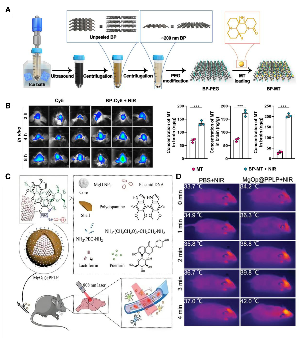
Photothermal-assisted nanomedicine for BBB modulation and brain delivery. (A) Schematic illustration of the preparation of the black phosphorus-based multifunctional nanoplatform BP-MT. (B) In vivo fluorescence imaging of C57BL/6 mice after administration of free Cy5 or BP-Cy5 with near-infrared (NIR) irradiation, together with quantitative analysis of matrine (MT) accumulation in the brain at 2, 4, and 8 h after treatment with free MT or BP-MT plus NIR irradiation (n = 3). Compared with the MT group: P < 0.05, *P < 0.01, and **P < 0.001[145]. Copyright 2022, Elsevier B.V. All rights reserved. (C) Schematic illustration of the synthesis of MgOp@PPLP and its NIR-triggered therapeutic procedure. (D) Representative infrared thermal images of mice exposed to 808 nm laser irradiation (0.8 W/cm2) after administration of PBS or MgOp@PPLP[146]. Copyright 2022, Wiley-VCH GmbH.
Other photothermal nanoplatforms have extended this concept by combining BBB modulation with gene-related or mitochondria-protective therapies. A representative example is the MgOp@PPLP system, which uses MgO nanoparticles as a core, polydopamine (PDA) as a photothermal framework, and an anti-SNCA plasmid as a therapeutic component, with additional PEG, puerarin, and lactoferrin modifications to improve hydrophilicity, antioxidant activity, and brain targeting (Figure 5C). Under 808 nm NIR irradiation, the PDA component generated a controlled temperature increase that promoted BBB permeability and enhanced brain delivery of the nanomedicine (Figure 5D)[146]. Similarly, a zeolitic imidazolate framework 8-coated Prussian blue nanocomposite loaded with quercetin has been developed to exploit the photothermal properties of Prussian blue for BBB modulation while simultaneously delivering a neuroprotective small molecule. These studies highlight the versatility of photothermal approaches, which can support the delivery of small molecules, nucleic acid cargos, and multifunctional combination systems[142].
Despite these promising results, photothermal BBB modulation remains at a relatively early stage compared with more established approaches such as FUS-assisted barrier opening. One important limitation is the restricted tissue penetration depth of light, which constrains the efficiency and clinical practicality of NIR-triggered strategies, especially for deep brain targets relevant to PD[147]. In addition, achieving a therapeutically useful increase in BBB permeability without inducing excessive endothelial damage or off-target thermal injury requires careful control of irradiation parameters and photothermal material properties. The long-term biosafety, reproducibility, and spatial precision of these systems also remain to be fully established[148]. Therefore, photothermal strategies should currently be viewed as an innovative but still developing class of BBB-modulating technologies, with particular value as multifunctional nanomedicine platforms rather than as mature standalone solutions for PD brain delivery.
6. Nanomedicine strategies for bypassing the BBB
In contrast to BBB-crossing approaches, bypass strategies seek to reduce dependence on transvascular transport across the classical BBB by exploiting alternative anatomical routes or local delivery methods. Rather than negotiating directly with endothelial transport systems or transiently modulating barrier permeability, these approaches attempt to access the CNS through pathways such as the nasal cavity, meningeal-associated routes, or direct intracranial administration[149–151]. In PD, bypass strategies are particularly attractive for therapeutic cargos that are difficult to deliver efficiently through systemic circulation, including macromolecules, nucleic acids, and other formulations that require higher local exposure. At the same time, bypassing the BBB does not eliminate the challenges of brain delivery; instead, it shifts the main constraints toward factors such as delivery flux, regional distribution, repeatability, invasiveness, and clinical feasibility[152,153]. Based on these principles, current nanomedicine-based bypass strategies can be broadly categorized into intranasal delivery, meningeal-associated pathways, skull–meninges-related routes, and convection-enhanced or other local intracranial delivery approaches.
6.1 Intranasal delivery
Intranasal delivery is one of the most extensively investigated strategies for bypassing the BBB in brain-targeted therapy. Its central appeal lies in the possibility of delivering therapeutic agents from the nasal cavity to the CNS through direct anatomical connections, thereby reducing reliance on systemic circulation and classical transvascular BBB transport. Compared with oral or parenteral administration, intranasal delivery offers several potential advantages, including noninvasiveness, ease of administration, relatively rapid onset, and the possibility of limiting peripheral exposure. In PD, these features are particularly attractive for therapeutic agents that require repeated dosing or that exhibit insufficient brain exposure after conventional administration[19,154].
The nose-to-brain transport of drugs is thought to occur mainly through direct pathways associated with the olfactory and trigeminal systems. After deposition on the upper nasal mucosa, therapeutic agents may access the olfactory bulb and then distribute to deeper brain regions along olfactory-associated routes or reach the brainstem and other CNS areas through trigeminal nerve-related pathways (Figure 6A)[155,158]. Additional contribution from perineural or cerebrospinal fluid-adjacent spaces has also been proposed[155]. Although part of the administered dose may still enter the systemic circulation through nasal capillaries, the principal value of intranasal delivery lies in its capacity to provide a more direct route to the brain and potentially improve intracerebral exposure while reducing systemic side effects[159].
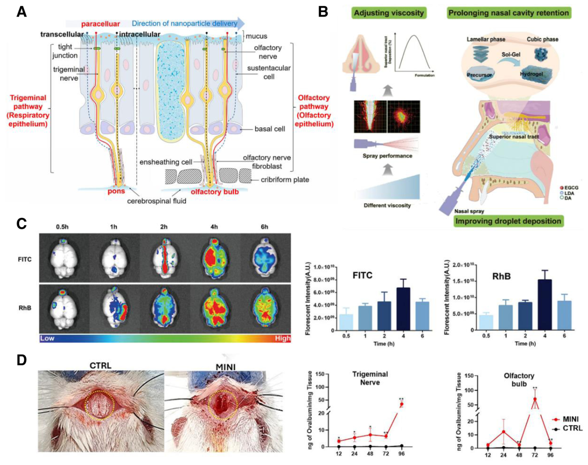
Intranasal nanomedicine delivery to the brain. (A) Schematic illustration of the major nose-to-brain transport pathways for nanoparticles, including trigeminal and olfactory routes [155]. Copyright 2023, American Chemical Society. (B) Schematic illustration of an in situ gel nasal spray designed for nose-to-brain delivery in a PD model. By adjusting formulation viscosity and inducing sol–gel phase transition after administration, this strategy improves upper nasal cavity deposition and prolongs mucosal retention to facilitate the codelivery of multiple drugs. (C)Representative whole-brain fluorescence imaging and semiquantitative analysis of surrogate drug distribution following intranasal administration of a lyotropic liquid crystal (LLC)-based in situ gel spray. Fluorescent probes, fluorescein isothiocyanate (FITC) and rhodamine B (RhB), were employed as surrogates for levodopa (LDA) and epigallocatechin gallate (EGCG), respectively[156]. Copyright 1999–2026, John Wiley & Sons, Inc. or related companies. (D) Schematic and representative images of a minimally invasive nasal infusion (MINI) strategy using a programmable infusion pump for intranasal delivery in mice. Quantification of ovalbumin levels in the trigeminal ganglion and olfactory bulb demonstrates enhanced regional brain delivery compared with control treatment [157]. Copyright 2024, The Authors
However, its practical performance is strongly constrained by multiple anatomical and physiological barriers within the nasal cavity. The nasal mucosa imposes multiple barriers, including mucus entrapment, epithelial resistance, local enzymatic degradation, mucociliary clearance, and the limited accessible area of the olfactory region. These factors can substantially reduce drug retention, absorption, and reproducibility of delivery to the brain. Nanocarriers substantially improve the performance of intranasal delivery by addressing multiple physiological barriers[19]. They can: (i) enhance mucosal penetration through surface charge or mucoadhesive polymers; (ii) protect encapsulated drugs from enzymatic degradation in the nasal cavity; (iii) prolong residence time by avoiding rapid mucociliary clearance; and (iv) provide sustained or controlled release after reaching the olfactory epithelium. By contrast, free drugs administered intranasally often suffer from poor absorption, rapid clearance, and low bioavailability. Thus, the value of nanomedicine in intranasal delivery is not merely in the route itself, but in the ability of nanocarriers to overcome the inherent limitations of the nasal mucosa[154,160,161]. Accordingly, current efforts to improve intranasal delivery have focused on 3 closely related aspects: formulation design, cargo optimization, and device engineering[161].
A representative example is an in situ gel nasal spray based on a lyotropic liquid crystal system for codelivering levodopa and epigallocatechin gallate. This formulation remains a low-viscosity precursor before administration, which facilitates atomization, but rapidly undergoes a sol–gel transition after reaching the olfactory mucosa, thereby forming a mucoadhesive depot that prolongs residence time and improves local retention (Figure 6B). By optimizing rheological behavior and deposition characteristics, this system was designed to preferentially target the upper nasal cavity, where olfactory nerve-associated transport is most relevant (Figure 6C). Such a strategy illustrates how nanostructured formulations can improve not only drug stability and residence, but also spatial delivery precision within the nasal cavity[156].
Other nanomedicine-based intranasal approaches further demonstrate the versatility of this route. Carrier-free self-assembled nanodrugs based on quercetin and PEG, as well as solid lipid nanoparticles encapsulating geraniol–ursodeoxycholic acid conjugates, have both been reported to enhance nose-to-brain delivery and produce neuroprotective effects in PD-related models. In addition, a smart biomimetic nanoplatform based on kudzu-derived exosomes (hPH-RNPEC) has been developed, which, upon intranasal administration, can penetrate the nasal mucosa, home to inflammatory sites, and achieve dual targeting of neurons and microglia. It codelivers miRNA and curcumin to synergistically alleviate mitochondrial damage and neuroinflammation, thereby remodeling the brain microenvironment in PD and promoting neural repair[162]. These examples suggest that intranasal nanomedicine is not limited to reformulating classical drugs, but may also serve as a flexible platform for antioxidant, antiinflammatory, and multitarget therapeutic strategies relevant to PD pathology[163,164].
In addition to formulation optimization, device innovation is increasingly recognized as a key determinant of intranasal delivery performance[165]. One notable example is the minimally invasive nasal infusion strategy, which combines transnasal surgical access with a programmable infusion system to achieve controlled drug administration to the submucosal region of the olfactory mucosa. In experimental studies, this approach enabled efficient delivery of a model protein to multiple brain regions while maintaining very low peripheral blood levels, highlighting the importance of administration precision in improving the reliability of nose-to-brain transport (Figure 6D)[157]. Although such methods are more intervention-intensive than conventional nasal sprays, they underscore a critical point: successful intranasal brain delivery depends not only on the nanocarrier itself, but also on where and how the formulation is deposited.
Overall, intranasal delivery represents a highly attractive BBB-bypassing strategy for PD because it offers a nonvascular route to the brain and is well suited to repeated administration. However, its actual performance depends on overcoming fundamental constraints in nasal deposition, mucosal retention, transport flux, and regional distribution[166,167]. Thus, the future of intranasal nanomedicine in PD is likely to depend on the coordinated development of advanced formulations and precision delivery devices, rather than on carrier design alone.
6.2 Meningeal lymphatic-associated delivery pathways
Meningeal lymphatic-associated delivery pathways have emerged as a novel and conceptually important route for bypassing the BBB. For a long time, the CNS was regarded as largely isolated from classical lymphatic drainage; however, the discoveries of the glymphatic system and meningeal lymphatic vessels (MLVs) have substantially revised this view[168,169]. It is now understood that cerebrospinal fluid and interstitial solutes can be exchanged and cleared through perivascular and meningeal lymphatic networks, ultimately draining toward the deep cervical lymph nodes[170,171]. This anatomical and functional organization establishes a “brain–lymph–immune” axis that connects CNS fluid homeostasis, waste clearance, and immune surveillance, while also suggesting a previously underappreciated route for drug and nanoparticle trafficking to the brain[173,174].
As an anatomical bypass, the MLV pathway allows therapeutic agents to reach the CNS without crossing the classical BBB. In principle, free drugs, antibodies, or even small molecules can enter this pathway after appropriate administration (eg, cervical subcutaneous injection). However, this route alone suffers from inherent limitations: free molecules are rapidly degraded by enzymes in the interstitial fluid and lymphatic vessels, they are easily cleared by phagocytic cells in lymph nodes, and they cannot achieve sustained release or codelivery of multiple agents. Therefore, simply using the MLV pathway as a passive conduit does not guarantee efficient and safe brain delivery[175].
Nanomedicine addresses these limitations by providing distinct added value that differentiates a nanocarrier-based approach from generic CNS delivery along the MLV route. First, encapsulation protects labile biologics from enzymatic degradation, allowing them to survive the journey through the lymphatic network[175,176]. Second, nanocarriers can be engineered with surface ligands or membrane coatings to actively evade phagocytic clearance by immune cells within the lymph nodes, thereby improving transport efficiency and prolonging exposure[177]. Third, nanoparticles enable codelivery of multiple therapeutic agents in a single formulation, which is difficult to achieve with free drugs[178]. Fourth, the nanocarrier itself can contribute therapeutic functions beyond passive cargo transport, such as ROS scavenging, immunomodulation, or repair of dysfunctional MLVs[18]. Consequently, while the MLV route is an anatomical bypass, nanomedicine transforms it from a passive clearance pathway into an actively engineerable delivery and therapeutic platform.
Several representative studies support this concept. In a PD mouse model, cervical subcutaneous administration of Cu2₋ₓSe nanoparticles was reported to repair MLV dysfunction induced by α-synuclein preformed fibrils and to improve behavioral and pathological phenotypes. Mechanistically, these nanoparticles were associated with upregulation of VEGF-C/VEGFR3 signaling, promotion of lymphangiogenesis, and restoration of meningeal lymphatic drainage. Such findings suggest that MLV-targeted nanomedicine may serve not only as a delivery route, but also as a therapeutic strategy aimed at correcting disease-related lymphatic impairment[18].
Biomimetic designs have further expanded the potential of this pathway. Because immune cell-rich lymphatic environments may promote nanoparticle clearance, membrane-camouflaged systems have been developed to improve transport efficiency and biological performance. For example, natural killer cell membrane-coated curcumin-loaded liposomes were reported to enhance delivery through the MLV-associated route while simultaneously reducing phagocytic clearance, scavenging ROS, and adsorbing α-synuclein[177]. In another study, an engineered hybrid membrane-coated nanoparticle combining copper–selenium nanomaterials with fused neuronal and macrophage-derived membrane components was used after cervical subcutaneous injection to target the brain through meningeal lymphatic pathways. The resulting system functioned both as a delivery vehicle and as an immunomodulatory nanoplatform, reducing inflammatory signaling and improving pathological outcomes in PD models[172].
This route may also offer practical advantages for reformulating existing therapies. One study encapsulated levodopa into tannic acid/polyvinyl alcohol nanoparticles and delivered it via the MLV-associated pathway, reporting reduced peripheral adverse effects together with improved central dopaminergic outcomes[179]. Such results are noteworthy because they suggest that meningeal lymphatic-associated delivery may not be limited to novel biological drugs or advanced nanoplatforms, but may also provide a means of improving the therapeutic index of established PD medications.
At present, MLV-associated brain delivery is best regarded as an emerging and still-evolving strategy rather than a fully established translational route. The exact transport dynamics, quantitative delivery efficiency, anatomical selectivity, and reproducibility of this route are not yet fully established, especially in larger animals and humans. In addition, the relationship between nanoparticle transport through lymphatic-associated pathways and final distribution within the brain parenchyma remains incompletely defined[180]. Therefore, meningeal lymphatic-associated delivery should currently be viewed as a highly promising but still developing bypass strategy, with particular value in expanding the conceptual landscape of brain-targeted nanomedicine and potentially linking drug delivery to restoration of CNS lymphatic homeostasis.
6.3 Skull–meninges vascular or microchannel-associated pathways
In addition to more established BBB-bypassing routes such as intranasal delivery and meningeal lymphatic-associated pathways, recently identified skull–meninges vascular or microchannel-associated structures have introduced a new conceptual framework for brain-targeted delivery. These channels connect the calvarial bone marrow with the meninges and appear to function as specialized conduits for communication between extracranial hematopoietic niches and the CNS border environment[181,182]. Under physiological conditions, they are thought to support the trafficking of immune cells from the skull bone marrow to the dura mater and thereby participate in meningeal immune surveillance. In pathological settings, these channels may also mediate bidirectional signaling between injured brain tissues and cranial bone marrow compartments, potentially guiding immune cell recruitment to sites of CNS damage[183].
From a drug delivery perspective, this anatomical connection is intriguing because it may provide an alternative route for transporting therapeutic systems toward CNS border regions without relying on classical transvascular passage across the BBB. Building on this concept, researchers have proposed a skull bone marrow–meningeal channel delivery strategy in which drug-loaded nanoparticles are introduced directly into the calvarial bone marrow cavity. After local uptake by endogenous immune cells, these therapeutic cargos may then be conveyed through the skull–meninges channel network toward lesion-associated meningeal or parenchymal regions[20,182]. This route is particularly notable because it does not simply bypass the BBB in a passive manner, but instead potentially harnesses physiological cell trafficking pathways as a form of guided delivery.
Preclinical studies have provided early support for the feasibility of this concept. For example, albumin-based nanoparticles delivered through the cranial bone marrow route were reported to achieve substantial enrichment at brain lesion sites even at very low doses, suggesting that this pathway may offer high delivery efficiency when coupled to endogenous migratory mechanisms. Preliminary human observations have also suggested a basic safety profile for local skull bone marrow administration, further increasing interest in this strategy as a possible precision brain delivery route[184,185]. These findings are important because they suggest that skull–meninges-associated pathways could expand the current landscape of CNS drug delivery beyond conventional vascular, nasal, or meningeal lymphatic routes.
At the same time, this approach remains highly exploratory, and several important questions remain unresolved. The anatomical variability, transport kinetics, cell-type specificity, and ultimate parenchymal distribution of therapeutics delivered through these channels are not yet fully defined. It is also unclear to what extent this pathway can support repeated administration, broad regional drug distribution, or delivery of different cargo classes beyond selected nanoparticle systems[20]. Moreover, because this route relies at least in part on immune cell-mediated trafficking and lesion-related signaling, its performance may be strongly influenced by disease state, inflammatory context, and patient-specific biology[186]. Therefore, skull–meninges vascular or microchannel-associated delivery should currently be regarded as an emerging and conceptually innovative bypass strategy, with particular value in reshaping how brain delivery routes are understood rather than as an established translational platform for PD therapy.
6.4 Convection-enhanced delivery and local intracranial administration
Convection-enhanced delivery (CED) and related local intracranial administration strategies represent the most direct approach to bypassing the BBB. Unlike systemic or anatomical-route-based delivery methods, these techniques rely on stereotactic placement of infusion devices into target brain regions, allowing therapeutic agents to be introduced directly into the parenchyma or its immediate extracellular environment. In CED, continuous low-flow infusion generates a pressure gradient that drives bulk convective movement of the infused agent through the interstitial space, thereby enabling broader and more controlled distribution than would be achieved by passive diffusion alone[187]. This feature makes CED particularly attractive for PD therapies involving large molecules, viral vectors, neurotrophic factors, or other cargos that are poorly suited to systemic brain delivery[188].
In PD, the most prominent applications of CED have focused on neurotrophic factor-based neuroprotection and gene therapy. Neurotrophic factors such as GDNF have long been considered highly promising because of their capacity to support dopaminergic neuron survival and repair, yet their therapeutic translation has been constrained by delivery barriers and insufficient target-site exposure[189]. CED offers a potential solution by enabling high local concentrations within disease-relevant structures, such as the putamen or related basal ganglia regions. Similarly, gene therapy strategies have employed CED to deliver viral vectors carrying therapeutic genes directly into the brain, with the goal of achieving sustained local expression of beneficial proteins rather than repeated systemic administration[190].
Although CED can be performed with free drugs, nanocarriers provide distinct added value. Encapsulation of fragile biologics (eg, nucleic acids) in nanoparticles protects them from enzymatic degradation during infusion and prolongs their activity in the brain parenchyma[191]. Moreover, nanocarriers can be engineered to achieve sustained local release, thereby reducing the need for repeated infusions[192]. Optimized particle size and surface properties can also minimize backflow along the catheter tract, a common limitation of CED with free solutions[193]. For example, 1 study employed engineered polyethylene glycol-poly (β-amino ester) nanoparticles (PEG-PBAE NPs) carrying human GBA1 plasmid DNA to achieve localized, nonviral GBA1 gene therapy via intracranial CED. In a mouse model of PD induced by α-synuclein PFFs, this strategy achieved efficient and sustained GBA1 transgene expression in the substantia nigra, significantly reduced α-synuclein aggregation, protected tyrosine hydroxylase-positive dopaminergic neurons, alleviated neuroinflammation, and improved motor function metrics such as grip strength, rod test, and open field performance. Compared with viral vectors, PEG-PBAE NPs offer the advantages of low immunogenicity, repeatable administration, and low cost, providing a potential strategy for shifting PD treatment from symptomatic management toward disease modification[194].
Furthermore, the practical performance of CED depends critically on how accurately the infused agent can be distributed within the brain tissue. A notable example is the use of AAV2-GDNF delivered by CED to the bilateral putamen in patients with PD. Although the infusion was stereotactically targeted to the intended structure, subsequent evaluation revealed that GDNF expression extended beyond the putamen into adjacent regions such as the caudate nucleus. This finding underscores a central issue in CED-based therapy: the actual intraparenchymal spread of therapeutic agents may not remain confined to the intended anatomical target, but may instead follow low-resistance pathways such as perivascular spaces or tissue interfaces[188]. Thus, while CED bypasses the BBB very effectively, it introduces a different challenge—namely, how to achieve predictable, regionally appropriate distribution within a heterogeneous brain environment.
This challenge is compounded by several additional factors that affect clinical translation. Drug distribution during CED can vary substantially because of interindividual anatomical differences, tissue anisotropy, local pressure gradients, catheter placement, and reflux along the catheter tract. Conventional single-port catheters may produce uneven distribution, incomplete target coverage, or backflow of infusate, particularly when delivering large biologics or viral particles. These limitations have motivated substantial interest in device innovation as a key determinant of CED success rather than a secondary technical detail[187,195].
Recent developments in catheter engineering illustrate how delivery hardware can reshape the feasibility of local intracranial administration. For example, multiporous perfusion catheters fabricated using precision laser drilling have been developed to improve the uniformity of intratissue convection and suppress reflux. By incorporating a permeable segment with multiple outlet pores, these devices are intended to distribute infusate more evenly and to better match the structural properties of brain tissue. Experimental studies have suggested that such catheters can deliver model therapeutics spanning a broad size range, from small molecules to viral-vector-scale particles, with high transmission efficiency and reduced clogging, while also improving intraparenchymal distribution during higher-flow infusions. These findings indicate that catheter design may be just as important as therapeutic cargo selection in determining the eventual success of CED-based therapy[196].
Overall, CED and local intracranial administration provide a uniquely powerful route for PD treatment because they can bypass the BBB most directly and support the delivery of cargos that are otherwise extremely difficult to administer to the brain. At the same time, their translational value depends not only on the biological efficacy of the infused therapy, but also on the precision, reproducibility, and safety of the infusion system itself. For this reason, the future of CED in PD will likely depend on the integration of optimized therapeutic cargos with advanced catheter technology, real-time imaging guidance, and individualized treatment planning[197,198].
7. Comparative evaluation of crossing and bypass strategies
Crossing and bypass strategies represent 2 fundamentally different solutions to the same delivery problem in PD. BBB-crossing approaches attempt to negotiate directly with the endothelial barrier by exploiting endogenous transport pathways, enhancing transcytosis, or transiently increasing BBB permeability. Their major advantage is that they are generally compatible with systemic administration and may preserve broader access to deep brain regions without the need for direct intracranial intervention. However, their performance is often constrained by limited transport efficiency, endothelial retention, off-target uptake, and the difficulty of achieving quantitatively meaningful drug accumulation in the brain parenchyma[165,199]. By contrast, bypass strategies reduce reliance on classical transvascular BBB transport by exploiting alternative anatomical routes or local administration. These approaches may provide more direct brain access for selected cargos, but they typically introduce a different set of limitations, including restricted delivery flux, uneven regional distribution, procedural invasiveness, or route-specific variability[200]. As summarized in Table 1, the relative value of each strategy depends less on whether it is formally categorized as “crossing” or “bypassing” and more on its compatibility with cargo type, target region, dosing pattern, and acceptable procedural complexity.
| Strategy | Typical cargos | Key strengths | Main limitations | Repeat dosing potential | Invasiveness |
|---|---|---|---|---|---|
| Carrier-mediated transport[201] | Selected small molecules; compact nanocarriers | Uses endogenous nutrient transport pathways; compatible with systemic delivery | Limited capacity; substrate competition; less suitable for large or complex cargos | Moderate to high | Low |
| Receptor-mediated transport[85] | Proteins, antibodies, nucleic acids, multifunctional nanoplatforms | Strong BBB-targeting rationale; adaptable ligand design; suitable for complex cargos | Endothelial retention; off-target uptake; receptor heterogeneity | Moderate to high | Low |
| Adsorptive-mediated transcytosis[202] | Cationic nanocarriers; selected macromolecules | Does not rely on a single receptor; broadly applicable design principle | Nonspecific binding; lower selectivity; toxicity risk | Moderate | Low |
| Biomimetic delivery[43] | RNA drugs, proteins, sensitive multifunctional cargos | Improved circulation, immune evasion, and biointerface compatibility | High complexity; unclear mechanisms; poor standardization | Moderate | Low |
| Focused ultrasound-assisted BBB opening[131] | Biologics, neurotrophic factors, enzymes, gene vectors, nanoparticles | Region-specific and noninvasive BBB opening; enables poorly penetrant cargos | Parameter-sensitive; safety and reproducibility require tight control | Moderate | Low to moderate |
| Photothermal BBB modulation[9] | Small molecules, nucleic acids, multifunctional nanosystems | Integrates BBB modulation with stimulus-responsive release | Limited light penetration; thermal safety concerns; low clinical maturity | Low to moderate | Low |
| Intranasal delivery[154] | Small molecules, peptides/proteins, some nucleic acids | Noninvasive and repeatable; reduces systemic exposure | Limited flux; variable deposition; mucosal barriers | High | Low |
| Meningeal lymphatic-associated pathways[175] | Nanoparticles; immunomodulatory systems; selected drugs/biologics | Bypasses classical BBB; may link delivery with clearance and immune modulation | Transport dynamics and efficiency remain incompletely defined | Moderate | Low to moderate |
| Skull–meninges microchannel-associated pathways[203] | Selected nanoparticles; cell-associated systems | Conceptually novel; potential lesion-oriented delivery | Highly exploratory; unclear kinetics and generalizability | Unknown to low | Moderate |
| CED/local intracranial administration[187] | Large biologics, viral vectors, neurotrophic factors | Most direct BBB bypass; high local exposure | Invasive; distribution variability; catheter dependence | Low to moderate | High |
From a therapeutic perspective, the relative value of these 2 strategy groups depends strongly on cargo type and treatment goal. BBB-crossing systems may be better suited to therapies that require repeated systemic administration or broad integration into existing pharmacological regimens, especially when the cargo can be effectively incorporated into ligand-modified or multifunctional nanoplatforms[50,204]. In contrast, bypass strategies may be particularly attractive for fragile macromolecules, high-local-dose therapies, or formulations that show extremely poor systemic brain exposure. For example, intranasal delivery offers a noninvasive and repeatable route, but its performance is limited by mucosal barriers and deposition efficiency[205]. Meningeal lymphatic-associated and skull–meninges-related routes expand the conceptual landscape of brain delivery, yet their transport dynamics and translational reproducibility remain less established[20,180]. Local intracranial approaches such as CED provide the most direct BBB bypass, but their success depends heavily on catheter design, distribution control, and procedural precision[206].
Overall, neither crossing nor bypass strategies should be viewed as universally superior. Instead, their utility should be judged according to the therapeutic cargo, the target brain region, the required dosing pattern, and the acceptable level of procedural complexity. In PD, where treatment needs range from chronic small-molecule management to biologics, gene therapy, and neurorestorative intervention, the most effective future approach is unlikely to be a single dominant route. Rather, progress will depend on developing cargo-matched and clinically realistic delivery strategies that balance access, precision, repeatability, and safety.
8. Translational challenges and future perspectives
Despite the remarkable preclinical performance of many multifunctional nanoplatforms described above, their translation into clinically viable therapies for PD faces substantial hurdles that are often insufficiently addressed in proof-of-concept studies. A critical gap exists between robust preclinical efficacy and clinical reality: although preclinical studies in PD models have demonstrated superior brain exposure, target engagement, and behavioral improvements compared with free drugs, the number of CNS-directed nanomedicine candidates that have advanced into human trials remains extremely limited. Several interconnected barriers collectively contribute to this translational bottleneck.
Manufacturing scalability, batch consistency, and long-term storage stability represent the primary foundational obstacles for clinical translation of nanomedicines. Most sophisticated nanoplatforms, particularly those incorporating biomimetic membranes, multiligand decorations, stimuli-responsive components, or complex drug combinations, are typically synthesized at the milligram scale using nonstandardized laboratory procedures that are often incompatible with large-scale Good Manufacturing Practice requirements. Scaling up production by several orders of magnitude to achieve the kilogram quantities required for animal safety testing and clinical trials frequently necessitates significant process changes, which can easily alter critical quality attributes such as particle size, surface charge, and drug loading capacity, leading to substantial batch-to-batch variability or even loss of core functionality. For biomimetic systems, the sourcing of membranes from cell cultures introduces additional variability in membrane composition and bioactivity, and source control and manufacturing standardization remain largely unresolved issues. These manufacturing challenges are not merely technical details; they fundamentally determine whether a nanoplatform can be produced consistently, cost-effectively, and in sufficient quantities to enter clinical evaluation. A realistic assessment must recognize that the structural complexity of nanoplatforms often inversely correlates with translational feasibility: while complex, multifunctional designs are valuable for exploring biological mechanisms and demonstrating proof of concept, the priority for clinical translation should be formulations that are mechanistically clear, technically robust, and practically scalable[207,208].
Rigorous quantification of successful delivery and target precision constitutes a core biological challenge constraining the translation of nanomedicine for PD. Many preclinical studies report only overall brain accumulation enhancement, improved behavioral outcomes, or reduced pathological burden, but the actual amount of therapeutic cargo reaching the relevant brain parenchyma (eg, the substantia nigra pars compacta and striatal projection regions), the specific cell populations exposed, and the duration of effective retention at target sites often remain poorly defined. For PD, therapeutic benefit depends not only on whether the drug enters the brain, but critically on whether it can be precisely delivered to key targets such as dopaminergic neurons in the substantia nigra pars compacta and their striatal projection areas. Currently, standardized and quantitatively robust frameworks for evaluating nanomedicine biodistribution, target engagement, and regional pharmacokinetics are largely absent[209]. This absence not only hampers crossstudy comparisons but also prevents the establishment of clear dose–response relationships between preclinical efficacy and actual delivered doses. Furthermore, inconsistency in the characterization and reporting of nanomaterial physicochemical properties further complicates quantitative delivery assessment and represents a major reason why preclinical outcomes are difficult to replicate in clinical trials[210].
Long-term safety and biocompatibility represent another major translational barrier for PD nanomedicines, particularly critical for chronic neurodegenerative diseases that require sustained or repeated dosing. A clinically valuable delivery system must not only be effective after a single administration but must also remain well-tolerated under long-term repeated use. This concern applies to both BBB-crossing and bypass strategies[211]. Different types of delivery systems carry distinct safety risks: ligand-modified or cationic nanoplatforms may increase off-target uptake in peripheral organs, trigger immunogenic responses, or induce cerebrovascular endothelial stress[212]; physical BBB-opening methods such as FUS and photothermal modulation must carefully balance delivery efficiency against the risk of neurovascular injury[213]; and local intracranial administration routes remain constrained by procedural invasiveness and the potential risks associated with uneven drug distribution[214]. Notably, most current preclinical safety studies only assess acute toxicity following single administration and are conducted in young, healthy animals, whereas PD patients are typically elderly with preexisting vascular dysfunction and neuroinflammation, factors that significantly alter BBB behavior and tissue responses to nanomaterials. This underscores the critical importance of long-term safety evaluation in disease-relevant animal models. For nonbiodegradable inorganic nanomaterials, cationic carriers, and membrane-coated systems, the cumulative toxicity, long-term tissue retention, and potential chronic neuroinflammatory risks following repeated administration require more systematic characterization[215].
Regulatory uncertainty and clinical design complexity further complicate the translation of PD nanomedicines. Currently, global regulatory agencies lack harmonized guidelines specifically tailored to complex nanomedicines, making it difficult for developers to clearly navigate the development pathway and regulatory requirements. The significant heterogeneity in BBB permeability across PD patients, across different brain regions, and across disease stages complicates patient stratification, dosing regimen design, and endpoint selection in clinical trials. Moreover, demonstrating therapeutic benefit requires not only evidence of successful brain delivery but also confirmation of target engagement and clinically meaningful functional improvement, endpoints that are often difficult to measure reliably in small early phase trials. The poor reproducibility of preclinical outcomes in clinical trials, partly attributable to nonstandardized characterization and reporting of nanomaterial physicochemical properties and biological effects, has been recognized as a major contributor to translational failure[216,217].
Looking forward, the next stage of development in PD nanomedicine should move toward cargo-matched, disease-stage-aware, and clinically realistic delivery design. No single delivery route is likely to suit all therapeutic scenarios. Instead, small molecules, biologics, nucleic acids, and gene-based therapies may each require different combinations of carrier design, delivery route, and treatment schedule. At the same time, greater emphasis should be placed on image-guided delivery, real-time monitoring, patient stratification, and integration with precision devices or minimally invasive administration technologies. Ultimately, the most impactful future strategies will likely be those that balance therapeutic performance with practical scalability, safety, and regulatory tractability—approaches that achieve reliable brain delivery while remaining compatible with the real-world demands of PD treatment.
9. Conclusions
PD remains constrained not only by the limited availability of disease-modifying therapies, but also by the persistent difficulty of achieving effective brain delivery. In this context, nanomedicine offers a versatile platform for improving the therapeutic potential of both conventional drugs and emerging biologics. Current strategies broadly fall into 2 categories: those designed to cross the BBB and those intended to bypass it, each with distinct advantages and limitations. Rather than identifying a universally optimal route, future progress in PD treatment will depend on matching delivery strategies to specific cargos, target regions, and clinical scenarios. Ultimately, the success of nanomedicine in PD will rely on achieving a practical balance between delivery efficiency, safety, manufacturability, and translational feasibility.
Acknowledgements
This work was financially supported by the National Natural Science Foundation of China (Grant Nos. 82130059, 82572396, and 82402458), the Natural Science Foundation of Jiangsu Province (Grant No. BK20255001), Suzhou Fundamental Research Project (Grant No. SJC2023001), and Priority Academic Program Development of Jiangsu Higher Education Institutions (PAPD). The funders had no role in study design, data collection, analysis, interpretation, or manuscript writing, and placed no restrictions on submission for publication.
Conflicts of interests
The authors declare that they have no conflicts of interest.
Author contributions
Xiao Xiao, Keyang Xu, and Kexin Ni contributed equally to this work. They conceived and organized the review, performed the literature search, and wrote the original draft. Yongzhen Su, Yadan Shi, Mengyao Wu, Xiuqi Hu, and Jianxian Ge contributed to visualization, data curation, and manuscript revision. Jianfeng Zeng supervised the project, acquired funding, and reviewed and edited the manuscript. All authors discussed and approved the final manuscript.
References
- Armstrong MJ, Okun MS. Diagnosis and treatment of Parkinson disease: a review. JAMA. 2020;323:548–560. DOI PubMed
- Irmady K, Przedborski S. Progression in Parkinson’s disease. Cold Spring Harb Perspect Med. 2026;16:a041641.
- Su D, Cui Y, He C, et al. Projections for prevalence of Parkinson’s disease and its driving factors in 195 countries and territories to 2050: modelling study of Global Burden of Disease Study 2021. BMJ. 2025;388:e080952. DOI PubMed
- Blauwendraat C, Nalls MA, Singleton AB. The genetic architecture of Parkinson’s disease. Lancet Neurol. 2020;19:170–178. DOI PubMed
- Vijiaratnam N, Simuni T, Bandmann O, et al. Progress towards therapies for disease modification in Parkinson’s disease. Lancet Neurol. 2021;20:559–572. DOI PubMed
- Stocchi F, Bravi D, Emmi A, et al. Parkinson disease therapy: current strategies and future research priorities. Nat Rev Neurol. 2024;20:695–707. PubMed
- Tan EK, Chao YX, West A, et al. Parkinson disease and the immune system - associations, mechanisms and therapeutics. Nat Rev Neurol. 2020;16:303–318. PubMed
- Morris HR, Spillantini MG, Sue CM, et al. The pathogenesis of Parkinson’s disease. Lancet. 2024;403:293–304. DOI PubMed
- Cheng G, Liu Y, Ma R, et al. Anti-Parkinsonian therapy: strategies for crossing the blood-brain barrier and nano-biological effects of nanomaterials. Nanomicro Lett. 2022;14:105.
- Hang Z, Zhou L, Xing C, et al. The blood-brain barrier, a key bridge to treat neurodegenerative diseases. Ageing Res Rev. 2023;91:102070.
- Rabanel JM, Delbreil P, Banquy X, et al. Periphery-confined particulate systems for the management of neurodegenerative diseases and toxicity: avoiding the blood-brain-barrier challenge. J Control Release. 2020;322:286–299. DOI PubMed
- Luo Q, Yang J, Yang M, et al. Utilization of nanotechnology to surmount the blood-brain barrier in disorders of the central nervous system. Mater Today Bio. 2025;31:101457.
- Kong J, Chu R, Ma S, et al. Treatment and early diagnosis of Parkinson’s disease: opportunities of nanotechnology. Coord Chem Rev. 2025;544:216959.
- Wu D, Chen Q, Chen X, et al. The blood-brain barrier: structure, regulation, and drug delivery. Signal Transduct Target Ther. 2023;8:217.
- Terstappen GC, Meyer AH, Bell RD, et al. Strategies for delivering therapeutics across the blood-brain barrier. Nat Rev Drug Discov. 2021;20:362–383. DOI PubMed
- Shen H, Aggarwal N, Cui B, et al. Engineered commensals for targeted nose-to-brain drug delivery. Cell. 2025;188:1545–1562.e16. DOI PubMed
- Pedder JH, Sonabend AM, Cearns MD, et al. Crossing the blood-brain barrier: emerging therapeutic strategies for neurological disease. Lancet Neurol. 2025;24:246–260. DOI PubMed
- Han M, Han Y, Jiang Z, et al. Ultrasmall inorganic nanoparticles repair damaged meningeal lymphatic vessels to boost Parkinson’s disease therapy. Proc Natl Acad Sci U S A. 2025;122:e2503434122.
- Kiran P, Debnath SK, Neekhra S, et al. Designing nanoformulation for the nose-to-brain delivery in Parkinson’s disease: advancements and barrier. Wiley Interdiscip Rev Nanomed Nanobiotechnol. 2022;14:e1768.
- Kang JH, Ko YT. Intraosseous administration into the skull: potential blood-brain barrier bypassing route for brain drug delivery. Bioeng Transl Med. 2023;8:e10424.
- Mao Q, Qin WZ, Zhang A, et al. Recent advances in dopaminergic strategies for the treatment of Parkinson’s disease. Acta Pharmacol Sin. 2020;41:471–482. DOI PubMed
- Riederer P, Strobel S, Nagatsu T, et al. Levodopa treatment: impacts and mechanisms throughout Parkinson’s disease progression. J Neural Transm (Vienna). 2025;132:743–779.
- Simuni T, Okun MS. Adjunctive therapies in Parkinson disease-have we made meaningful progress? JAMA Neurol. 2022;79:119–120. DOI PubMed
- Sahoo LK, Holla VV, Batra D, et al. Comparison of effectiveness of trihexyphenidyl and levodopa on motor symptoms in Parkinson’s disease. J Neural Transm (Vienna). 2020;127:1599–1606. PubMed
- Rascol O, Fabbri M, Poewe W. Amantadine in the treatment of Parkinson’s disease and other movement disorders. Lancet Neurol. 2021;20:1048–1056. DOI PubMed
- Barak Y, Wittenberg N, Naor S, et al. Clozapine in elderly psychiatric patients: tolerability, safety, and efficacy. Compr Psychiatry. 1999;40:320–325. DOI PubMed
- Hariz M, Blomstedt P. Deep brain stimulation for Parkinson’s disease. J Intern Med. 2022;292:764–778. DOI PubMed
- Sharma VD, Patel M, Miocinovic S. Surgical treatment of Parkinson’s disease: devices and lesion approaches. Neurotherapeutics. 2020;17:1525–1538.
- Parmar M, Grealish S, Henchcliffe C. The future of stem cell therapies for Parkinson disease. Nat Rev Neurosci. 2020;21:103–115. DOI PubMed
- Skidmore S, Barker RA. Challenges in the clinical advancement of cell therapies for Parkinson’s disease. Nat Biomed Eng. 2023;7:370–386. DOI PubMed
- Barker RA, Buttery PC. The future of gene therapy for Parkinson’s disease. Lancet Neurol. 2025;24:476–477.
- Ji W, Li Y, Peng H, et al. Self-catalytic small interfering RNA nanocarriers for synergistic treatment of neurodegenerative diseases. Adv Mater. 2022;34:e2105711.
- Wang Y, Pang J, Wang Q, et al. Delivering antisense oligonucleotides across the blood-brain barrier by tumor cell-derived small apoptotic Bodies. Adv Sci (Weinh). 2021;8:2004929.
- Masoudi Asil S, Ahlawat J, Guillama Barroso G, Narayan M. Nanomaterial based drug delivery systems for the treatment of neurodegenerative diseases. Biomater Sci. 2020;8:4109–4128. PubMed
- Jankovic J, Tan EK. Parkinson’s disease: etiopathogenesis and treatment. J Neurol Neurosurg Psychiatry. 2020;91:795–808. DOI PubMed
- Han L, Jiang C. Evolution of blood-brain barrier in brain diseases and related systemic nanoscale brain-targeting drug delivery strategies. Acta Pharm Sin B. 2021;11:2306–2325.
- Chamundeeswari M, Jeslin J, Verma ML. Nanocarriers for drug delivery applications. Environ Chem Lett. 2019;17:849–865.
- Shankar J, G KM, Wilson B. Potential applications of nanomedicine for treating Parkinson’s disease. J Drug Delivery Sci Technol. 2021;66:102793.
- Madhubala D, Mahato R, Khan MR, et al. Neurotrophin peptidomimetics for the treatment of neurodegenerative diseases. Drug Discov Today. 2024;29:104156.
- Naimi N, Seyedmirzaei H, Hassannejad Z, Soltani Khaboushan A. Advanced nanoparticle strategies for optimizing RNA therapeutic delivery in neurodegenerative disorders. Biomed Pharmacother. 2024;175:116691.
- Pineiro-Alonso L, Rubio-Prego I, Lobyntseva A, et al. Nanomedicine for targeting brain Neurodegeneration: critical barriers and circadian rhythm Considerations. Adv Drug Deliv Rev. 2025;222:115606.
- Wang W, Zhao Z, Zhang Z, et al. Delivery of small interfering RNA by hydrogen sulfide-releasing nanomotor for the treatment of Parkinson’s disease. J Control Release. 2025;377:648–660. DOI PubMed
- Wang S, Yang L, He W, et al. Cell membrane camouflaged biomimetic nanoparticles as a versatile platform for brain diseases treatment. Small Methods. 2025;9:e2400096.
- Faria P, Pacheco C, Moura RP, et al. Multifunctional nanomedicine strategies to manage brain diseases. Drug Deliv Transl Res. 2023;13:1322–1342.
- Dong-Chen X, Yong C, Yang X, et al. Signaling pathways in Parkinson’s disease: molecular mechanisms and therapeutic interventions. Signal Transduct Target Ther. 2023;8:73.
- Liu Z, Xiang S, Chen B, et al. Parkinson disease -targeted nanocapsules for synergistic treatment: combining dopamine replacement and neuroinflammation mitigation. Adv Sci (Weinh). 2024;11:e2404717.
- Li R, Li Y, Mu M, et al. Multifunctional nanoprobe for the delivery of therapeutic siRNA and real-time molecular imaging of Parkinson’s disease biomarkers. ACS Appl Mater Interfaces. 2021;13:11609–11620.
- Forgham H, Chang Y, Wang Y, et al. The evolution of nanomedicine: the rise of next-generation nanomaterials in cancer nanomedicine. Sci Adv. 2025;11:eadx1576.
- Karnwal A, Sharma V, Kumar G, et al. Transforming medicine with nanobiotechnology: nanocarriers and their biomedical applications. Pharmaceutics. 2024;16:1114.
- Qin L, Wang X, Liang T, et al. Research progress of blood-brain barrier penetrating and brain diseases therapy by natural biopolymer - based nanomedicine delivery systems. Mater Today Bio. 2026;37:102849.
- Quader S, Kataoka K, Cabral H. Nanomedicine for brain cancer. Adv Drug Deliv Rev. 2022;182:114115.
- Kim D, Yoo JM, Hwang H, et al. Graphene quantum dots prevent α-synucleinopathy in Parkinson’s disease. Nat Nanotechnol. 2018;13:812–818.
- Oz T, Alwani A, Kamińska A, Jachimska B, et al. Characterization and evaluation of the ability of graphene quantum dots to affect α-synuclein aggregation in synucleinopathy models. Sci Technol Adv Mater. 2026;27:2662693.
- Gambaryan PY, Kondrasheva IG, Severin ES, et al. Increasing the efficiency of Parkinson’s disease treatment using a poly(lactic-co-glycolic acid) (PLGA) based L-DOPA delivery system. Exp Neurobiol. 2014;23:246–252. DOI PubMed
- Pardridge WM. Treatment of Parkinson’s disease with biologics that penetrate the blood-brain barrier via receptor-mediated transport. Front Aging Neurosci. 2023;15:1276376.
- Gao Y, Zhai L, Chen J, et al. Focused ultrasound-mediated cerium-based nanoreactor against Parkinson’s disease via ROS regulation and microglia polarization. J Control Release. 2024;368:580–594. DOI PubMed
- Zhang S, Li M, Li Y, et al. Mitochondria-targeted nanovesicles for ursodeoxycholic acid delivery to combat neurodegeneration by ameliorating mitochondrial dysfunction. J Nanobiotechnology. 2025;23:202.
- Eleftheriadou D, Kesidou D, Moura F, et al. Redox-responsive nanobiomaterials-based therapeutics for neurodegenerative diseases. Small. 2020;16:e1907308. PubMed
- Islam Y, Ehtezazi P, Cashmore A, et al. The inclusion of a matrix metalloproteinase-9 responsive sequence in self-assembled peptide-based brain-targeting nanoparticles improves the efficiency of nanoparticles crossing the blood-brain barrier at elevated MMP-9 levels. J Pharm Sci. 2021;110:1349–1364.
- Beygi M, Oroojalian F, Azizi-Arani S, et al. Multifunctional nanotheranostics for overcoming the blood–brain barrier. Adv Funct Mater. 2024;34:2310881.
- Rust R, Yin H, Achón Buil B, Sagare AP, Kisler K. The blood-brain barrier: a help and a hindrance. Brain. 2025;148:2262–2282. DOI PubMed
- Ding S, Khan AI, Cai X, et al. Overcoming blood-brain barrier transport: advances in nanoparticle-based drug delivery strategies. Mater Today (Kidlington). 2020;37:112–125.
- Knox EG, Aburto MR, Clarke G, et al. The blood-brain barrier in aging and neurodegeneration. Mol Psychiatry. 2022;27:2659–2673. DOI PubMed
- Friedman A, Prager O, Serlin Y, et al. Dynamic modulation of the blood-brain barrier in the healthy brain. Nat Rev Neurosci. 2025;26:749–764.
- Pandit R, Chen L, Götz J. The blood-brain barrier: physiology and strategies for drug delivery. Adv Drug Deliv Rev. 2020;165-166:1–14. DOI PubMed
- Ramirez-Velez I, Belardi B. Storming the gate: new approaches for targeting the dynamic tight junction for improved drug delivery. Adv Drug Deliv Rev. 2023;199:114905. DOI PubMed
- Li J, Zheng M, Shimoni O, et al. Development of novel therapeutics targeting the blood-brain barrier: from barrier to carrier. Adv Sci (Weinh). 2021;8:e2101090.
- Ali A, Sohail Arshad M, Ahmad Khan M, Chang MW, Ahmad Z. Recent advances towards overcoming the blood-brain barrier. Drug Discov Today. 2023;28:103735. DOI PubMed
- Brunner J, Ragupathy S, Borchard G. Target specific tight junction modulators. Adv Drug Deliv Rev. 2021;171:266–288. DOI PubMed
- Yokoyama R, Taharabaru T, Nishida T, et al. Lactose-appended β-cyclodextrin as an effective nanocarrier for brain delivery. J Control Release. 2020;328:722–735.
- Neganova ME, Aleksandrova YR, Sukocheva OA, et al. Benefits and limitations of nanomedicine treatment of brain cancers and age-dependent neurodegenerative disorders. Semin Cancer Biol. 2022;86:805–833. DOI PubMed
- Lan G, Wang P, Chan RB, et al. Astrocytic VEGFA: an essential mediator in blood-brain-barrier disruption in Parkinson’s disease. Glia. 2022;70:337–353. DOI PubMed
- Singh G, Aran KR. Blood-brain barrier dysfunction in parkinson’s disease: mechanisms and emerging therapeutic strategies. Curr Neurovasc Res. 2026;23:20–37.
- Lau K, Kotzur R, Richter F. Blood-brain barrier alterations and their impact on Parkinson’s disease pathogenesis and therapy. Transl Neurodegener. 2024;13:37.
- Pan Y, Nicolazzo JA. Impact of aging, Alzheimer’s disease and Parkinson’s disease on the blood-brain barrier transport of therapeutics. Adv Drug Deliv Rev. 2018;135:62–74. DOI PubMed
- Chen Y, Sun X, Xi Y, et al. Pathology-directed drug delivery strategies: how to overcome blood–brain barrier for the treatment of brain diseases. Acta Pharm Sin B. 2025;16:2250–2281.
- Ronaldson PT, Davis TP. Blood-brain barrier transporters: a translational consideration for CNS delivery of neurotherapeutics. Expert Opin Drug Deliv. 2024;21:71–89.
- Culkins C, Adomanis R, Phan N, et al. Unlocking the gates: therapeutic agents for noninvasive drug delivery across the blood-brain barrier. Mol Pharm. 2024;21:5430–5454.
- Shi S, Ren H, Xie Y, et al. Engineering advanced nanomedicines against central nervous system diseases. Mater Today. 2023;69:355–392.
- Wu D, Qin M, Xu D, et al. A Bioinspired platform for effective delivery of protein therapeutics to the central nervous system. Adv Mater. 2019;31:e1807557.
- Liu L, Li M, Xu M, et al. Actively targeted gold nanoparticle composites improve behavior and cognitive impairment in Parkinson’s disease mice. Mater Sci Eng C Mater Biol Appl. 2020;114:111028. DOI PubMed
- Alo B, Lamers C. Crossing barriers: advancements in macromolecular therapeutics for neurodegenerative diseases and strategies to overcome the blood-brain barrier. ACS Pharmacol Transl Sci. 2025;8:2353–2383.
- Whelan R, Hargaden GC, Knox AJS. Modulating the blood-brain barrier: a comprehensive review. Pharmaceutics. 2021;13:1980.
- Vargas R, Martinez-Martinez N, Lizano-Barrantes C, et al. Advancing through the blood-brain barrier: mechanisms, challenges and drug delivery strategies. ADMET DMPK. 2025;13:2988.
- Ding L, Kshirsagar P, Agrawal P, et al. Crossing the blood-brain barrier: innovations in receptor- and transporter-mediated transcytosis strategies. Pharmaceutics. 2025;17:706.
- Willson J. Transferrin’ across the blood-brain barrier. Nat Rev Drug Discov. 2020;19:444.
- Tan Q, Zhao S, Xu T, et al. Inorganic nano-drug delivery systems for crossing the blood–brain barrier: advances and challenges. Coord Chem Rev. 2023;494:215344.
- Hu B, Fang H, Huang Z, et al. An upconversion nanoplatform based multi-effective theatment for Parkinson’s disease. Chem Eng J. 2023;465:142959.
- Xiong S, Li Z, Liu Y, et al. Brain-targeted delivery shuttled by black phosphorus nanostructure to treat Parkinson’s disease. Biomaterials. 2020;260:120339. DOI PubMed
- Sela M, Poley M, Mora-Raimundo P, et al. Brain-targeted liposomes loaded with monoclonal antibodies reduce alpha-synuclein aggregation and improve behavioral symptoms in Parkinson’s disease. Adv Mater. 2023;35:e2304654. PubMed
- Fan Z, Jin H, Tan X, et al. ROS-responsive hierarchical targeting vehicle-free nanodrugs for three-pronged Parkinson’s disease therapy. Chem Eng J. 2023;466:143245.
- Xie L, Lin H, Lv L, et al. Rhynchophylline-encapsulating core-shell nanoparticles to overcome blood-brain-barrier and inhibit drug efflux for efficient anti-Parkinson therapy. Appl Mater Today. 2023;30:101715.
- Peng H, Li Y, Ji W, et al. Intranasal administration of self-oriented nanocarriers based on therapeutic exosomes for synergistic treatment of Parkinson’s disease. ACS Nano. 2022;16:869–884. DOI PubMed
- Liu Y, Luo J, Liu Y, et al. Brain-targeted biomimetic nanodecoys with neuroprotective effects for precise therapy of Parkinson’s disease. ACS Cent Sci. 2022;8:1336–1349.
- Fu J, Zhao Y, Hu R, et al. Activatable NIR-IIb nanosensor for visualizing brain HClO to monitor Parkinson’s disease. ACS Sens. 2026;11:2554–2564.
- Kucharz K, Kristensen K, Johnsen KB, et al. Post-capillary venules are the key locus for transcytosis-mediated brain delivery of therapeutic nanoparticles. Nat Commun. 2021;12:4121.
- Zhang L, Mao Z, Li W, et al. Promoting drug delivery to the brain by modulating the transcytosis process across the blood-brain barrier. ACS Appl Mater Interfaces. 2025;17:27030–27048.
- Baghirov H. Receptor-mediated transcytosis of macromolecules across the blood-brain barrier. Expert Opin Drug Deliv. 2023;20:1699–1711. DOI PubMed
- Stordy B, Zhang Y, Sepahi Z, et al. Conjugating ligands to an equilibrated nanoparticle protein corona enables cell targeting in serum. Chem Mater. 2022;34:6868–6882.
- Boland N, Cammack E, Scheideler O, et al. Abstract A026: discovery and development of a transferrin receptor antibody panel for efficient blood-brain barrier delivery as part of a multispecific antibody. Cancer Res. 2026;86:A026–A026.
- Johnsen KB, Bak M, Melander F, et al. Modulating the antibody density changes the uptake and transport at the blood-brain barrier of both transferrin receptor-targeted gold nanoparticles and liposomal cargo. J Control Release. 2019;295:237–249. DOI PubMed
- Gao X, Xu J, Yao T, et al. Peptide-decorated nanocarriers penetrating the blood-brain barrier for imaging and therapy of brain diseases. Adv Drug Deliv Rev. 2022;187:114362.
- Liu HJ, Xu P. Strategies to overcome/penetrate the BBB for systemic nanoparticle delivery to the brain/brain tumor. Adv Drug Deliv Rev. 2022;191:114619. DOI PubMed
- Nájera-Maldonado L, Parra-González M, Peralta-Cuevas E, et al. Cracking the blood-brain barrier code: rational nanomaterial design for next-generation neurological therapies. Pharmaceutics. 2025;17:1169.
- Tsou Y-H, Zhang X-Q, Zhu H, et al. Drug delivery to the brain across the blood–brain barrier using nanomaterials. Small. 2017;13:1701921.
- Ghouri MD, Saleem J, Ren J, et al. Nanomaterials-mediated structural and physiological modulation of blood brain barrier for therapeutic purposes. Adv Mater Interfaces. 2022;9:2101391.
- Wang Y, Wang B, Su F, et al. Functionalized biomimetic nanoplatforms breaching the blood–brain barrier: a targeted therapeutic strategy for cerebral ischemia–reperfusion injury. Materials Design. 2025;260:115154.
- Hersh AM, Alomari S, Tyler BM. Crossing the blood-brain barrier: advances in nanoparticle technology for drug delivery in neuro-oncology. Int J Mol Sci . 2022;23:4153. DOI
- Zheng F, Pang Y, Li L, et al. Applications of nanobodies in brain diseases. Front Immunol. 2022;13:978513.
- Zhu L, Zhong Y, Wu S, et al. Cell membrane camouflaged biomimetic nanoparticles: focusing on tumor theranostics. Mater Today Bio. 2022;14:100228.
- Alvarez-Lorenzo C, Ramirez-Romero A, Peixoto D, et al. Biomimetic cell membrane-coated scaffolds for enhanced tissue regeneration. Adv Mater. 2025;37:e2507084.
- Oroojalian F, Beygi M, Baradaran B, et al. Immune cell membrane-coated biomimetic nanoparticles for targeted cancer therapy. Small. 2021;17:e2006484. PubMed
- Sun Y, Kong J, Ge X, et al. An antisense oligonucleotide-loaded blood-brain barrier penetrable nanoparticle mediating recruitment of endogenous neural stem cells for the treatment of Parkinson’s disease. ACS Nano. 2023;17:4414–4432. DOI PubMed
- Huang Z, Hu B, Xiang B, et al. Biomimetic biomembrane encapsulation and targeted delivery of a nitric oxide release platform for therapy of Parkinson’s disease. ACS Biomater Sci Eng. 2023;9:2545–2557.
- Liu H, Han Y, Wang T, et al. Targeting microglia for therapy of Parkinson’s disease by using biomimetic ultrasmall nanoparticles. J Am Chem Soc. 2020;142:21730–21742. DOI PubMed
- Liu L, Li Y, Peng H, et al. Targeted exosome coating gene-chem nanocomplex as “nanoscavenger” for clearing α-synuclein and immune activation of Parkinson’s disease. Sci Adv. 2020;6:eaba3967.
- Xu H, Li P, Wang S, et al. Nanozyme-integrated curcumin exosome biomimetic nanosystems traverse the blood–brain barrier for anti-inflammatory Parkinson’s therapy. Materials Design. 2026;265:115899.
- Li Z, Zhu J, Wang Y, et al. In situ apolipoprotein E-enriched corona guides dihydroartemisinin-decorating nanoparticles towards LDLr-mediated tumor-homing chemotherapy. Asian J Pharm Sci. 2020;15:482–491.
- Hou Y, Wu R, Zhou Y, et al. Macrophage membrane-coated nanoparticles in inflammatory diseases: from bioinspired design to translational potential. J Nanobiotechnology. 2025;24:37.
- Xu S, Yang H, Minev B, et al. Biomimetic and personalized nanovaccines in cancer immunotherapy: design innovations, translational challenges, and future directions. J Adv Res. 2026:S2090-1232(26)00096-2. doi:10.1016/j.jare.2026.01.070.
- Raviv SA, Mhajne R, Ben-Eliezer M, Gross Lev T, Zinger A. Selective membrane protein enrichment enables defined biomimetic nanoparticles for endothelial targeting. Small. 2026;22:e13548.
- Yang YP, Nicol CJB, Chiang MC. A review of the neuroprotective properties of exosomes derived from stem cells and exosome-coated nanoparticles for treating neurodegenerative diseases and stroke. Int J Mol Sci . 2025;26:3915.
- Lipsman N, Hynynen K, Chen R, et al. Transcranial focused ultrasound in the human brain. Neuron. 2026;114:601–621.
- Zhao P, Wu T, Tian Y, et al. Recent advances of focused ultrasound induced blood-brain barrier opening for clinical applications of neurodegenerative diseases. Adv Drug Deliv Rev. 2024;209:115323.
- Na YC, Chang WS, Jung HH, et al. Unilateral magnetic resonance-guided focused ultrasound pallidotomy for Parkinson disease. Neurology. 2015;85:549–551. DOI PubMed
- LeWitt PA, Lipsman N, Kordower JH. Focused ultrasound opening of the blood-brain barrier for treatment of Parkinson’s disease. Mov Disord. 2019;34:1274–1278.
- Aman Y. Promising potential of focused ultrasound therapy in Parkinson’s disease. Nature Aging. 2023;3:369–369.
- Gorick CM, Breza VR, Nowak KM, et al. Applications of focused ultrasound-mediated blood-brain barrier opening. Adv Drug Deliv Rev. 2022;191:114583. DOI PubMed
- Schoen S Jr, Kilinc MS, Lee H, et al. Towards controlled drug delivery in brain tumors with microbubble-enhanced focused ultrasound. Adv Drug Deliv Rev. 2022;180:114043. DOI PubMed
- Jaberi KR, Haghighi MR, Aligholi H, et al. Focused ultrasound-mediated nanocarrier delivery across the blood-brain barrier for neurodegenerative diseases. Biomed Pharmacother. 2026;201:119622.
- Wang J, Li Z, Pan M, et al. Ultrasound-mediated blood-brain barrier opening: an effective drug delivery system for theranostics of brain diseases. Adv Drug Deliv Rev. 2022;190:114539. DOI PubMed
- Chan MH, Chen W, Li CH, et al. An advanced in situ magnetic resonance imaging and ultrasonic theranostics nanocomposite platform: crossing the blood-brain barrier and improving the suppression of glioblastoma using iron-platinum nanoparticles in nanobubbles. ACS Appl Mater Interfaces. 2021;13:26759–26769.
- Kwak G, Grewal A, Slika H, et al. Brain nucleic acid delivery and genome editing via focused ultrasound-mediated blood-brain barrier opening and long-circulating nanoparticles. ACS Nano. 2024;18:24139–24153. DOI PubMed
- Lin CY, Lin YC, Huang CY, et al. Ultrasound-responsive neurotrophic factor-loaded microbubble- liposome complex: preclinical investigation for Parkinson’s disease treatment. J Control Release. 2020;321:519–528. DOI PubMed
- Meng Y, Pople CB, Huang Y, et al. Putaminal recombinant glucocerebrosidase delivery with magnetic resonance-guided focused ultrasound in Parkinson’s disease: a phase I study. Mov Disord. 2022;37:2134–2139. DOI PubMed
- Legon W, Strohman A. Low-intensity focused ultrasound for human neuromodulation. Nature Rev Method Prim. 2024;4:91.
- Toccaceli G, Barbagallo G, Peschillo S. Low-intensity focused ultrasound for the treatment of brain diseases: safety and feasibility. Theranostics. 2019;9:537–539.
- Henn MC, Smith HD, Lopez Ramos CG, et al. A systematic review of focused ultrasound for psychiatric disorders: current applications, opportunities, and challenges. Neurosurg Focus. 2024;57:E8. PubMed
- Li C, Cheng Y, Li D, et al. Antitumor applications of photothermal agents and photothermal synergistic therapies. Int J Mol Sci. 2022; 23:7909. DOI
- Liang P, Mao L, Dong Y, et al. Design and application of near-infrared nanomaterial-liposome hybrid nanocarriers for cancer photothermal therapy. Pharmaceutics. 2021; 13:2070.
- Liang K, Yang L, Kang J, et al. Improving treatment for Parkinson’s disease: harnessing photothermal and phagocytosis-driven delivery of levodopa nanocarriers across the blood-brain barrier. Asian J Pharm Sci. 2024;19:100963. DOI PubMed
- Stamp MEM, Halwes M, Nisbet D, et al. Breaking barriers: exploring mechanisms behind opening the blood-brain barrier. Fluids Barriers CNS. 2023;20:87.
- Liu Y, Bhattarai P, Dai Z, et al. Photothermal therapy and photoacoustic imaging via nanotheranostics in fighting cancer. Chem Soc Rev. 2019;48:2053–2108. DOI PubMed
- Peng F, Zhao F, Shan L, et al. Black phosphorus nanosheets-based platform for targeted chemo-photothermal synergistic cancer therapy. Colloids Surf B Biointerfaces. 2021;198:111467.
- Cheng G, Li Z, Liu Y, et al. “Swiss Army Knife” black phosphorus-based nanodelivery platform for synergistic antiparkinsonian therapy via remodeling the brain microenvironment. J Control Release. 2023;353:752–766. DOI PubMed
- Gao Y, Cheng Y, Chen J, et al. NIR-assisted MgO-based polydopamine nanoparticles for targeted treatment of Parkinson’s disease through the blood-brain barrier. Adv Healthc Mater. 2022;11:e2201655.
- Wang S, Shi H, Wang L, et al. Photostable small-molecule NIR-II fluorescent scaffolds that cross the blood-brain barrier for noninvasive brain imaging. J Am Chem Soc. 2022;144:23668–23676. DOI PubMed
- Niu H, Xue X, Xu M, et al. Molecular windows into the brain: advances in BBB-penetrable NIR fluorescent imaging probes. ACS Appl Mater Interfaces. 2025;17:47835–47856.
- Kandimalla KK. Drug delivery strategies to cross, target, or bypass the blood-brain barrier. Mol Pharm. 2025;22:6395–6397.
- Kiani L. Immune gateways bypass blood-brain barrier for drug delivery in stroke. Nat Rev Neurol. 2026;22:138.
- Wang X, Yin Y, Zhou H, et al. Drug delivery pathways to the central nervous system via the brain glymphatic system circumventing the blood-brain barrier. Exploration (Beijing). 2025;5:20240036.
- Correia AC, Monteiro AR, Silva R, et al. Lipid nanoparticles strategies to modify pharmacokinetics of central nervous system targeting drugs: crossing or circumventing the blood-brain barrier (BBB) to manage neurological disorders. Adv Drug Deliv Rev. 2022;189:114485. DOI PubMed
- Qu Z, Luo J, Li Z, et al. Advancements in strategies for overcoming the blood-brain barrier to deliver brain-targeted drugs. Front Aging Neurosci. 2024;16:1353003.
- Jiang Y, Pan X, Yu T, et al. Intranasal administration nanosystems for brain-targeted drug delivery. Nano Res. 2023;16:13077–13099.
- Kou D, Gao Y, Li C, et al. Intranasal pathway for nanoparticles to enter the central nervous system. Nano Lett. 2023;23:5381–5390. DOI PubMed
- Xia X, Zhong Z, Wei C, et al. Lyotropic liquid crystalline based nasal spray for improved Parkinson’s treatment: enhanced superior nasal tract deposition and antioxidation strategy. Adv Funct Mater. 2025;35:2411426.
- Di Francesco V, Chua AJ, Davoudi E, et al. Minimally invasive nasal infusion (MINI) approach for CNS delivery of protein therapeutics: a case study with ovalbumin. J Control Release. 2024;372:674–681.
- Ryan R, Leslie MN, He P, et al. Intranasal and inhaled delivery systems for targeting circadian dysfunction in neurodegenerative disorders, perspective and future outlook. Adv Drug Deliv Rev. 2025;220:115575.
- Quintana DS, Guastella AJ, Westlye LT, et al. The promise and pitfalls of intranasally administering psychopharmacological agents for the treatment of psychiatric disorders. Mol Psychiatry. 2016;21:29–38. DOI PubMed
- Ahmad N, Shaikh MF, Ansari MT, et al. Nanoformulations in epilepsy therapy - a systematic review of emerging strategies in status epilepticus. Daru. 2025;33:31.
- Huang Q, Chen Y, Zhang W, et al. Nanotechnology for enhanced nose-to-brain drug delivery in treating neurological diseases. J Control Release. 2024;366:519–534. DOI PubMed
- Xu Y, Zhao JY, Xu XY, et al. Engineered biomimetic nanorobots orchestrate targeted nose-to-brain delivery to resolve neuron-glia entanglement against Parkinson’s disease. Small. 2026;22:e13394.
- Zhao D, Tian C, Cheng M, et al. Carrier-free quercetin nanomedicine blocks NLRP3 deubiquitination and TXNIP recruitment for Parkinson’s disease therapy. Chem Eng J. 2023;464:142697.
- de Oliveira Junior ER, Truzzi E, Ferraro L, et al. Nasal administration of nanoencapsulated geraniol/ursodeoxycholic acid conjugate: towards a new approach for the management of Parkinson’s disease. J Control Release. 2020;321:540–552. DOI PubMed
- Qiao L, Du X, Wang H, et al. Research progress on the strategies for crossing the blood-brain barrier. Mol Pharm. 2024;21:4786–4803. DOI PubMed
- Drath I, Richter F, Feja M. Nose-to-brain drug delivery: from bench to bedside. Transl Neurodegener. 2025;14:23.
- Xu K, Duan S, Wang W, et al. Nose-to-brain delivery of nanotherapeutics: transport mechanisms and applications. Wiley Interdiscip Rev Nanomed Nanobiotechnol. 2024;16:e1956.
- Kim MW, Kipnis J. Glymphatics and meningeal lymphatics unlock the brain-immune code. Immunity. 2025;58:1040–1051. DOI PubMed
- Ahn JH, Cho H, Kim JH, et al. Meningeal lymphatic vessels at the skull base drain cerebrospinal fluid. Nature. 2019;572:62–66. DOI PubMed
- Iliff JJ, Wang M, Liao Y, et al. A paravascular pathway facilitates CSF flow through the brain parenchyma and the clearance of interstitial solutes, including amyloid β. Sci Transl Med. 2012;4:147ra11.
- Louveau A, Smirnov I, Keyes TJ, et al. Structural and functional features of central nervous system lymphatic vessels. Nature. 2015;523:337–341. DOI PubMed
- Zheng Q, Gao Y, Han M, et al. Inhibiting immune crosstalk by modulation of the intracellular function and extracellular environment of diseased microglia to boost Parkinson’s disease therapy. ACS Nano. 2025;19:19177–19197.
- Lan YL, Wang H, Chen A, et al. Update on the current knowledge of lymphatic drainage system and its emerging roles in glioma management. Immunology. 2023;168:233–247. DOI PubMed
- Hu X, Deng Q, Ma L, et al. Meningeal lymphatic vessels regulate brain tumor drainage and immunity. Cell Res. 2020;30:229–243. DOI PubMed
- Wen N, Xiao X, Lu H, et al. Immuno-oncological interactions between meningeal lymphatics and glioblastoma: from mechanisms to therapies. Theranostics. 2025;15:6983–7000.
- Liu Z, Zhang Y, Li H, et al. Furan-derived lipid nanoparticles for transporting mRNA to the central nervous system. J Am Chem Soc. 2025;147:16007–16017. DOI PubMed
- Liu J, Gao D, Hu D, et al. Delivery of biomimetic liposomes via meningeal lymphatic vessels route for targeted therapy of Parkinson’s disease. Research (Wash D C). 2023;6:0030.
- Yang X, Meng R, Liu X, et al. Meningeal lymphatic system-driven nanodelivery unlocks ursolic acid’s pleiotropic benefits to optimize multiple sclerosis treatment. ACS Nano. 2026;20:4782–4798.
- Nie T, He Z, Zhu J, et al. Non-invasive delivery of levodopa-loaded nanoparticles to the brain via lymphatic vasculature to enhance treatment of Parkinson’s disease. Nano Res. 2021;14:2749–2761.
- Salvador AFM, Abduljawad N, Kipnis J. Meningeal lymphatics in central nervous system diseases. Annu Rev Neurosci. 2024;47:323–344. PubMed
- Gadani SP, Calabresi PA. The calvaria stands alone: unique aspects of the skull bone marrow-meninges border. Cell. 2023;186:3524–3526.
- Mazzitelli JA, Pulous FE, Smyth LCD, et al. Skull bone marrow channels as immune gateways to the central nervous system. Nat Neurosci. 2023;26:2052–2062. DOI
- Nguyen RH, Kubes P. Bespoke brain immunity. Science. 2021;373:396–397. DOI PubMed
- Gao X, Liu X, Wang N, et al. Nanoparticles hijack calvarial immune cells for CNS drug delivery and stroke therapy. Cell. 2026;189:1341–1355.e17. DOI PubMed
- Liu W, Yang M, Wang N, et al. Intracalvariosseous injection: an approach for central nervous system drug delivery through skull bone marrow with a preclinical research in stroke. EBioMedicine. 2025;112:105568.
- Eme-Scolan E, Gomes M, Bani A, et al. Remodeling of skull bone channels regulates immune infiltration into the meninges during neuroinflammation. Immunity. 2026;59:116–128.e7. DOI PubMed
- Mehta AM, Sonabend AM, Bruce JN. Convection-enhanced delivery. Neurotherapeutics. 2017;14:358–371.
- Munjal V, Akhter AS, Rocco MT, et al. Bilateral putaminal convection of adeno-associated virus vector serotype 2 glial cell line-derived neurotrophic factor gene therapy in Parkinson’s disease may provide glial cell line-derived neurotrophic factor transgene expression to caudate nucleus: potential role of perivascular and other low resistance leakage Pathways. Neurosurgery. 2025;98:1241–1246.
- Luz M, Allen PC, Bringas J, et al. Intermittent convection-enhanced delivery of GDNF into rhesus monkey putamen: absence of local or cerebellar toxicity. Arch Toxicol. 2018;92:2353–2367. DOI PubMed
- Merola A, Van Laar A, Lonser R, et al. Gene therapy for Parkinson’s disease: contemporary practice and emerging concepts. Expert Rev Neurother. 2020;20:577–590. DOI PubMed
- Mitrach F, Schmid M, Toussaint M, et al. Amphiphilic anionic oligomer-stabilized calcium phosphate nanoparticles with prospects in siRNA delivery via convection-enhanced delivery. Pharmaceutics. 2022;14:326.
- He Q, Zhang S, Wang Y, et al. An optical/magnetic imaging nanoplatform-mediated convection-enhanced delivery of oxaliplatin for glioma treatment. Nanoscale. 2025;17:17636–17646. PubMed
- Amirrashedi M, Jensen AI, Tang Q, et al. The influence of size on the intracranial distribution of biomedical nanoparticles administered by convection-enhanced delivery in minipigs. ACS Nano. 2024;18:17869–17881.
- Kwatra M, Kwak G, Li H, et al. Polymeric nanoparticle-mediated GBA1 gene therapy is neuroprotective in a preclinical model of Parkinson’s disease. Drug Deliv Transl Res. 2026;16:894–910.
- Wang Y, Zhang A, Cao L, et al. Evaluation of drug distributions in brain tumor therapy: an in silico comparative study of multi-side-hole and standard end-hole catheters in convection-enhanced delivery. Int J Pharm. 2025;685:126230.
- Brady ML, Grondin R, Zhang Z, et al. In-vitro and in-vivo performance studies of a porous infusion catheter designed for intraparenchymal delivery of therapeutic agents of varying size. J Neurosci Methods. 2022;378:109643.
- Parvar SJ, Wong CI, Lewis A, et al. Convection-enhanced delivery for brain malignancies: technical parameters, formulation strategies and clinical perspectives. Adv Drug Deliv Rev. 2025;224:115657.
- Faraji AH, Rajendran S, Jaquins-Gerstl AS, et al. Convection-enhanced delivery and principles of extracellular transport in the brain. World Neurosurg. 2021;151:163–171.
- Jiao Y, Yang L, Wang R, et al. Drug delivery across the blood-brain barrier: a new strategy for the treatment of neurological diseases. Pharmaceutics. 2024;16:1611.
- Xu D, Song XJ, Chen X, et al. Advances and future perspectives of intranasal drug delivery: a scientometric review. J Control Release. 2024;367:366–384. DOI PubMed
- Li X, Tsibouklis J, Weng T, et al. Nano carriers for drug transport across the blood-brain barrier. J Drug Target. 2017;25:17–28. DOI PubMed
- Li G, Li Z, Sun Y, et al. Advances in CNS drug delivery strategies to cross the blood-brain barrier. Chin Chem Lett. 2026;37:111524.
- Zhao S, Xu ML, Tao W. Catching a ride: nanoparticles bypass the blood-brain barrier. Neuron. 2026;114:1537–1539.
- Pediaditakis I, Tsakali S, Lemieux MR, et al. Pharmacological modulation of the blood-brain barrier: mechanisms, therapeutic strategies, and emerging technologies. Pharmacol Rev. 2026;78:100118.
- Wang Z, Xiong G, Tsang WC, et al. Nose-to-brain delivery. J Pharmacol Exp Ther. 2019;370:593–601. DOI PubMed
- Lueshen E, Tangen K, Mehta AI, et al. Backflow-free catheters for efficient and safe convection-enhanced delivery of therapeutics. Med Eng Phys. 2017;45:15–24.
- Tong F, Wang Y, Gao H. Progress and challenges in the translation of cancer nanomedicines. Curr Opin Biotechnol. 2024;85:103045.
- Kelly OM, Hanna AR, Byrne AK, et al. Towards clinical translation of nanomedicines: formulation scale-up and model systems. Adv Drug Deliv Rev. 2026;234:115862.
- Qian Y, Chang X, Lv C, et al. Peptide-based brain distribution improvement strategies for nano delivery systems in CNS diseases treatment. Nano Res. 2025;18:94907916.
- Wang G, Wang N, Xia X, et al. Clinical translation of nanomedicines for brain diseases: current challenges and future directions. J Control Release. 2026;389:114426.
- Szabat-Iriaka B, Borgne M Le. Brain safety concerns of nanomedicines: the need for a specific regulatory framework. Drug Discov Today. 2021;26:2502–2507.
- Joshi R, Missong H, Mishra J, et al. Nanotheranostics revolutionizing neurodegenerative diseases: from precision diagnosis to targeted therapies. J Drug Delivery Sci Technol. 2023;89:105067.
- Xie Y, Hu J, Lei W, et al. Prediction of vascular injury by cavitation microbubbles in a focused ultrasound field. Ultrason Sonochem. 2022;88:106103.
- Tam VH, Sosa C, Liu R, et al. Nanomedicine as a non-invasive strategy for drug delivery across the blood brain barrier. Int J Pharm. 2016;515:331–342. DOI PubMed
- Lee S, Nyamzaya K, Han J, et al. Membrane fusion-inspired nanomaterials: emerging strategies for infectious disease and cancer diagnostics. Adv Healthc Mater. 2026;15:e2500485.
- Younis MA, Tawfeek HM, Abdellatif AAH, et al. Clinical translation of nanomedicines: challenges, opportunities, and keys. Adv Drug Deliv Rev. 2022;181:114083. DOI PubMed
- Joyce P, Allen CJ, Alonso MJ, et al. A translational framework to DELIVER nanomedicines to the clinic. Nat Nanotechnol. 2024;19:1597–1611.