Zinc gallate nanoclusters-mediated enhanced metalloptosis in melanoma
Authors
Ran Ma, Jiajia Liu, Zi Mei, Yanglong Hou, Shuren Wang*
- aSchool of Materials Science and Engineering, Peking University, Beijing, China
- bDepartment of Infection and Immunity, Luxembourg Institute of Health, Esch-surAlzette, Luxembourg
- cSchool of Materials, Shenzhen Campus of Sun Yat-sen University, Shenzhen, China
- dState Key Laboratory of Optoelectronic Materials and Technologies, Sun Yat-Sen University, Guangzhou, China.
* Correspondence: Address: Shuren Wang. School of Materials Science and Engineering, Peking University, Beijing, 100871, China. Email: shurenwang_wsr@pku.edu.cn (S. Wang).
MedMat · 2026 · Vol. 3 · No. 2 · pp. 217-226
Abstract
As one of the most malignant tumors, metastatic melanoma essentially requires the regulation of the lysosomal cation channel, transient receptor potential mucolipin channel 1 (TRPML1), which is also involved in the participation of various ions, especially zinc ions. This perfectly aligns with the advanced therapeutic approach based on nanomaterials, because nanomaterials can easily integrate multiple metal elements to disrupt the internal homeostasis of various ions, eventually inhibiting the cell growth of metastatic melanoma. Hence, based on our previously proposed metalloptosis strategy, herein, zinc gallate-based nanoclusters (ZGOCs) containing both zinc and gallium were developed, capable of inducing metalloptosis. Responded to a weak-acid tumor microenvironment, ZGOCs could effectively release gallium ions that triggered ferroptosis through depleting glutathione and increasing lipid peroxidation levels. Moreover, the released zinc ions co-treated with ML-SA5, a TRPML-specific agonist, further promoted lysozincrosis. In addition, ZGOCs exhibited rechargeable afterglow luminescence, which facilitated in vivo clearer diagnostic images with high contrast for more accurate detection of tumor boundaries. Collectively, ZGOCs in combination with ML-SA5 significantly suppressed tumor growth with favorable biosafety, providing a promising metalloptosis-based strategy for the treatment of metastatic melanoma.
Translations
Long abstracts in additional languages. The English article is the version of record.
中文zh-Hans
转移性黑色素瘤作为最具侵袭性的恶性肿瘤之一,其治疗面临巨大挑战。该疾病的发生与溶酶体阳离子通道——瞬时受体电位粘附素样蛋白1(TRPML1)的调节密切相关,该通道参与多种离子的转运,尤其是锌离子。现有的纳米材料疗法为这一病理机制提供了理想的干预窗口,因为纳米载体能够整合多种金属元素,通过破坏细胞内各种离子的稳态来抑制肿瘤细胞的生长。本研究旨在开发一种基于“金属死亡”(metalloptosis)策略的新型治疗手段,利用双金属协同作用精准靶向黑色素瘤的代谢弱点,以克服传统疗法的局限性并提高治疗效果。
为此,我们设计并合成了锌镓酸盐纳米团簇(ZGOCs),该材料同时富含锌和镓两种关键金属元素。这种独特的纳米结构设计旨在响应肿瘤微环境的弱酸性特征,实现药物的智能释放与协同作用。在制备过程中,通过精确控制合成条件确保了纳米团簇的稳定性及其生物相容性。作为核心治疗载体,ZGOCs不仅承载了诱导细胞死亡所需的活性成分,还集成了可充电余辉发光特性,使其兼具诊断与治疗的双重功能,为后续的临床转化奠定了坚实的材料学基础。
实验结果表明,在弱酸性肿瘤微环境中,ZGOCs能够有效释放镓离子,通过消耗谷胱甘肽并显著增加脂质过氧化水平来触发铁死亡(ferroptosis)。与此同时,释放出的锌离子与TRPML特异性激动剂ML-SA5协同作用,进一步促进了溶酶体锌毒性(lysozincrosis)的发生。这种双重机制的联合作用导致细胞内金属稳态彻底崩溃,从而高效诱导黑色素瘤细胞的程序性死亡。此外,ZGOCs表现出的可充电余辉发光特性显著提升了体内成像的对比度,使得肿瘤边界的检测更加清晰准确,为实时监测治疗效果提供了强有力的光学工具支持。
综上所述,ZGOCs与ML-SA5联合应用展现出显著的抑瘤效果和良好的生物安全性,为转移性黑色素瘤的治疗提供了一种极具前景的金属死亡策略。尽管本研究在体外和体内模型中验证了其有效性,但仍需进一步探索其在复杂生理环境中的长期稳定性及潜在的免疫反应机制。未来的工作将聚焦于优化纳米团簇的靶向递送效率,并开展更大规模的临床前安全性评估,以推动这一创新疗法从实验室走向临床应用,最终改善黑色素瘤患者的生存预后和生活质量。
Françaisfr
Le mélanome métastatique, l'une des tumeurs malignes les plus agressives, nécessite une régulation précise du canal cationique lysosomal TRPML1 (transient receptor potential mucolipin channel 1), impliqué dans le transport de divers ions, notamment le zinc. Les approches thérapeutiques avancées basées sur les nanomatériaux offrent un alignement parfait avec cette cible biologique, car ces matériaux peuvent intégrer plusieurs éléments métalliques pour perturber l'homéostasie ionique interne et inhiber la croissance des cellules tumorales. L'objectif de ce travail est d'établir une stratégie novatrice fondée sur le « metalloptose » (métalloptose), exploitant cette synergie entre les nanomatériaux et la régulation ionique pour contrer l'invasion du mélanome métastatique avec une précision accrue.
Sur la base de notre approche antérieure, nous avons développé des agrégats nanostructurés à base de gallate de zinc (ZGOCs), contenant simultanément du zinc et du gallium. Ces nanoclasters sont conçus pour répondre spécifiquement au microenvironnement tumoral faiblement acide en libérant sélectivement leurs ions constitutifs. La synthèse a permis d'obtenir une structure stable capable de déclencher la metalloptose, combinant ainsi l'action cytotoxique des métaux avec les propriétés optiques uniques du matériau. Cette conception ingénieuse permet non seulement une action thérapeutique ciblée mais intègre également un mécanisme de détection optique pour le suivi en temps réel.
Les résultats principaux montrent que sous l'effet du pH acide tumoral, les ZGOCs libèrent des ions gallium qui déclenchent la ferroptose par épuisement du glutathion et augmentation significative de la peroxydation lipidique. Parallèlement, les ions zinc libérés, en synergie avec le ML-SA5 (un agoniste spécifique de TRPML), promeuvent efficacement la lysozincrose. Cette double action conduit à une rupture complète de l'homéostasie cellulaire et inhibe fortement la croissance tumorale. De plus, les ZGOCs présentent une luminescence après- Glow rechargeable qui facilite des images diagnostiques in vivo d'un contraste élevé, permettant une détection précise des limites tumorales grâce à un signal optique persistant.
En conclusion, l'association de ZGOCs avec le ML-SA5 offre une stratégie prometteuse basée sur la metalloptose pour traiter le mélanome métastatique, démontrant une suppression efficace de la tumeur tout en maintenant une biosécurité favorable. Bien que ces résultats soient encourageants, des limitations subsistent concernant l'optimisation à long terme et les interactions complexes avec le système immunitaire in vivo. Les travaux futurs devront se concentrer sur l'amélioration de la spécificité du ciblage et l'évaluation approfondie de la toxicité chronique dans des modèles animaux plus avancés avant toute translation clinique potentielle, afin d'élargir l'application thérapeutique de cette approche innovante pour les patients atteints de mélanome métastatique.
Españoles
El melanoma metastásico, como uno de los tumores malignos más agresivos, requiere esencialmente la regulación del canal catiónico lisosomal TRPML1 (transient receptor potential mucolipin channel 1), el cual participa en el transporte de diversos iones, especialmente zinc. Este mecanismo se alinea perfectamente con enfoques terapéuticos avanzados basados en nanomateriales, ya que estos pueden integrar múltiples elementos metálicos para alterar la homeostasis iónica interna y finalmente inhibir el crecimiento celular del melanoma metastásico. El objetivo de este estudio es desarrollar una estrategia basada en la «metaloptosis» utilizando nanoclusters a base de galato de zinc (ZGOCs) que contengan tanto zinc como galio, diseñados para inducir muerte celular programada específica y superar las limitaciones de los tratamientos convencionales actuales.
En este trabajo, se desarrollaron ZGOCs capaces de responder al microambiente tumoral débilmente ácido mediante la liberación controlada de sus componentes activos. Estos nanoclusters fueron sintetizados integrando zinc y galio en una estructura estable que facilita su acción sinérgica. El diseño aprovecha las propiedades químicas únicas del material para activarse selectivamente en el entorno patológico, asegurando que los iones metálicos se liberen donde sea más necesario. Además de su función terapéutica, la inclusión de características ópticas avanzadas permite una integración diagnóstica sin precedentes dentro de un solo sistema nanométrico.
Los hallazgos principales demuestran que bajo condiciones ácidas tumorales, los ZGOCs liberan iones galio que desencadenan ferroptososis al agotar el glutatión y aumentar significativamente los niveles de peroxidación lipídica. Simultáneamente, los iones zinc liberados, en combinación con ML-SA5 (un agonista específico de TRPML), promueven la lisozincrosis, exacerbando el daño celular. Esta doble vía conduce a una ruptura efectiva del equilibrio iónico y suprime notablemente el crecimiento tumoral. Adicionalmente, ZGOCs exhiben luminiscencia fosforescente recargable que facilita imágenes diagnósticas in vivo con alto contraste, permitiendo una detección más precisa de los límites tumorales para un seguimiento óptico optimizado.
En conclusión, la combinación de ZGOCs con ML-SA5 ofrece una estrategia prometedora basada en metaloptosis para el tratamiento del melanoma metastásico, logrando suprimir el crecimiento tumoral con favorable biosseguridad. Aunque los resultados son alentadores, existen limitaciones relacionadas con la optimización a largo plazo y las interacciones inmunológicas complejas que requieren mayor investigación. El trabajo futuro se centrará en mejorar la eficiencia de entrega dirigida y realizar evaluaciones exhaustivas de seguridad preclínica antes de cualquier posible traslación clínica, asegurando así el desarrollo seguro y efectivo de esta innovación terapéutica para pacientes con melanoma metastásico avanzado.
日本語ja
転移性黒色腫は最も悪性の腫瘍の一つであり、その治療にはリソゾーム陽イオンチャネルであるTRPML1(transient receptor potential mucolipin channel 1)の調節が本質的に必要です。このチャネルは亜鉛を含む多様なイオンの関与にも関わっており、ナノ材料を用いた先進的な治療アプローチと完全に一致します。ナノ材料は複数の金属元素を容易に統合し、細胞内の各種イオンの恒常性を破壊することで転移性黒色腫の細胞増殖を抑制できるからです。本研究では、この「メタロプトーシス(metalloptosis)」戦略に基づき、亜鉛とガリウムを含む亜鉛ガレートベースのナノクラスター(ZGOCs)を開発し、効果的なメタロプトーシスを誘導することを目指しました。
本アプローチでは、弱酸性の腫瘍微小環境に応答して設計されたZGOCsを用いました。この材料は亜鉛とガリウムの両元素を含有しており、ナノクラスター構造として合成されました。その設計思想は、腫瘍部位でのみ活性化する能動的な薬剤放出メカニズムにあり、外部刺激や内部環境の変化に対して反応して治療効果を最大化します。ZGOCsの製造プロセスでは、生体適合性を確保しつつ、診断と治療を統合した多機能ナノ材料としての特性が維持されるよう精密に制御されました。これにより、従来の単一メカニズム療法を超える複合的な抗腫瘍効果が期待されます。
主要な知見として、ZGOCsは弱酸性の環境下でガリウムイオンを効果的に放出し、グルタチオンの枯渇と脂質過酸化レベルの上昇を通じてフェロプトーシスを誘導することが確認されました。さらに、放出された亜鉛イオンがTRPML特異的アゴニストであるML-SA5と共に作用することで、リソゾーム锌毒性(lysozincrosis)を促進し、細胞死を引き起こします。この相乗効果により腫瘍成長は著しく抑制されます。また、ZGOCsは充電可能な後発光特性を示し、体内での診断画像のコントラストが向上して腫瘍境界のより正確な検出が可能となりました。これは治療効果を視覚的に追跡する上で極めて重要な利点です。
本研究成果は、ZGOCsとML-SA5を併用することで転移性黒色腫の治療に有望なメタロプトーシス戦略を提供し、良好な生体安全性を保ちつつ腫瘍成長を抑制できることを示しています。しかしながら、このアプローチの限界として、長期的な体内動態や免疫系への影響についてはさらなる検討が必要です。今後の研究では、より複雑な生理環境における安定性の評価や、臨床応用に向けた大規模な前臨床試験の実施が求められます。これらの課題を克服することで、本戦略は黒色腫患者の治療予後と生活の質(QOL)の向上に大きく貢献する可能性があります。
العربيةar
يُعد الميلانوما النقيلي أحد أكثر الأورام الخبيثة عدوانية، ويتطلب علاجه بشكل جوهري تنظيم قناة الكاتيون الليزوزومية (TRPML1)، التي تشارك في نقل أيونات متعددة خاصة الزنك. يتوافق هذا تمامًا مع النهج العلاجي المتقدم القائم على المواد النانوية، حيث يمكن لهذه المواد دمج عناصر معدنية متعددة لتعطيل التوازن الأيوني الداخلي والحد من نمو خلايا الميلانوما النقيلي. يهدف هذا البحث إلى تطوير استراتيجية جديدة تعتمد على «الموت المعدني» (metalloptosis) باستخدام عناقيد نانوية قائمة على غالات الزنك (ZGOCs) تحتوي على كل من الزنك والغاليوم، بهدف استغلال هذه الآلية البيولوجية الدقيقة لعلاج الميلانوما النقيلي بفعالية أعلى.
في هذا العمل، تم تطوير ZGOCs القادرة على الاستجابة للبيئة الحمضية الضعيفة في الورم عن طريق إطلاق أيونات الغاليوم والزنك بشكل انتقائي. صُممت هذه العناقيد النانوية لتتفاعل مع الظروف الفسيولوجية الخاصة بالورم، مما يضمن تحرير المكونات العلاجية عند الحاجة القصوى. تم تصميم المواد بدقة لضمان استقرارها وفعاليتها في تحفيز الموت المعدني للخلايا السرطانية. بالإضافة إلى ذلك، تمتلك ZGOCs خصائص بصرية فريدة تتيح دمج التشخيص والعلاج في نظام واحد متكامل، مما يعزز من قدرتها على استهداف الورم بدقة متناهية.
أظهرت النتائج الرئيسية أن ZGOCs تحرر أيونات الغاليوم بكفاءة عند التعرض للحموضة التورمية، مما يحفز عملية الموت الخلوي المبرمج (ferroptosis) عبر استنفاد الجلوتاثيون وزيادة مستويات بيروكسيد الدهون. وبالإضافة إلى ذلك، تعمل الأيونات المنبعثة من الزنك بالتآزر مع ML-SA5 (وهو محدد لمستقبل TRPML) لتعزيز ظاهرة السمية الليزوزومية للزنك (lysozincrosis). هذا المزيج يؤدي إلى انهيار التوازن الداخلي للخلايا السرطانية ويثبط نمو الورم بشكل ملحوظ. كما أظهرت ZGOCs خاصية التألق القابل لإعادة الشحن، مما يسهل الحصول على صور تشخيصية داخل الجسم الحي ذات تباين عالٍ للكشف الدقيق عن حدود الأورام.
ختامًا، توفر ZGOCs مع ML-SA5 استراتيجية واعدة قائمة على الموت المعدني لعلاج الميلانوما النقيلي، حيث تثبط نمو الورم بشكل كبير مع الحفاظ على سلامة بيولوجية جيدة. ومع ذلك، لا تزال هناك تحديات تتعلق بالتحسين طويل الأمد والتفاعلات المناعية المعقدة التي تتطلب مزيدًا من البحث المستقبلي. ستركز الأعمال القادمة على تحسين كفاءة التوصيل الموجه وتقييم السلامة في نماذج حيوانية أكثر تعقيدًا قبل الانتقال إلى التجارب السريرية، لضمان تطوير هذا النهج العلاجي الجديد بشكل آمن وفعال لتحسين نتائج علاج المرضى المصابين بالميلانوما النقيلي.
Keywords
Full Text
1. Introduction
Compared to primary melanoma, metastatic melanoma often calls for more efficient and precise treatment due to the higher mortality rates and more severe suffering. Therefore, identifying appropriate targets is particularly important for the precise treatment of metastatic melanoma[1]. Dependent on the pathological features of metastatic melanoma, the abnormality of lysosomes is crucial to promote the development of metastatic melanoma, which is also regulated by the lysosomal cation channel, transient receptor potential mucolipin channel 1 (TRPML1)[2]. TRPML1 is a kind of permeable cation channel involved in various metal ions, such as calcium ions (Ca2+), ferrous ions (Fe2+), and zinc ions (Zn2+)[3]. Expectedly, disrupting the homeostasis of these metal ions is one of the effective strategies for suppressing metastatic melanoma. For example, TRPML1-specific synthetic agonists trigger lysosomal Zn2+ release, leading to rapid metastatic melanoma cell death, while curcumin targets Ca2+ signaling pathways, enhancing endoplasmic reticulum stress and killing melanoma cells[4–5–6]. However, the ion homeostasis disruption effects induced by these small molecule drugs are relatively limited, and they often face significant challenges in clinical applications, such as short circulation times, poor targeting specificity, and limited functional versatility, which restrict their overall efficacy[7]. These limitations highlight the need for more advanced therapeutic approaches to improve treatment outcomes. Nanomaterials (NMs) possess unique physicochemical properties that allow for the integration of targeted delivery, controlled release, and multimodal therapeutic functionalities, potentially enhancing the synergistic effects of traditional therapies[8–9–10]. Hence, NMs-mediated advanced treatments can effectively overcome the above-mentioned limitations by incorporating multiple metal elements into a single carrier, aligning with the principles of the metalloptosis strategy[11].
Metalloptosis is our newly-proposed concept that describes programmed cell death (PCD) induced by metal ions, including well-established forms such as ferroptosis, cuproptosis, and calcium overload, which are closely linked to the levels of iron (Fe), copper (Cu), and calcium (Ca) ions in cells[11]. Additionally, other metal ions, such as zinc (Zn), manganese (Mn), and sodium (Na), can also trigger PCD through more general signaling pathways, and these processes are collectively categorized under metalloptosis[11]. Metalloptosis presents several advantages, including high specificity, reduced resistance, and diverse mechanisms of action, which have generated increasing interest in its potential application in cancer therapy[12,13]. Several design principles are essential for the development of NMs in this emerging field[11]. These include rational metal selection based on valence-dependent redox activity and cytotoxicity, and incorporation of controlled-release mechanisms responsive to tumor-specific characteristics such as pH, reactive oxygen species (ROS), and glutathione (GSH)[14]. For example, by engineering iron-based nanoparticles (NPs) into smart nanocapsules, they could be programmed to respond to acidic or hyperthermic stimuli, thereby enabling precise activation of the ferroptosis pathway[15]. However, previously reported NMs that induce metalloptosis still have some inevitable limitations. For instance, ferroptosis, a form of lipid peroxidation (LPO)-dependent, iron-driven PCD, exhibits high specificity for cancer cells with aberrant iron metabolism or compromised antioxidant defenses[16,17]. However, the effectiveness of ferroptosis-inducing NMs may be hindered by their short shelf life, as they tend to aggregate, which can reduce their stability and performance[18]. To address this issue and ensure consistent therapeutic efficacy, it is crucial to synthesize NMs with stable particle sizes, uniform morphology, and good biocompatibility, thereby maximizing their potential to induce ferroptosis effectively. Moreover, the release of lysosomal Zn ions can trigger a nonapoptotic cell death pathway in melanoma cells, referred to as “lysozincrosis”[4]. In metastatic melanoma cells, the expression of TRPML1 is significantly upregulated. Treatment of these cells with TRPML1-specific synthetic agonists (ML-SAs, such as ML-SA5) can rapidly induce lysosome-dependent, Zn2+-mediated necrotic cell death, accompanied by mitochondrial dysfunction and adenosine triphosphate (ATP) depletion[4]. While lysozincrosis shows promising effects against metastatic melanoma cells, further investigation into its mechanisms and interactions with other forms of metalloptosis is crucial for enhancing its therapeutic potential.
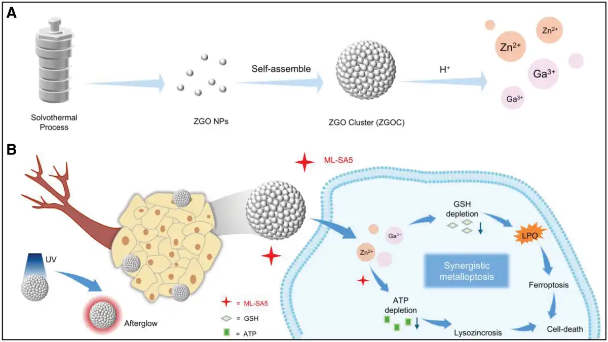
Herein, we synthesized a kind of zinc gallate-based NPs containing both gallium (Ga) ions and zinc ions to enhance metalloptosis for metastatic melanoma therapy. By introducing trace amounts of Cr³⁺, the resulting ZnGa₂O₄:Cr (simplified as ZGO) NPs also exhibited a persistent near-infrared (λ = 650–750 nm) afterglow emission, which is advantageous for high-sensitivity tumor imaging without background fluorescence interference[19]. Furthermore, by assembling individual ZGO NPs into well-defined clusters (ZGOCs), we obtained nanostructures with uniform morphology, optimized size, and favorable surface charge, thereby promoting prolonged blood circulation and enhanced tumor accumulation[20]. In addition, such clustering also improved the persistent luminescence (PL) of ZGOCs to a certain extent, further strengthening their potential for more precise imaging-guided cancer therapy with a high signal-to-noise ratio. After being engulfed by melanoma cells, ZGOCs were effectively degraded and released gallium ions and zinc ions, which induced ferroptosis and promoted lysozincrosis, respectively. Specifically, ZGOCs administration resulted in obvious GSH depletion and LPO increment, triggering ferroptosis. Besides, the co-treatment of ZGOCs and ML-SA5, a TRPML-specific agonist, significantly reduced cellular ATP levels, subsequently enhancing zinc ions-mediated lysozincrosis. In summary, we presented an enhanced metalloptosis strategy of synergistical ferroptosis and lysozincrosis based on ZGOCs, holding promise for the efficient treatment of melanoma in the future (Figure 1).
Figure 1.
Schematic illustration of ZGOCs-induced metalloptosis for melanoma therapy. (A) Preparation of ZGOCs. (B) Proposed mechanism of ZGOCs in combination with ML-SA5 in melanoma cells, enabling synergistic metalloptosis induction and afterglow-based imaging applications. ZGOCs, zinc gallate-based nanoclusters.
2. Experimental
2.1 Materials and reagents
All purchased information of materials and reagents were as follows: Zn(NO3)2·6H2O (XILONG Scientific Company); Ga(NO3)3·xH2O and Sodium dodecylsulfate (J&K Chemicals); Cr(NO3)3·9H2O (Aladdin); ML-SA5 (Target Molecule Corp.); NaOH (Energy Chemical); Absolute ethanol and Cyclohexane (Beijing Tong Guang Fine Chemical Company); Oleic acid (OA, Alfa Aesar); 5-FAM (Xi’an Ruixi Biological Technology Company); Deferoxamine (DFO) mesylate (Abcam); Chloroquine (CQ), Z-VAD-fmk and Necrostatin-1 (HARVEYBIO); Lipid peroxide colorimetric assay kit (Elabscience Biotechnology Co., Ltd.); Calcein-AM/PI double stain kit and Reduced glutathione content assay kit (Beijing Solarbio Science & Technology Co., Ltd.); ATP content assay kit (Shanghai Acmec Biochemical Technology Co., Ltd.); N,N,N,N-Tetrakis(2-pyridylmethyl)-ethylenediamine (Shanghai Macklin Biochemical Technology Co., Ltd.); DAPI and ROS assay kit (Beyotime); Cell counting kit-8 (CCK-8) (LABLEAD); Dulbecco’ s modified Eagle medium (DMEM), PBS and trypsin-EDTA (Shanghai XP Biomed Ltd.); fetal bovine serum (FBS) and penicillin-streptomycin (Invitrogen). The following primary antibodies were purchased from HUABIO: Beta Actin (1:5000, EM21002), glutathione peroxidase 4 (GPX4) (1:1000, ET1706-45).
2.2 Methods
2.2.1 Preparation of ZGO NPs and ZGOCs
ZGO NPs were synthesized as follows. Typically, 0.297 g of Zn(NO3)2·6H2O, 0.511 g of Ga(NO3)3·xH2O, and 1.6 mg of Cr(NO3)3·9H2O were dissolved in a mixture formed by 0.6 g of NaOH, 6 mL of OA, 8 mL of deionized water, and 18 mL of ethanol under vigorous stirring. The mixture was then transferred to a 50 mL teflon-lined autoclave and heated at 210 °C for 16 h. After cooling to room temperature, the resulting ZGO NPs were precipitated, isolated by centrifugation, washed alternately with ethanol and cyclohexane for 3 cycles, and finally redispersed in cyclohexane for further synthesis.
ZGOCs were synthesized with some modifications by following a previous protocol[21]. Sodium dodecylsulfate (28 mg) was first dissolved in 10 mL of deionized water. Then, 10 mg of ZGO NPs in 1 mL of cyclohexane was added to the solution. The mixture was emulsified using ultrasonic treatment, and the cyclohexane was evaporated by heating at 70 °C with vigorous stirring for 4 hours, assembling the ZGO NPs into ZGOCs. After cooling to room temperature, the products were collected, purified by repeated centrifugation, and redispersed. The final products were stored in water or PBS for future use.
2.2.2 Characterization of ZGO NPs and ZGOCs
The morphology and element mapping of the ZGO NPs and ZGOCs were examined using transmission electron microscopy (TEM, FEI Tecnai T20, HT-7700, 200 kV). X-ray diffraction (XRD) patterns were obtained with a Rigaku DMAX-2400 diffractometer using Cu Kα radiation (λ = 0.15405 nm). The hydrodynamic sizes of ZGOCs were measured with a dynamic light scattering analyzer (Zetasizer Nano ZS-90, Malvern, England). Fourier transform infrared (FT-IR) spectra were collected by a Bruker Frontier spectrometer (VERTEX 70v). PL observation of ZGO NPs and ZGOCs was measured using an In Vivo Imaging System (IVIS Luminn XRMS, Series III).
2.2.3 Degradation behavior analysis
ZGOCs were dispersed in PBS solutions with pH levels of 7.4, 6.5, and 5.4, then placed in a 37 °C shaker at 150 rpm for 8 h. Afterward, 50 μL of the solution was placed on a copper grid for TEM analysis.
2.2.4 Cell culture and cell viability study
NIH3T3 and B16F10 cell lines were obtained from the Cancer Institute and Hospital of the Chinese Academy of Medical Sciences. NIH3T3 and B16F10 cell lines were cultured in DMEM medium containing 10% FBS and 1% penicillin-streptomycin in a humidified environment at 37 °C with 5% CO2.
For the cell viability study, cells were incubated with varying concentrations of ZGOCs or ML-SA5 in DMEM at 37 °C and 5% CO2 for 24 h. Cell viability was then assessed using the CCK-8 assay. Additionally, the protective effect of different cell death inhibitors on ZGOCs-treated B16F10 cells was evaluated by incubating the cells with different inhibitors for 24 h, followed by cell viability measurement using the CCK-8 assay.
2.2.5 In vitro ferroptosis assay
To evaluate the ability of ZGOCs to induce ferroptosis, B16F10 cells were incubated and treated with either PBS (control) or ZGOCs. The cells were then collected to measure GSH and LPO levels using commercial kits. ROS levels were assessed with a ROS assay kit, and fluorescence images were captured using fluorescence microscopy.
2.2.6 Animal experiments
Female C57BL/6J mice (8 weeks old) were purchased from Beijing Vital River Laboratory Animal Technology Co., Ltd. All animal experiments were conducted following guidelines approved by the Institutional Animal Care and Use Committee of Peking University, with an assigned approval number: MSE-HouYL-1.
2.2.7 In vivo tumor treatment effect evaluation
Subcutaneous melanoma-bearing C57BL/6J mice were randomly divided into 4 groups: (1) control (saline), (2) ML-SA5, (3) ZGOCs, and (4) ZGOCs + ML-SA5. ZGOCs (10 mg/kg, 200 µL) were injected intravenously, and ML-SA5 was administered intraperitoneally (5 mg/kg), following the timeline shown in Figure 6A. During the treatment period, tumor volumes and body weights were monitored and recorded at specific time points. After treatment, tumors from each group were collected for histopathological analysis, including hematoxylin and eosin (H&E) staining and immunohistochemical (IHC) analysis.
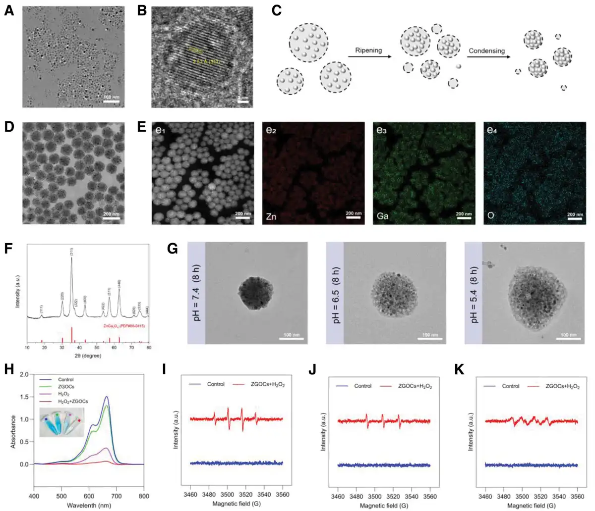
Figure 2.
Preparation and characterization of ZGOCs. (A) TEM characterization of the ZGO NPs. (B) HR-TEM image of ZGO NPs. (C) Illustration of the forming process of ZGOCs through a microemulsion-based approach. (D) TEM image and (E) EDS mappings of ZGOCs. (F) XRD pattern of ZGOCs. (G) TEM images of ZGOCs after incubation in PBS with pH values of 7.4, 6.5, and 5.4 (37 °C) for 8 hours. (H) UV-Vis absorbance spectra and photo (inset) of MB solutions with different treatments. (I–K) EPR spectra of ZGOCs treated with H₂O₂ in the presence of different spin-trapping agents. EDS, Energy dispersive spectrometry; EPR, electron paramagnetic resonance; MB, methylene blue; NP, nanoparticles; TEM, transmission electron microscopy; XRD, X-ray diffraction; ZGO, ZnGa₂O₄; ZGOCs, zinc gallate-based nanoclusters.
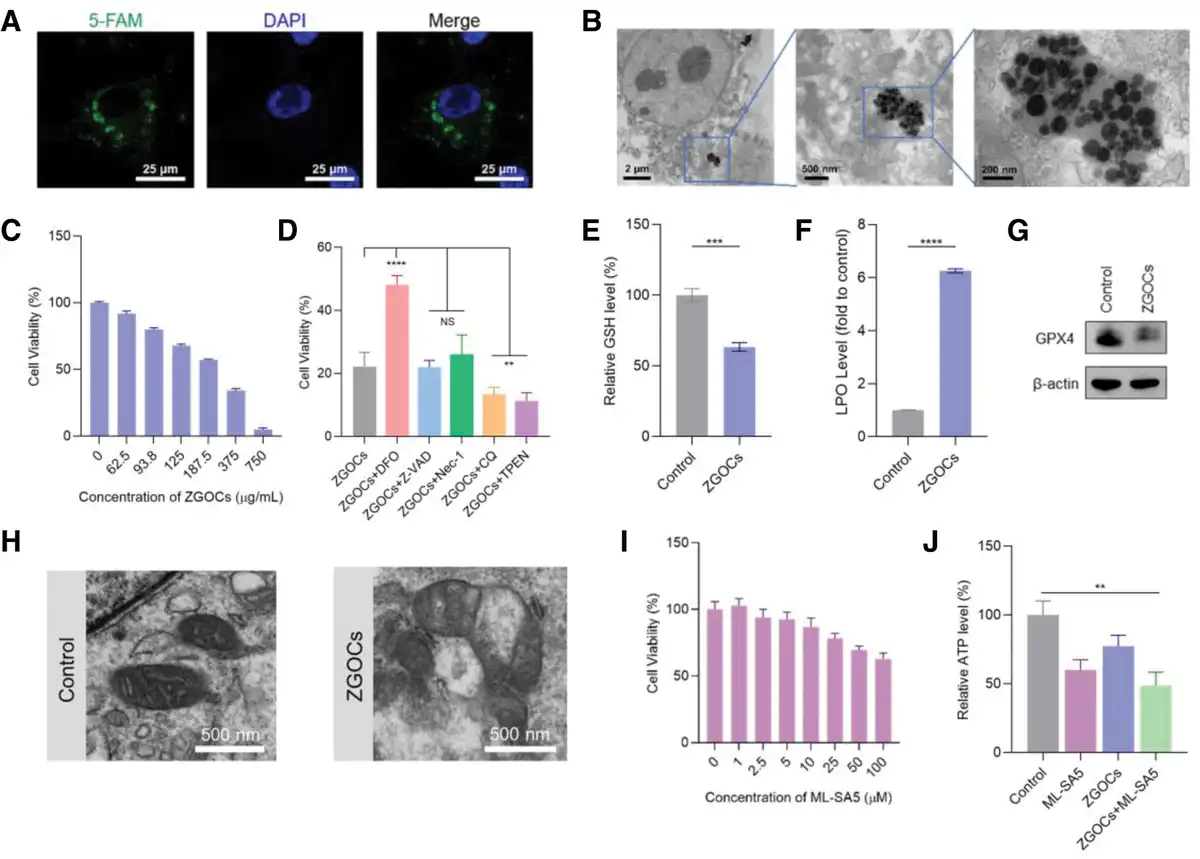
Figure 3.
Antitumor effects and relative mechanism of ZGOCs. (A) CLSM images showing cellular uptake of 5-FAM-labeled ZGOCs in B16F10 cells, where blue represents cell nuclei, green represents NPs. (B) Bio-TEM images of B16F10 cells treated with ZGOCs. (C) Cell viability data of B16F10 cells when treated with ZGOCs at the indicated concentration for 24 h (n = 3). (D) Cell viability in B16F10 cells after being treated with ZGOCs with or without different cell death inhibitors (n = 3). The detection of (E) GSH levels (n = 3) and (F) LPO levels (n = 4) in B16F10 cells after ZGOCs treatments. (G) Western blot of GPX4 expression after ZGOCs treatments. (H) Bio-TEM images of the cross-sections of B16F10 cells after different treatments. (I) Cell viability of B16F10 cells treated with different concentrations of ML-SA5 for 24 h (n ≥ 3). (J) Relative ATP of B16F10 cells after different treatments (n = 3). Data in (C–F) and (I–J) are presented as mean ± SD. P values: *P < 0.05, ** P < 0.01, and ***P < 0.001, ****P < 0.0001. CLSM, confocal laser scanning microscopy; GPX4, glutathione peroxidase 4; GSH, glutathione; LPO, lipid peroxidation; NPs, nanoparticles; NS, no significance; TEM, transmission electron microscopy; ZGOCs, zinc gallate-based nanoclusters.
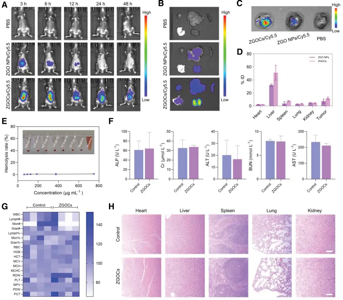
Figure 4.
Biodistribution and biocompatibility assessment of ZGOCs. (A) Fluorescence images at the indicated time of tumor-bearing mice after intravenous injection with PBS, ZGO NPs, and ZGOCs. (B, C) Representative in vivo fluorescence images of (B) major organs and (C) tumors at 48 h after treatment of different treatment groups. (D) Biodistribution of ZGO NPs or ZGOCs in tumor-bearing mice after 48 hours postinjection. (E) Hemolysis assay of ZGOCs at different concentrations. Saline was set as the negative control, and distilled water was set as the positive control. (F) Serum biochemistry analysis (n = 3), (G) routine blood assay (n = 4), and (H) H&E staining images of major organs from mice treated with saline (control) and ZGOCs for 7 days. Scale bar: 200 µm. Data in (D–F) are presented as mean ± SD. H&E, hematoxylin and eosin; NPs, nanoparticles; ZGO, ZnGa₂O₄; ZGOCs, zinc gallate-based nanoclusters.
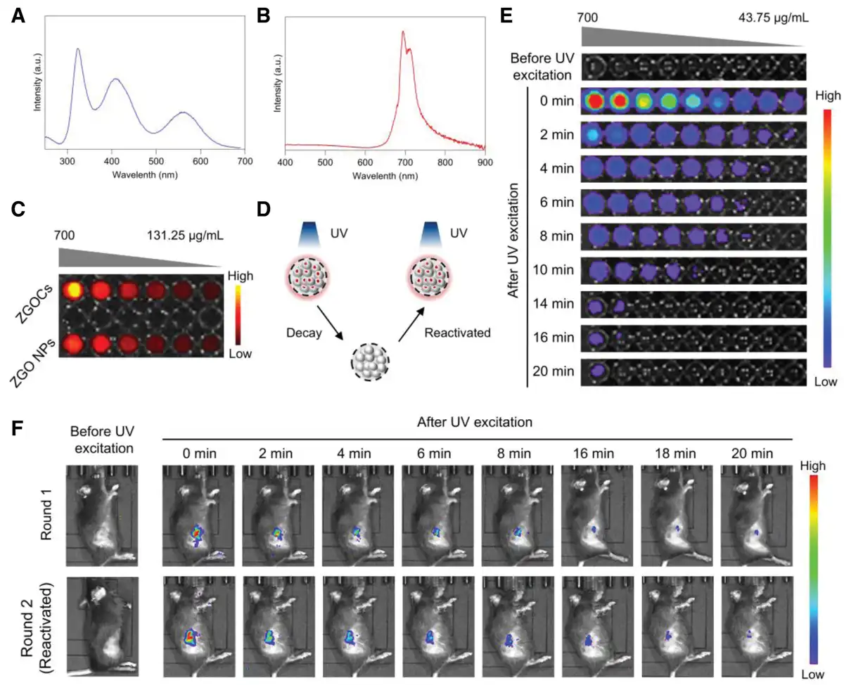
Figure 5.
In vitro and in vivo afterglow imaging performance of ZGO NPs and ZGOCs. (A) Excitation spectrum of ZGO NPs by monitoring the Cr3+ emission at 695 nm. (B) Emission spectrum of ZGO NPs under UV excitation at 260 nm. (C) PL images of ZGO NPs and ZGOCs after UV excitation at different concentrations. (D) Schematic illustration showing the rechargeable and UV-activated PL in ZGOCs. (E) Luminescence images of ZGOCs solution at different concentrations, recorded at various time points after excitation. (F) Luminescence images of tumor-bearing mice after intratumoral injection of ZGOCs, recorded at various time points postexcitation and reactivated. NPs, nanoparticles; PL, persistent luminescence; ZGO, ZnGa₂O₄; ZGOCs, zinc gallate-based nanoclusters.
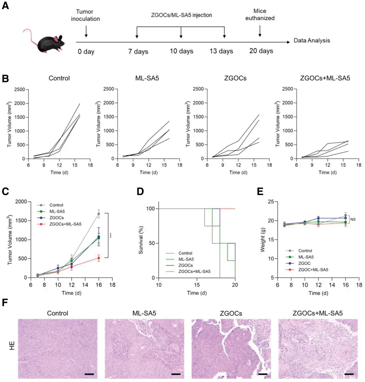
Figure 6.
In vivo therapeutic effect of ZGOCs. (A) Schematic treatment protocol of the mice with different treatments (n = 4). (B) Individual tumor growth curves, (C) average tumor growth curves, and (D) survival rate of mice after different treatments (n = 4). (E) Body weight of tumor-bearing mice under various treatments (n = 4). (F) H&E staining images of tumor tissues at the end of various treatments. Scale bar: 200 µm. Data in (C, E) are presented as mean ± SEM. P values: *P < 0.05, ** P < 0.01, and ***P < 0.001. H&E, hematoxylin and eosin; NS, not significant; ZGOCs, zinc gallate-based nanoclusters.
2.2.8 Histological evaluation
The isolated major organs and tumors were fixed with 4% paraformaldehyde. The tissues were embedded in paraffin and sectioned at 5 mm; H&E staining was performed for histological evaluation. The slides were observed under an optical microscope.
2.2.9 Immunohistochemistry
The paraffin sections of tumor tissues in mice were prepared at the end of the treatment. IHC was performed as described previously[22]. Primary antibodies used for IHC staining included anti-Ki-67 (1:500; Bioss, BSM-52455R) and anti-Solute Carrier Family 7 Member 11 (SLC7A11, also known as xCT) (1:200; Affinity Biosciences, DF12509).
2.2.10 Statistical analysis
Results in this work were presented as mean values ± SD or mean values ± SEM, which were described in the corresponding figure legends. The student’s two-tailed nonpaired t-test was used to determine significance between treatment and control groups in all experiments. P < 0.05 was considered statistically significant; *P < 0.05, **P < 0.01, ***P < 0.001, and ****P < 0.0001. NS, not significant.
3. Result and discussion
3.1 Synthesis and characterization
ZGO NPs were synthesized using a simple solvothermal liquid-solid-solution method, involving the reaction of metal salts in a mixture of NaOH, ethanol, and OA. Due to the presence of OA ligands on the surface, the resulting ZGO NPs could be well-dispersed in nonpolar solvents such as cyclohexane (CYH), which enabled the formation of clusters in subsequent steps. TEM images demonstrated that ZGO NPs exhibited excellent dispersion and uniformity, with a particle size of approximately 10 nm (Figure 2A, B).
Subsequently, the organic phase (CYH) containing OA-modified ZGO NPs was mixed with an aqueous solution containing the surfactant sodium dodecylsulfate (SDS) to form an oil-in-water microemulsion via ultrasonic treatment. The low-boiling organic solvent CYH was then evaporated through vigorous stirring, successfully yielding ZGOCs. During this process, emulsions formed by ultrasonication underwent ripening, where solvent-swollen micelles were generated. As the solvent evaporated, clusters and SDS micelles formed (Figure 2C)[21]. TEM images revealed that the assembled ZGO clusters maintained a uniform morphology (Figure 2D). It is important to note that the feeding ratio of ZGO NPs during synthesis was critical. A reduced amount of ZGO NPs resulted in many unassembled monomers in the product (Supplementary Figure S1, https://links.lww.com/MEDMAT/A7). The hydrophobic van der Waals interactions between the alkyl chains of SDS surfactant in the aqueous phase and the alkane chains of OA ligands on ZGO NPs surface (Supplementary Figure S2, https://links.lww.com/MEDMAT/A7) contributed to the good water solubility of the obtained ZGOCs[21]. The hydrodynamic diameter of ZGOCs in water was approximately 100 nm, with a polydispersity index of 0.075 (Supplementary Figure S3a, https://links.lww.com/MEDMAT/A7), indicating their excellent dispersibility and uniformity, which was advantageous for subsequent biological applications. The zeta potential of ZGOCs was measured to be −19 mV (Supplementary Figure S3b, https://links.lww.com/MEDMAT/A7), suggesting their favorable long-circulating behavior in vivo[23]. Energy dispersive spectrometry mapping and X-ray photoelectron spectroscopy together confirmed the presence of Zn, Ga, and O in ZGOCs (Figure 2E and Supplementary Figure S4, https://links.lww.com/MEDMAT/A7). The lack of Cr in the elemental mapping analysis arises from its trace doping concentration that could not be detected by the instrument[24]. The XRD patterns further confirmed the presence of a pure zinc gallate phase in ZGOCs (Figure 2F).
The degradation behavior of ZGOCs was studied by dispersing them in PBS with different pH values at 37 °C environments, simulating specific physiological conditions (Figure 2G). Remarkably, ZGOCs were stable at pH 7.4, indicating strong stability under physiological conditions, consistent with the results shown in Supplementary Figure S3c, https://links.lww.com/MEDMAT/A7. However, at pH 6.5 and 5.4, ZGOCs gradually degraded within 8 hours, accompanied by an increase in particle size and the release of ZGO NPs, demonstrating an acid-sensitive degradation process. Such pH-responsive degradation not only ensures the structural stability of ZGOCs during systemic circulation but also facilitates the release of active zinc and gallium ions in the acidic TME, which is expected to enhance their catalytic reactivity. Guided by this rationale, we next evaluated the catalytic activities of ZGOCs. Methylene blue (MB) was employed as a dye indicator to assess the generation of ROS. As shown in the UV-vis absorbance spectra (Figure 2H), the treatment of MB with ZGOCs in the presence of hydrogen peroxide (H₂O₂) led to the most pronounced decrease in absorbance compared with other groups, indicating the superior catalytic performance of ZGOCs. Furthermore, electron paramagnetic resonance spectroscopy confirmed the production of multiple ROS species, including hydroxyl radicals, superoxide anions, and singlet oxygen, when ZGOCs were incubated with H₂O₂ in the presence of different spin-trapping agents (Figure 2I–K).
3.2. In vitro anticancer activity and mechanism
We chose metastatic melanoma cell lines B16F10 and investigated the cellular uptake behavior of ZGOCs. After incubating 5-FAM-labeled ZGOCs with B16F10 cells for 8 hours, significant fluorescence was observed in the cells using confocal laser scanning microscopy, confirming efficient intracellular internalization (Figure 3A). Additionally, cellular bio-TEM images (Figure 3B) further supported the phagocytosis of ZGOCs by tumor cells, laying the foundation for subsequent metalloptosis-based therapy.
To assess the cytotoxicity of ZGOCs on B16F10 cells, we used the CCK-8 assay. After 24 h of incubation, ZGOCs exhibited a concentration-dependent cytotoxic effect on B16F10 cells (Figure 3C). Notably, no significant toxicity of ZGOCs was observed in normal cells NIH3T3 (Supplementary Figure S5, https://links.lww.com/MEDMAT/A7). To further investigate the specific mechanism of cell death, various cell death inhibitors were selected for validation. Specifically, the apoptosis inhibitor Z-VAD-fmk (Z-VAD), the necroptosis inhibitor necrostatin-1 (Nec-1), the autophagy inhibitor CQ, and the lysosomal inhibitor N,N,N,N-Tetrakis(2-pyridylmethyl)-ethylenediamine (a Zn2+ chelator) showed no rescue effects on ZGOCs-induced cell death. However, the ferroptosis inhibitor DFO reversed the cytotoxic effect of ZGOCs to a certain extent, indicating that ZGOCs induced B16F10 cell death primarily through ferroptosis. This finding was also consistent with recent research, which suggested that gallium ions could also induce oxidative stress and trigger ferroptosis via indirect mechanisms[25]. We further measured GSH levels and the deactivation of GSH-related GPX4 under ZGOCs treatments. The results showed that ZGOCs could strongly deplete GSH, and after treatment, GPX4 activity was significantly reduced (Figure 3E, G). The resulting GSH depletion and GPX4 inactivation further hindered the clearance of LPO (Figure 3F), indicating that ZGOCs could effectively suppress antioxidant defenses, thereby promoting ferroptosis.
To assess intracellular oxidative stress, we used 2,7-dichlorofluorescein diacetate (DCFH-DA) as a ROS probe. After 8 hours of ZGOCs treatment, a strong green fluorescence signal from DCF was observed in B16F10 cells, indicating elevated intracellular oxidative stress (Supplementary Figure S6, https://links.lww.com/MEDMAT/A7). Bio-TEM images also revealed that ZGOCs caused mitochondrial damage, as evidenced by the disruption of the mitochondrial cristae (Figure 3H).
Previous studies have shown that TRPML-specific synthetic agonists, such as ML-SA5 and ML-SA8, can induce rapid lysosomal Zn²⁺-dependent necrotic cell death, termed lysozincrosis, in metastatic melanoma cells like M12 or MeWo[4]. Surprisingly, we found that ML-SA5 alone did not exhibit significant cytotoxicity in B16F10 cells, and only at high concentrations after 24 hours did it show a certain level of toxicity (Figure 3I). However, when ZGOCs (100 µg/mL) were combined with ML-SA5 (1 µM) in B16F10 cells, a potent antitumor effect was observed, which far exceeded the effects of either ZGOCs or ML-SA5 alone, accompanied by a significant reduction in intracellular ATP levels (Figure 3J and Supplementary Figure S7, https://links.lww.com/MEDMAT/A7). We hypothesized that this synergistic effect arose from Zn ions released by ZGOCs, which served as an exogenous Zn²⁺ source and, upon ML1 activation, cooperatively triggered lysozincrosis, thereby efficiently killing B16F10 cells.
3.3. In vivo biodistribution and biocompatibility evaluation
To facilitate the subsequent in vivo antitumor applications of ZGOCs, we next investigated their biodistribution and biosafety. Compared with Cy5.5-labeled free ZGO NPs (ZGO NPs/Cy5.5), Cy5.5-labeled ZGOCs (ZGOCs/Cy5.5) exhibited prolonged blood circulation time, suggesting their improved physiological stability and reduced rapid clearance by the body (Figure 4A–B). The prolonged circulation time of ZGOCs, together with their size more suitable for the enhanced permeability and retention effect, thereby favoring efficient tumor accumulation (Figure 4C)[26]. Moreover, as shown in Figure 4B, D, ZGOCs were predominantly metabolized through the liver and kidneys, which is consistent with the conventional metabolic pathways of NMs and indicates their feasibility for safe clearance from the body[26].
The biocompatibility of ZGOCs was further evaluated both in vitro and in vivo. Initially, different concentrations of ZGOCs were co-incubated with mouse erythrocytes, and no significant hemolysis (hemolysis ratio < 5%) or coagulation was observed compared with the control group, demonstrating excellent hemocompatibility (Figure 4E and Supplementary Figure S8, https://links.lww.com/MEDMAT/A7). Subsequently, in vivo safety assessments were carried out in female C57BL/6J mice. Specifically, mice received intravenous injections of either ZGOCs or saline (control) and were sacrificed on day 7 or 14 postinjection to evaluate both short-term and longer-term biosafety. Serum biochemistry and routine blood assays (Figure 4F–G and Supplementary Figure S9a, b, https://links.lww.com/MEDMAT/A7) revealed no significant differences in all tested parameters between the ZGOCs and control groups. In addition, H&E staining of major organs—including the heart, liver, spleen, lungs, and kidneys—revealed no pathological alterations or tissue damage at day 7 or 14 (Figure 4H and Supplementary Figure S9c, https://links.lww.com/MEDMAT/A7), further confirming the favorable short- and long-term safety profile of ZGOCs.
Taken together, these results confirmed that ZGOCs possessed favorable pharmacokinetics, efficient tumor-targeting capability, and excellent biocompatibility, highlighting their promise as a safe and effective nanoplatform for future melanoma therapy.
3.4. In vitro and in vivo persistent luminescence performance
We further investigated the in vitro and in vivo afterglow imaging performance of ZGO NPs due to their intrinsic PL capability. The excitation and emission spectra of ZGO NPs are shown in Figure 5A, B. Under UV excitation, ZGO NPs exhibited a broad emission band spanning the red to near-infrared (NIR) region (600–800 nm) with a pronounced peak at 695 nm. Interestingly, the formation of ZGO clusters (ZGOCs) resulted in an enhanced and prolonged PL intensity (Figure 5C), which can be ascribed to the effective suppression of surface defect–mediated nonradiative transitions within the densely packed cluster structure. In addition, the clustered configuration may partially shield the particles from water-induced quenching, thereby further sustaining the PL[19]. As shown in Figure 5E, following 10 min of UV excitation, ZGOCs displayed concentration-dependent afterglow performance, with the highest concentration group exhibiting decay times of approximately 20 min. Moreover, ZGOCs demonstrated rechargeable afterglow properties: upon re-excitation with UV light after initial decay, the afterglow intensity was fully restored without noticeable signal attenuation (Figure 5D and Supplementary Figure S10, https://links.lww.com/MEDMAT/A7).
The NIR emission of ZGOCs confers distinct advantages for in vivo applications, including deeper tissue penetration and minimized autofluorescence interference, which are critical for high-contrast imaging. Indeed, as illustrated in Figure 5F, in B16F10 tumor-bearing mice, intratumoral injection of ZGOCs enabled detectable afterglow within the tumor site for nearly 20 min, providing a sufficient imaging window for real-time tumor visualization and potential guidance for therapeutic interventions. Moreover, this afterglow can be reactivated on demand by subsequent UV excitation, demonstrating its rechargeable capability.
3.5. In vivo synergistic anticancer evaluation
Encouraged by the efficacy of ZGOCs in inducing metalloptosis and their biosafety, we conducted an in vivo therapeutic experiment to evaluate whether ZGOCs in combination with ML-SA5 could inhibit tumor growth. Subcutaneous melanoma models were established by inoculating B16F10 cells into C57BL/6J mice and allowing the tumors to grow for 7 days. The tumor-bearing mice were then randomly divided into 4 groups: saline (control), ML-SA5 alone, ZGOCs alone, and the combination of ZGOCs with ML-SA5. Following the treatment protocols shown in Figure 6A, it was evident that the tumor size in the “ZGOCs+ML-SA5” group was significantly smaller compared with the other groups (Figure 5B, C), indicating a more potent metalloptosis-inducing effect. The survival curves in Figure 5D further confirmed the synergistic effect of ZGOCs and ML-SA5. Throughout the treatment period, no significant weight loss was observed in the mice treated with ZGOCs, ML-SA5, or their combination, compared with the saline control group (Figure 5E). Consistent with the tumor inhibition results, H&E staining of tumor sections from the ZGOCs and ML-SA5 combination group showed distinct features of necrotic cells, such as cell shrinkage, loss of cell–cell contacts, and condensed chromatin (Figure 5F). Moreover, IHC of Ki-67, a marker of cell proliferation, and xCT, a key cystine/glutamate antiporter that suppresses ferroptosis, further confirmed that the combination treatment induced the most pronounced inhibition of tumor growth and activation of ferroptosis (Supplementary Figure S11, https://links.lww.com/MEDMAT/A7). Overall, the synergistic action of ZGOCs and ML-SA5 effectively induced metalloptosis in melanoma-bearing mice, leading to significant tumor suppression and achieving a favorable therapeutic outcome.
4. Conclusions
For an enhanced metalloptosis strategy disrupting ion homeostasis in metastatic melanoma, ZGOCs combining with ML-SA5 were successfully developed for the treatment of melanoma. The synthesized ZGOCs exhibited uniform morphology, good stability, and strong metalloptosis-inducing capabilities. These nanoclusters were responsive to the acidic TME, where they degraded to release gallium and Zn ions. On one hand, these metal ions triggered GSH depletion, ROS production, and GPX4 deactivation in B16F10 cells, ultimately leading to a sharp increase in LPO levels for inducing ferroptosis. On the other hand, the released Zn ions synergized with ML-SA5, a TRPML1 agonist, to potentiate lysozincrosis, ultimately enhancing melanoma cell death. This dual pathway of ferroptosis and lysozincrosis was further validated in vivo, where the combination of ZGOCs and ML-SA5 effectively suppressed tumor growth with favorable biosafety.
In parallel, the unique optical properties of ZGOCs provided additional diagnostic advantages. Unlike conventional fluorescent probes that require continuous external excitation, ZGOCs exhibited long-lasting, rechargeable afterglow emission. The cluster formation further suppressed nonradiative transitions, thereby prolonging the duration and intensity of the afterglow. Importantly, the emission lies in the NIR window, which matches well with the superficial growth characteristics of melanoma, enabling deeper tissue penetration, minimized autofluorescence, and real-time tumor tracking. These features highlight ZGOCs as a dual-functional platform that integrates therapeutic efficacy with high-contrast afterglow imaging, thereby advancing precise melanoma theranostics.
It is also worth noting that this clustering strategy is not limited to single ZGO-based systems. ZGO NPs could also be hybridized with other functional NMs, such as gold or iron carbide (Fe₂C) NPs, further expanding the bio-application range of this strategy (Supplementary Figure S12, https://links.lww.com/MEDMAT/A7). For example, integrating gold or Fe2C NPs may endow the hybrid clusters with strong photothermal or magnetothermal conversion efficiency, thereby enabling synergistic thermal ablation in combination with metalloptosis. In parallel, the incorporation of these functional components also enhances diagnostic capabilities—gold NPs can serve as excellent computed tomography contrast agents, while Fe₂C NPs offers magnetic resonance imaging potential—supporting multimodal imaging-guided therapy.
Acknowledgements
This work was financially supported by the National Natural Science Foundation of China (Grant Nos. 52201198, 22575006, 52027801, and 92263203), Beijing Natural Science Foundation-Haidian Primitive Innovation Joint Fund (Grant No. L232015), the National Key R&D Program of China (Grant No. 2022YFA1203902), the China-Germany Collaboration Project (Grant No. M-0199), the Fundamental Research Funds for the Central Universities, Peking University, The Clinical Medicine Plus X-Young Scholars Project of Peking University, and the Fundamental Research Funds for the Central Universities (Grant Nos. PKU2024LCXQ014 and PKU2025PKULCXQ040).
Conflicts of interest
The authors declare that they have no conflicts of interest.
Data availability statement
The data that support the findings of this study are available from the corresponding author upon reasonable request.
Author Contribution
Shuren Wang supervised and administered the project. Yanglong Hou, Shuren Wang, and Ran Ma conceived and designed the experiments. Ran Ma, Jiajia Liu, and Zi Mei performed the experiments, collected the data, analyzed, and interpreted the data. Ran Ma and Shuren Wang wrote the paper. All the authors discussed the results and reviewed the paper at all stages.
References
- [1] Welsh SJ, Rizos H, Scolyer RA, et al. Resistance to combination BRAF and MEK inhibition in metastatic melanoma: where to next? Eur J Cancer (Oxford, England: 1990). 2016;62:76–85.
- [2] Kasitinon SY, Eskiocak U, Martin M, et al. TRPML1 promotes protein homeostasis in melanoma cells by negatively regulating MAPK and mTORC1 signaling. Cell Rep. 2019;28(9):2293–2305.e9.
- [3] Dong X-P, Cheng X, Mills E, et al. The type IV mucolipidosis-associated protein TRPML1 is an endolysosomal iron release channel. Nature. 2008;455(7215):992–996.
- [4] Du W, Gu M, Hu M, et al. Lysosomal Zn2+ release triggers rapid, mitochondria-mediated, non-apoptotic cell death in metastatic melanoma. Cell Rep. 2021;37(3):109848.
- [5] Bakhshi J, Weinstein L, Poksay KS, et al. Coupling endoplasmic reticulum stress to the cell death program in mouse melanoma cells: effect of curcumin. Apoptosis. 2008;13(7):904–914.
- [6] Xu L, Peng M, Gao T, et al. Nanoenabled intracellular metal ion homeostasis regulation for tumor therapy. Adv Sci. 2024;11(7):e2306203.
- [7] Ma R, Hou Y, Wang S. Combatting cancer with tiny intelligent warriors: Stimuli-responsive micro/nanorobots in chemotherapeutic delivery. Sci China Mater. 2024;67(8):2469–2476.
- [8] Ma R, Hou Y, Wang S. Imaging-guided cancer therapy based on multifunctional magnetic nanoparticles. Chin J Chem. 2023;41(19):2557–2573.
- [9] Wang S, Wang Z, Li Z, et al. Amelioration of systemic antitumor immune responses in cocktail therapy by immunomodulatory nanozymes. Sci Adv. 2022;8(21):eabn3883.
- [10] Wang J, Sun Z, Wang S, et al. Biodegradable ferrous sulfide-based nanocomposites for tumor theranostics through specific intratumoral acidosis-induced metabolic symbiosis disruption. J Am Chem Soc. 2022;144(43):19884–19895.
- [11] Wang S, Ma R, Mei Z, et al. Metalloptosis: metal ions-induced programmed cell death based on nanomaterials for cancer therapy. MedMat. 2024;1(1):6–26.
- [12] Ko MJ, Min S, Hong H, et al. Magnetic nanoparticles for ferroptosis cancer therapy with diagnostic imaging. Bioact Mater. 2024;32:66–97.
- [13] Gu J, Guo C, Ruan J, et al. From ferroptosis to cuproptosis, and calcicoptosis, to find more novel metals-mediated distinct form of regulated cell death. Apoptosis. 2024;29(5):586–604.
- [14] Wang J, Zhang R, Gao S, et al. Degradable nanoregulators based on ultra-small ferrous sulfide for photoacoustic/magnetic resonance imaging-guided tumor starvation and ferroptosis. Adv Healthc Mater. 2025;14(19):e2500560.
- [15] Wang J, Fang Z, Zhao C, et al. Intelligent size-switchable iron carbide-based nanocapsules with cascade delivery capacity for hyperthermia-enhanced deep tumor ferroptosis. Adv Mater. 2024;36(9):e2307006.
- [16] Dixon SJ, Lemberg KM, Lamprecht MR, et al. Ferroptosis: an iron-dependent form of nonapoptotic cell death. Cell. 2012;149(5):1060–1072.
- [17] Lei G, Zhuang L, Gan B. Targeting ferroptosis as a vulnerability in cancer. Nat Rev Cancer. 2022;22(7):381–396.
- [18] Shen Z, Song J, Yung BC, et al. Emerging strategies of cancer therapy based on ferroptosis. Adv Mater. 2018;30(12):e1704007.
- [19] Fan W, Lu N, Xu C, et al. Enhanced afterglow performance of persistent luminescence implants for efficient repeatable photodynamic therapy. ACS Nano. 2017;11(6):5864–5872.
- [20] Davis ME, Chen Z, Shin DM. Nanoparticle therapeutics: an emerging treatment modality for cancer. Nat Rev Drug Discovery. 2008;7(9):771–782.
- [21] Bai F, Wang D, Huo Z, et al. A versatile bottom-up assembly approach to colloidal spheres from nanocrystals. Angew Chem Int Ed. 2007;46(35):6650–6653.
- [22] Wang S, Ma K, Chen L, et al. TAZ promotes cell growth and inhibits celastrol-induced cell apoptosis. Biosci Rep. 2016;36(5):e00386.
- [23] Blanco E, Shen H, Ferrari M. Principles of nanoparticle design for overcoming biological barriers to drug delivery. Nat Biotechnol. 2015;33(9):941–951.
- [24] Zhou Z, Zheng W, Kong J, et al. Rechargeable and LED-activated ZnGa2O4: Cr3+ near-infrared persistent luminescence nanoprobes for background-free biodetection. 10.1039/C7NR01209H. Nanoscale. 2017;9(20):6846–6853.
- [25] Peng X-X, Zhang H, Zhang R, et al. Gallium triggers ferroptosis through a synergistic mechanism. Angew Chem Int Ed. 2023;62(36):e202307838.
- [26] Xu M, Qi Y, Liu G, et al. Size-dependent in vivo transport of nanoparticles: implications for delivery, targeting, and clearance. ACS Nano. 2023;17(21):20825–20849.
This article is available under Open Access.
© 2026 The Authors