Exogenous electrical stimulation in soft tissue wounds healing: mechanism and devices
Authors
Jingwei Yu, Dawei Zhao, Yue Yuan, Minghao Zhou, Peng Li, Tengjiao Wang, Hongbo Wei*
- aState Key Laboratory of Oral & Maxillofacial Reconstruction and Regeneration, National Clinical Research Center for Oral Diseases, Shaanxi Engineering Research Center for Dental Materials and Advanced Manufacture, The Fourth Military Medical University, Xi’an, China
- bDepartment of Oral Implants, School of Stomatology, The Fourth Military Medical University, Xi’an, China
- cFrontiers Science Center for Flexible Electronics, Institute of Flexible Electronics, Institute of Biomedical Materials and Engineering, Northwestern Polytechnical University, Xi’an, China.
* Correspondence: Address: Hongbo Wei, Department of Oral Implants, School of Stomatology, State Key Laboratory of Oral & Maxillofacial Reconstruction and Regeneration, National Clinical Research Center for Oral Diseases, Shaanxi Engineering Research Center for Dental Materials and Advanced Manufacture, The Fourth Military Medical University, Xi’an, Shaanxi, China; Email: weihongbo@fmmu.edu.cn (H. Wei). Jingwei Yu and Dawei Zhao contributed equally to this work.
MedMat · 2026 · Vol. 3 · No. 2 · pp. 203-216
Abstract
Electrical stimulation can serve as a therapeutic modality, accelerating the healing of soft tissue wounds. However, endogenous electric fields are frequently found to be attenuated in chronic wounds. Consequently, exogenous electrical stimulation devices have been explored to supplement and enhance endogenous electric fields, thereby promoting faster and more robust healing of soft tissue injuries. This review outlines the generation of endogenous electric fields in wounds, the molecular mechanisms by which electric fields facilitate healing, and the roles of endogenous electric fields across various stages of tissue repair. It further highlights recent advancements in applying exogenous electrical stimulation to wound sites. Finally, we discuss the challenges and future directions for the widespread clinical implementation of exogenous electrical stimulation in soft tissue wound management.
Translations
Long abstracts in additional languages. The English article is the version of record.
中文zh-Hans
软组织伤口愈合是临床治疗中的关键挑战,而电刺激作为一种新兴的治疗手段展现出加速愈合的巨大潜力。然而,慢性伤口中普遍存在内源性电场衰减的问题,导致自然修复过程受阻。本文旨在系统综述外源性电刺激在补充和增强受损内源性电场方面的作用机制与应用前景。通过深入分析生物电信号在组织再生中的核心地位,本研究试图阐明为何引入外部电能干预能够克服慢性伤口的病理生理障碍,从而为开发更高效的伤口管理策略提供理论依据和实践指导。
作为一篇综述性文章,本文并未进行新的实验操作,而是对现有文献进行了系统的梳理与整合。研究框架涵盖了内源性电场在伤口处的生成机制、其促进愈合的分子生物学路径以及在不同修复阶段的具体功能。我们重点考察了近年来应用于伤口部位的外源性电刺激设备的技术进展,包括不同波形、频率和强度的参数设计及其生物物理效应。此外,文章还详细探讨了各类设备的临床适用性,分析了从实验室研究到临床应用转化过程中所面临的材料学挑战与工程限制。
综述发现,内源性电场在伤口愈合的炎症期、增殖期和重塑期中均发挥关键调节作用,而外源性电刺激能够有效弥补慢性伤口中缺失的生物电信号。分子机制研究表明,电场通过调控细胞迁移、血管生成及生长因子表达等途径显著加速组织修复过程。最新的外源性设备设计已能更精准地模拟生理性电场环境,从而在多种软组织损伤模型中展现出优于传统疗法的愈合效果。这些发现证实了电刺激疗法在恢复伤口微环境稳态方面的科学有效性,为理解生物电信号与组织再生的耦合机制提供了新的视角。
尽管外源性电刺激显示出巨大的临床转化潜力,但其广泛推广仍面临诸多挑战,包括设备标准化不足、长期安全性数据缺乏以及个体化治疗方案的优化需求等。本文指出,未来研究需聚焦于开发更智能、便携且生物相容性更好的新型刺激装置,并开展多中心临床试验以验证其疗效与安全性。此外,深入探索电刺激参数与伤口类型之间的特异性匹配关系将是实现精准医疗的关键方向。通过克服当前的技术与临床障碍,外源性电刺激有望成为软组织伤口管理中的常规治疗手段,显著改善患者预后并降低慢性伤口的社会负担。
Françaisfr
La cicatrisation des plaies de tissus mous constitue un défi clinique majeur, et la stimulation électrique émerge comme une modalité thérapeutique prometteuse capable d'accélérer ce processus. Cependant, les champs électriques endogènes sont fréquemment atténués dans les plaies chroniques, entravant ainsi le mécanisme naturel de réparation. L'objectif de cette revue est d'examiner comment la stimulation électrique exogène peut compléter et renforcer ces champs endogènes déficients pour favoriser une cicatrisation plus rapide et robuste des lésions tissulaires molles. En analysant les fondements biologiques de ce phénomène, nous cherchons à établir un cadre théorique solide justifiant l'intervention externe dans la gestion complexe des plaies persistantes.
Cette étude adopte une approche synthétique basée sur une revue exhaustive de la littérature scientifique existante plutôt que sur des expérimentations nouvelles. Le cadre d'analyse couvre la génération naturelle des champs électriques au niveau des blessures, les mécanismes moléculaires par lesquels ces champs facilitent la réparation tissulaire et leurs rôles spécifiques à travers les différentes phases du processus de guérison. Nous mettons particulièrement l'accent sur les avancées récentes dans le développement d'appareils de stimulation électrique exogène destinés aux sites de plaies, en évaluant leur conception technique, leurs paramètres opérationnels et leurs interactions avec la biologie tissulaire pour optimiser l'efficacité thérapeutique.
Les résultats principaux indiquent que les champs électriques endogènes jouent un rôle régulateur crucial durant toutes les étapes de la réparation tissulaire, depuis l'inflammation initiale jusqu'à la phase de remodelage final. La stimulation exogène s'avère efficace pour compenser l'atténuation pathologique observée dans les plaies chroniques en restaurant une signalisation électrique adéquate. Les mécanismes moléculaires identifiés incluent la modulation de la migration cellulaire, l'angiogenèse et l'expression des facteurs de croissance sous l'influence du champ électrique appliqué. Ces découvertes démontrent que les dispositifs modernes peuvent simuler avec précision les environnements électriques physiologiques, offrant ainsi une alternative thérapeutique supérieure aux méthodes conventionnelles pour divers types d'accidents tissulaires.
Bien que prometteuse, la mise en œuvre clinique généralisée de la stimulation électrique exogène se heurte à des défis significatifs tels que le manque de standardisation des protocoles et l'absence de données longitudinales sur la sécurité. Les perspectives futures soulignent la nécessité de développer des dispositifs plus intelligents et biocompatibles, ainsi que d'exécuter des essais cliniques multicentriques rigoureux pour valider leur efficacité à grande échelle. Il est également crucial d'affiner les paramètres de stimulation en fonction du type spécifique de plaie pour une approche personnalisée. En surmontant ces obstacles techniques et réglementaires, la stimulation électrique pourrait devenir un pilier standard dans la gestion des plaies de tissus mous, améliorant ainsi considérablement le pronostic des patients et réduisant l'impact sociétal des cicatrisations chroniques prolongées.
Españoles
La cicatrización de heridas en tejidos blandos representa un desafío clínico significativo, y la estimulación eléctrica emerge como una modalidad terapéutica capaz de acelerar este proceso. Sin embargo, los campos eléctricos endógenos se encuentran frecuentemente atenuados en las heridas crónicas, lo que obstaculiza el mecanismo natural de reparación. El objetivo de esta revisión es examinar cómo los dispositivos de estimulación eléctrica exógena pueden complementar y potenciar estos campos endógenos comprometidos para promover una curación más rápida y robusta de las lesiones tisulares blandas. Al analizar la importancia central de estas señales bioeléctricas, buscamos establecer un marco teórico sólido que justifique la intervención externa en el manejo complejo de heridas persistentes.
Como artículo de revisión, este estudio no realiza experimentos nuevos sino que se basa en una síntesis sistemática y exhaustiva de la literatura científica existente. El marco analítico abarca los mecanismos de generación de campos eléctricos endógenos en las lesiones, las rutas moleculares mediante las cuales estos campos facilitan la curación y sus roles específicos a través de las diversas etapas del proceso reparativo. Se pone un énfasis particular en los avances recientes en el desarrollo de dispositivos de estimulación eléctrica exógena destinados a sitios de heridas, evaluando su diseño técnico, parámetros operativos e interacciones con la biología tisular para optimizar la eficacia terapéutica.
Los hallazgos principales indican que los campos eléctricos endógenos desempeñan un papel regulador crucial durante todas las etapas de reparación tisular, desde la inflamación inicial hasta la fase final de remodelado. La estimulación exógena resulta efectiva para compensar la atenuación patológica observada en heridas crónicas al restaurar una señalización eléctrica adecuada. Los mecanismos moleculares identificados incluyen la modulación de la migración celular, angiogénesis y expresión de factores de crecimiento bajo la influencia del campo eléctrico aplicado. Estos descubrimientos demuestran que los dispositivos modernos pueden simular con precisión entornos eléctricos fisiológicos, ofreciendo así una alternativa terapéutica superior a los métodos convencionales para diversos tipos de lesiones tisulares.
Aunque prometedora, la implementación clínica generalizada de la estimulación eléctrica exógena enfrenta desafíos significativos como la falta de estandarización en protocolos y ausencia de datos longitudinales sobre seguridad. Las perspectivas futuras subrayan la necesidad de desarrollar dispositivos más inteligentes y biocompatibles, así como ejecutar ensayos clínicos multicéntricos rigurosos para validar su eficacia a gran escala. Es igualmente crucial refinar los parámetros de estimulación según el tipo específico de herida para un enfoque personalizado. Al superar estos obstáculos técnicos y regulatorios, la estimulación eléctrica podría convertirse en un pilar estándar en el manejo de heridas de tejidos blandos, mejorando así considerablemente el pronóstico de los pacientes y reduciendo el impacto social de las cicatrizaciones crónicas prolongadas.
日本語ja
軟組織創傷の治癒は臨床において重要な課題であり、電気刺激はその治療法として治癒を加速する可能性を示しています。しかしながら、慢性創傷では内因性電場が頻繁に減衰しており、自然な修復プロセスが阻害されるという問題があります。本稿の目的は、外因性電気刺激装置がこれらの損傷した内因性電場を補完・増強し、より速く堅牢な軟組織傷害の治癒を促進するメカニズムと応用可能性について体系的に概説することです。生物学的信号伝達におけるこの役割を理解することで、慢性創傷に対する新たな治療戦略の理論的基盤を提供することを意図しています。
本稿はレビュー論文として位置づけられており、新規の実験データではなく既存文献を統合・分析するアプローチを採用しています。調査枠組みには、創傷部位での内因性電場の生成メカニズム、治癒促進に関与する分子レベルの経路、および組織修復の各段階におけるその役割が含まれます。特に近年、創傷部位への適用が進む外因性電気刺激装置に関する技術的進展に焦点を当て、波形や周波数などのパラメータ設計とその生物物理学的効果について詳述しています。また、実験室研究から臨床応用へ移行する過程で直面している材料科学上の課題と工学制限についても議論を加えています。
主要な知見として、内因性電場が炎症期、増殖期、再構築期のすべての治癒段階において重要な調節機能を果たすことが確認されました。慢性創傷における病理的な減衰を補うため、外因性電気刺激は生物学的信号の欠如を効果的に埋める役割を果たします。分子メカニズムの研究では、電場が細胞遊走、血管新生、および成長因子発現などの経路を調節することで組織修復プロセスを著しく加速させることが示されています。最新の装置設計は生理的な電場環境をより精密に模倣できるようになり、従来の治療法よりも優れた治癒効果を示すケースが増えています。
外因性電気刺激には臨床転換の大きな可能性がありますが、その普及には依然として多くの課題が存在します。具体的には、機器の標準化不足や長期安全性データの欠如、個別化治療計画の最適化が必要とされています。今後の方向性としては、よりスマートで携帯性に優れ生体適合性の高い新型刺激装置の開発に注力し、多施設共同臨床試験を通じてその有効性と安全性を検証することが不可欠です。さらに、電気刺激パラメータと創傷タイプとの特異的関連性を解明することは精密医療の実現に向けた鍵となります。これらの技術的・臨床的障壁を克服することで、外因性電気刺激は軟組織創傷管理の標準治療法となり得ます。
العربيةar
يُعد التئام الجروح في الأنسجة الرخوة تحديًا سريريًا رئيسيًا، وتظهر التحفيز الكهربائي كوسيلة علاجية واعدة قادرة على تسريع هذه العملية. ومع ذلك، فإن المجالات الكهربائية الذاتية غالبًا ما تكون مُضعَفة في الجروح المزمنة، مما يعيق آلية الإصلاح الطبيعية. يهدف هذا الاستعراض إلى فحص كيفية قدرة أجهزة التحفيز الكهربائي الخارجي على تعويض وتعزيز هذه المجالات الذاتية المتضررة لتعزيز التئام أسرع وأكثر متانة لإصابات الأنسجة الرخوة. من خلال تحليل الأهمية المركزية للإشارات البيولوجية الكهربائية في تجديد الأنسجة، نسعى لتقديم أساس نظري قوي يبرر التدخل الخارجي في الإدارة المعقدة للجروح المستعصية.
باعتبارها ورقة مراجعة، لا تقوم هذه الدراسة بإجراء تجارب جديدة بل تعتمد على نهج تجميعي يقوم بمراجعة وتحليل شامل للأدبيات العلمية الحالية. يشمل الإطار التحليلي آليات توليد المجالات الكهربائية الذاتية في مواقع الجروح، والمسارات الجزيئية التي تسهل بها هذه المجالات عملية الشفاء، وأدوارها المحددة عبر مراحل مختلفة من الإصلاح النسيجي. نركز بشكل خاص على التطورات الحديثة في تطوير أجهزة التحفيز الكهربائي الخارجي المخصصة لمواقع الجروح، مع تقييم تصميمها التقني ومعاملاتها التشغيلية وتفاعلاتها مع بيولوجيا الأنسجة لتحسين الفعالية العلاجية.
تشير النتائج الرئيسية إلى أن المجالات الكهربائية الذاتية تلعب دورًا تنظيميًا حاسمًا خلال جميع مراحل إصلاح النسيج، بدءًا من الالتهاب الأولي وحتى مرحلة إعادة التشكيل النهائية. يُظهر التحفيز الخارجي فعاليته في تعويض الضعف المرضي الملاحظ في الجروح المزمنة عن طريق استعادة إشارة كهربائية مناسبة. وتشمل الآليات الجزيئية التي تم تحديدها تعديل هجرة الخلايا وتكوين الأوعية الدموية وتعبير عوامل النمو تحت تأثير المجال الكهربائي المطبق. تثبت هذه الاكتشافات أن الأجهزة الحديثة يمكنها محاكاة البيئات الكهربائية الفسيولوجية بدقة، مما يوفر بديلًا علاجيًا متفوقًا على الطرق التقليدية لأنواع مختلفة من إصابات الأنسجة.
على الرغم من الإمكانات الكبيرة للتحفيز الكهربائي الخارجي في التحول السريري، إلا أنه لا يزال يواجه تحديات كبيرة أمام تعميم استخدامه. وتشمل هذه التحديات نقص توحيد البروتوكولات ونقص البيانات طويلة الأمد حول السلامة والحاجة إلى تحسين خطط العلاج المخصصة. تشير التوجهات المستقبلية إلى ضرورة التركيز على تطوير أجهزة محفزة جديدة أكثر ذكاءً ومحمولة ومتوافقة حيويًا، وإجراء تجارب سريرية متعددة المراكز بشكل صارم للتحقق من فعاليتها وسلامتها على نطاق واسع. علاوة على ذلك، فإن استكشاف العلاقة المحددة بين معاملات التحفيز الكهربائي ونوع الجرح سيكون مفتاح تحقيق الطب الدقيق. من خلال التغلب على هذه العقبات التقنية والسريرية، يمكن أن يصبح التحفيز الكهربائي الخارجي إجراءً روتينيًا في إدارة جروح الأنسجة الرخوة.
Keywords
Full Text
1. Introduction
Soft tissues, such as skin and mucous membranes, constitute the outermost protective layer of the human body and are frequently susceptible to external mechanical damage. Owing to their abundant blood supply and the high proliferative capacity of their cellular components, these tissues typically exhibit rapid healing following injury. However, aging and the presence of chronic diseases, such as diabetes, significantly impair the intrinsic repair mechanisms of tissues, leading to the development of persistent chronic wounds, including pressure ulcers and diabetic foot ulcers. These wounds are often complicated by recalcitrant chronic infections, further hindering the healing process[1]. Current therapeutic strategies, including repetitive debridement, administration of antibiotics, and anti-inflammatory agents, are commonly employed to facilitate wound healing and control infections. Nevertheless, their efficacy remains suboptimal. The persistent pain associated with chronic wounds profoundly impacts patients’ physical and psychological well-being, while also imposing a substantial economic burden on healthcare systems globally. Notably, global expenditures on wound care reached approximately $2.8 billion in 2014, and the advanced wound care market is expected to exceed $22 billion by 2024[2].
Compared with these traditional approaches, the application of electrical stimulation therapy to promote soft tissue wound healing is gaining increasing attention. Electrotherapy, as a physiotherapeutic intervention, boasts a history spanning several centuries[3]. In recent years, driven by a deeper understanding of its cellular and molecular mechanisms and the rapid development of materials and electronics, it has returned to people’s vision. The foundational principles of electrotherapy trace back to the late 18th century, when Italian scientist Luigi Galvani discovered that electrical currents could induce contractions in frog legs[4]. Living cells utilize the flow of electrochemical ions to regulate physiological activities. The movement of charged ions across cell membranes provides bioelectric currents, closely related to an electrochemical gradient between intracellular and extracellular environments[5].
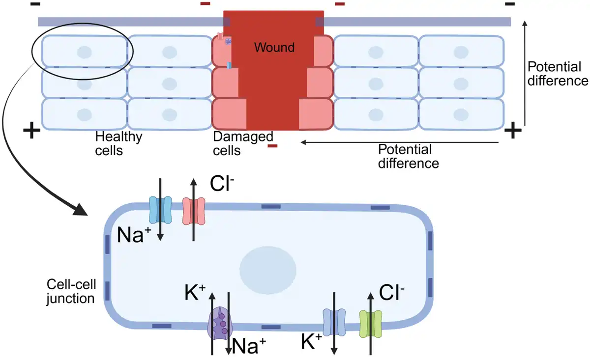
Much evidence suggests that bioelectrical phenomena are intricately linked to the wound healing and regeneration of animals and plants[6–7–8]. The epithelium of skin, cornea, and other places belongs to polarized epithelium, which can directionally transport ions and maintain transepithelial potentials (TEP) (10–60 mV), depending on ion channels or pumps and intercellular junctions[9]. When the epithelial barrier is compromised, the epithelial layer and the underlying tissue are directly connected by body fluids[10], resulting in a short circuit of the TEP. Consequently, the potential at the wound site becomes negative relative to the intact epithelium surrounding the wound. This potential gradient drives ionic currents toward the more negative site-wound, thereby establishing a transverse wound electric field (EF)[11]. These changes establish an ionic gradient across the cell membrane, driving ionic currents and generating a voltage gradient at the wound site[12]. Distant cells continuously supply ions to sustain the TEP, maintaining the current flow until wound closure and barrier restoration are achieved (Figure 1). The voltage gradient induces cellular-level physical effects, manifested as EFs, membrane potential, ion fluxes, and ionic concentration gradient. Then, cellular signal receptors, including ion channels, transporters, and EF-sensitive membrane receptors, detect the voltage gradient through the aforementioned physical effects. These receptors activate downstream signaling pathways, culminating in a transcriptional cascade that governs morphogenesis and tissue regeneration[8]. The wound EF, an endogenous EF generated by the wound itself during this process, provides cells with complex spatiotemporal information about the injury site and guides wound healing (For details, refer to Section 3). However, aging and pathological conditions can disrupt these bioelectrical processes, impair natural healing, and lead to chronic wounds.
Figure 1.
The endogenous electric field at the wound. Created with BioRender.com.
Exogenous electrical stimulation has demonstrated the ability to mimic the physiological EF, modulating diverse cellular behaviors and promoting faster, more effective wound healing[13]. Traditional electrotherapy relies on bulky external power supplies, which restrict its application due to limited portability and flexibility. However, recent advancements in materials science and electronics have led to the development of compact, intelligent, and minimally invasive exogenous electrical stimulation devices tailored to the specific requirements of wound environments. These innovations address critical medical needs by enabling targeted and efficient therapeutic interventions. This paper provides a concise overview of the generation of endogenous EFs, elucidates their roles and mechanisms in the wound healing process, and reviews recent progress in the application of exogenous EFs to enhance soft tissue wound healing over the past few years.
2. The molecular mechanism of the electric field in the wound healing process
EF stimulation modulates the activity of ion channels and membrane-bound receptors, triggering downstream intracellular signaling cascades. These cascades regulate critical cellular processes, including hyperpolarization, depolarization, migration, proliferation, and differentiation[14,15].
2.1. Ion channels, transporters, and EF-sensitive membrane receptors in EF
When cells are exposed to an EF, the applied field alters protein conformation by influencing the axial dipole moment of proteins[16]. This activation mechanism targets voltage-sensitive proteins, including ion channels, transporters, and EF-sensitive receptors.
Among ion channels, Ca²⁺ and Na⁺ channels are the most extensively studied. In cells expressing voltage-gated Ca²⁺ channels, the EF induces channel opening near the cathode, facilitating ion influx, elevating intracellular ion concentration and pH, and ultimately leading to membrane polarization[17]. Additionally, transporters such as Na⁺/K⁺-ATPase and Na⁺/H⁺ exchanger 3 are locally activated, triggering downstream intracellular signaling pathways. These processes are closely associated with the directed migration of certain cell types in response to EFs[18].
The EGF receptor is an EF-sensitive receptor that undergoes upregulation and asymmetric redistribution in response to both endogenous and exogenous EFs. Studies have demonstrated that the downstream epidermal growth factor (EGF) receptor–mitogen-activated protein kinase (MAPK) signaling pathway plays a critical role in EF-directed cell migration. Similarly, the acetylcholine receptor (AChR) redistributes under EF stimulation[19], and its activation facilitates cytoskeletal reorganization, cell polarization, and subsequent electrotactic responses[20]. Additionally, N-methyl-D-aspartate receptors (NMDARs), which are ligand-gated Ca²⁺-permeable ion channels, are activated by EF stimulation. This activation mediates the NMDAR/Ras-related C3 botulinum toxin substrate 1 (Rac1)/actin signaling pathway, ultimately promoting cell migration[21] (Figure 2).
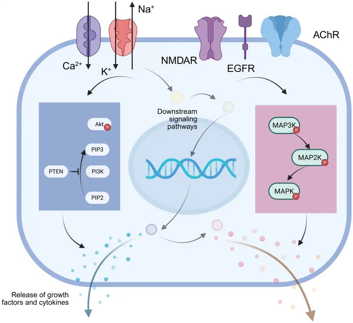
Figure 2.
The molecular mechanism of the electric field in the wound healing process. Created with BioRender.com.
Furthermore, electrical stimulation, depending on its voltage intensity and frequency, may also have effects on different plasma membranes, inducing reversible pore formation. These reversible pores, commonly known as electroporation, provide a mechanism for the delivery of biomolecules, including therapeutic drugs. The ability of symmetric alternating current EFs to guide cell migration in a frequency-dependent manner further suggests that nonprotein components of the cell membrane may play a critical role in modulating cellular behavior[22]. Moreover, studies have demonstrated that lipid rafts respond to EFs, promoting the polarization of membrane proteins and initiating intracellular signaling cascades[23].
2.2. Signaling pathway in electric field
EFs activate various intracellular signaling pathways by modulating ion channels, transporters, and EF-sensitive membrane receptors, which subsequently influence cellular activity[24]. Notably, the phosphatase and tensin homolog (PTEN)-phosphoinositide 3-kinase (PI3K) and MAPK pathways have been identified as critical regulators of these processes. Specifically, the PI3K–protein kinase B (Akt) pathway is a central regulator of EF-directed cell migration, while the MAPK pathway plays a significant role in modulating other cellular behaviors.
2.2.1. The PI3K–PTEN signaling pathway
The PI3K signaling pathway plays a critical role in regulating cell growth, metabolism, and survival. The canonical PI3K pathway activates the downstream Akt-mechanistic target of rapamycin complex 1 signaling cascade, while PTEN acts as a negative regulator of this pathway. The PTEN–PI3K axis is a key modulator of cellular behaviors, including migration, differentiation, and proliferation.
EF-induced directional cell migration, known as electrotaxis, has been extensively investigated. Studies on Dictyostelium cells have proposed a tug-of-war mechanism, suggesting the involvement of multiple signaling pathways in this process[25]. Sun et al. further advanced the “compass” model based on keratocyte fragments and cells, highlighting the essential role of the PI3K pathway in whole-cell orientation sensing[26].
Existing research results suggest that extracellular calcium levels may influence PI3K pathway activation[26,27]. Beyond migration, electrical stimulation has been shown to inhibit the transition of macrophages from pro-inflammatory M1 to anti-inflammatory M2 polarization via the PI3K–Akt pathway[28,29]. Additionally, it suppresses E-cadherin expression by inhibiting the PI3K/Akt/Snail pathway, thereby reducing keratinocyte differentiation, a process crucial for wound healing[30]. Furthermore, electrical stimulation promotes neural stem cell differentiation into neurons through the activation of the PI3K/Akt/glycogen synthase kinase-3 β/β-catenin pathway[31].
2.2.2. The MAPK signaling pathway
The MAPK pathway regulates cell growth, differentiation, stress responses, and inflammation through the sequential activation of MAPK, mitogen-activated protein kinase kinase (MEK), and MAPK kinase kinase. Numerous studies have demonstrated the pivotal involvement of the MAPK pathway in these processes.
Electrical stimulation can modulate various types of cells through the MAPK signaling pathway, thereby promoting the healing of diverse injuries. Electrical stimulation has been widely utilized in nerve regeneration and the reduction of postinjury nerve inflammation, playing a critical role in the recovery of nervous system function. For instance, exposure of PC12 mutant cells to 100 mA electrical stimulation for 30 minutes activates the MAPK/cAMP-response element binding protein pathway, significantly enhancing neural synapse growth[32]. However, prolonged electrical stimulation can induce cell death[32]. Additionally, electrical stimulation exerts anti-inflammatory and analgesic effects through the MAPK pathway, participates in postinjury protective mechanisms, regulates gastrointestinal function, and protects cardiovascular and cerebrovascular systems[33]. Research has also shown that electrical stimulation restores mitochondrial respiratory dysfunction via the p38 MAPK signaling pathway, thereby modulating cellular function[34].
Likewise, electrical stimulation plays a significant role in soft tissue wound healing mediated by the MAPK pathway. Furthermore, we recognize that the specific approach to electrical stimulation appears to be a determining factor in cellular responses. In microvascular endothelial cells, high-frequency (7.5 GHz), low-amplitude EFs induce phosphorylation of MEK and extracellular regulated protein kinases (ERK), leading to vascular endothelial growth factor (VEGF) release and capillary morphogenesis, without activating the MAPK/c-Jun N-terminal kinase or MAPK/p38 pathways[35]. Conversely, evidence suggests that the p38 MAPK signaling pathway in epithelial cells treated with a 1 V/cm EF, and 0.1-ms pulse width is involved in EF-induced p53 phosphorylation, potentially reducing infection-mediated inflammation[36]. Furthermore, microcurrents activate the MAPK pathway, promote cell migration and proliferation, and induce transforming growth factor-beta 1 (TGF-β1) secretion by fibroblasts and osteoblast-like cells, facilitating wound closure[37].
2.3. Growth factors and cytokines in electric field
Following the initiation of an EF-induced intracellular signaling cascade, the resulting effects propagate through the surrounding tissue via growth factors and cytokines, establishing a cell-to-cell regulatory network. This phenomenon is particularly pronounced in immune cells, such as macrophages. Exogenous EFs modulate macrophage phenotypes, influence their polarization, and subsequently reshape the immune microenvironment, thereby impacting the overall wound healing process. Furthermore, exogenous electrical stimulation induces fibroblasts to secrete fibroblast growth factor 2 and enhances VEGF protein synthesis in human umbilical vein endothelial cells (HUVECs). This activation of the MAPK/ERK signaling pathway promotes HUVEC migration, invasion, and angiogenesis[38]. In keratinocytes, exogenous electrical stimulation (100–200 mV/mm, 6 hours) increases EGF and VEGF secretion, while reducing interleukin-6 (IL-6) and interleukin-8 (IL-8) levels. These changes indirectly influence physiological processes, such as blood vessel formation, ultimately facilitating wound healing[39].
In summary, the molecular mechanism of EFs in wound healing is a multifaceted process initiated by the perturbation of voltage-sensitive proteins in the cell membrane. This activation of ion channels, transporters, and receptors triggers well-defined intracellular signaling cascades (e.g., MAPK, PI3K, Rac1) that govern essential cellular functions, most notably electrotaxis and proliferation. These cellular-level events culminate in significant physiological benefits, including enhanced angiogenesis, improved blood perfusion, and modulation of the immune response, which collectively accelerate wound closure.
3. The role of the electric field in stages of tissue healing
Wound healing is a highly organized and complex process involving a series of cellular and biochemical events (Figure 3). It is conventionally categorized into 3 overlapping phases: (1) hemostasis and inflammation, (2) proliferation, and (3) maturation or remodeling[40]. These 3 phases are highly overlapping and dynamically continuous with no absolute demarcation. The inflammatory response initiates during the hemostasis phase and persists throughout the entire healing process; its intensity and duration directly modulate the final repair outcome. Furthermore, the progressive restoration of tissue structural integrity is driven by the cascade regulation of cytokines across these phases, working synergistically to achieve complete repair.
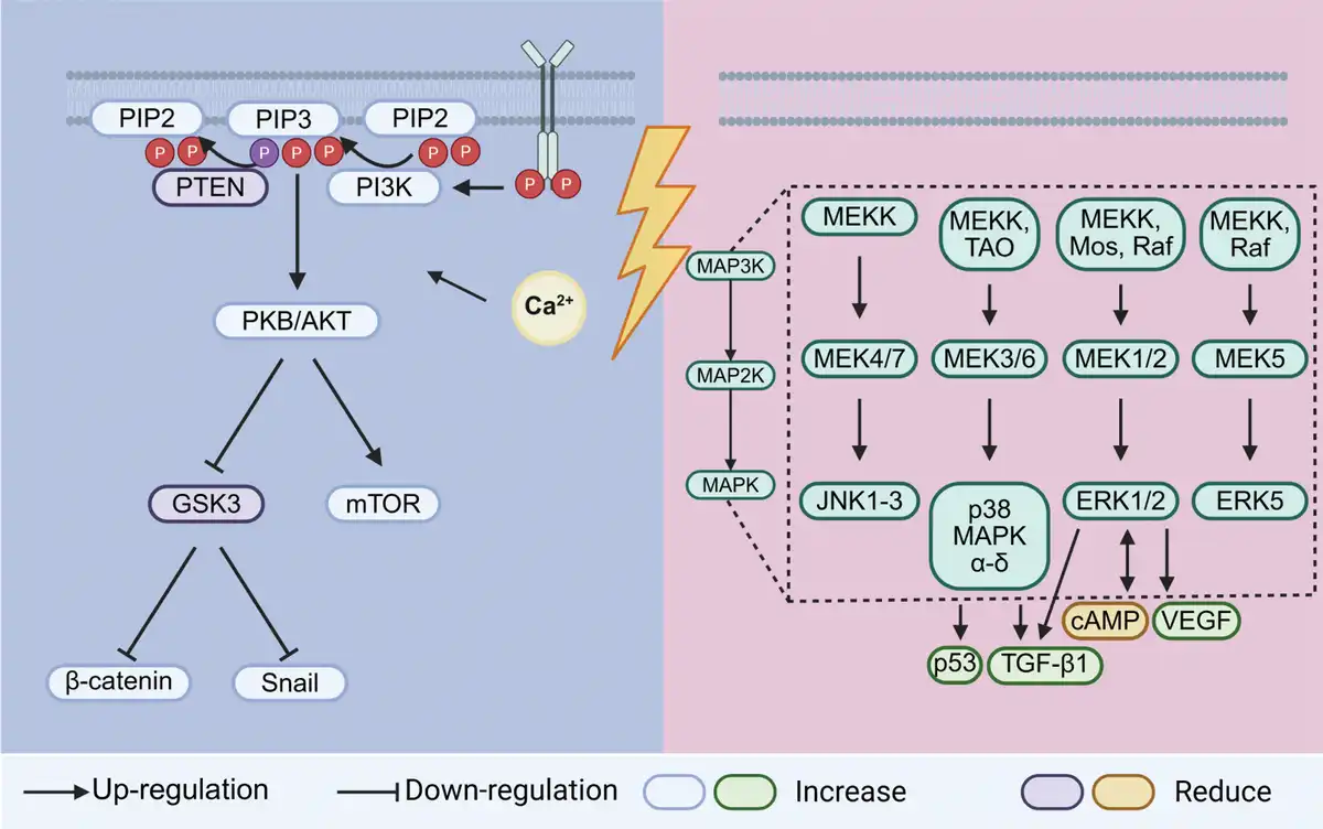
Figure 3.
The signaling pathway in the electric field. Created with BioRender.com.
3.1. Hemostasis and inflammation
Following injury, vascular rupture exposes subendothelial collagen to platelets, triggering platelet aggregation and initiating the intrinsic coagulation cascade. The resulting blood clots, along with locally released cytokines and growth factors, promote cell chemotaxis and tissue cell adhesion during the subsequent inflammatory phase. Inflammation is a critical stage of wound healing, characterized by a vascular-centric response. Upon injury, tissue-resident sentinel cells release inflammatory mediators, activating vascular responses and recruiting inflammatory cells to clear tissue debris and pathogens. This process is gradually replaced by tissue repair[41]. Notably, macrophage activation is essential for transitioning to the proliferative phase, as it also mediates angiogenesis[42].
The EF plays a pivotal role in modulating immune cell activity during the inflammatory process. In the initial phase of inflammation, endogenous EFs facilitate the directional migration of macrophages, lymphocytes, and neutrophils to the wound site. Notably, mast cells, which are often implicated in skin pathologies, exhibit a significant reduction in population under electrical stimulation during the acute inflammatory phase[43]. Furthermore, macrophages demonstrate an EF intensity-dependent anodal migration, with enhanced phagocytic activity under EF exposure, thereby accelerating the clearance of pathogens and necrotic tissue, and promoting soft tissue regeneration[44]. During the later stages of inflammation, electrical stimulation has been shown to attenuate immune cell infiltration and downregulate pro-inflammatory cytokine secretion, facilitating inflammation resolution. Of particular significance is the role of macrophage polarization in chronic inflammation. Recent studies indicate that electrical stimulation can induce macrophage polarization towards the M2 phenotype, thereby shifting the wound microenvironment from an inflammatory to a reparative state, effectively preventing chronic inflammation[45].
Simultaneously, the EF enhances vascular responsiveness, augmenting blood flow to the wound site, facilitates the migration of inflammatory cells and the transport of essential nutrients, thereby accelerating wound healing. Additionally, studies have demonstrated that electrical stimulation, particularly through the application of symmetrical biphasic pulsed currents, significantly attenuates posttraumatic edema[46,47].
3.2. Proliferation
During the proliferation phase, epithelial cells, fibroblasts, and endothelial cells play pivotal roles in tissue repair[41]. Epithelial cells at the wound margin initiate proliferation and extend projections to re-establish the protective barrier, thereby preventing fluid loss and bacterial infiltration. Activated platelets and macrophages secrete EGF and transforming growth factor-beta, which promote epithelial cell proliferation and chemotaxis[48]. Concurrently, VEGF, secreted by keratinocytes, macrophages, fibroblasts, platelets, and endothelial cells at the wound periphery, drives the formation of new capillaries. Fibroblasts migrate from adjacent tissues to the wound site, where they synthesize a provisional extracellular matrix comprising type 3 collagen, glycosaminoglycans, and fibronectin. Platelet-derived growth factor and EGF, released by platelets and macrophages, serve as key signaling molecules that stimulate fibroblast proliferation and activity.
The EF facilitates directional cell migration by establishing a stable voltage gradient at the wound site, which provides spatial guidance for the movement of surrounding cells. This process is primarily regulated by the PI3K–PTEN signaling pathway. Additionally, extensive research has demonstrated that exogenous electrical stimulation enhances cell proliferation, thereby accelerating wound healing. Specifically, electrical stimulation activates the TGF-β1/ERK/nuclear factor kappa-light-chain-enhancer of activated B-cell signaling pathway, inducing fibroblast proliferation and upregulating α-smooth muscle actin expression, which drives the differentiation of fibroblasts into contractile myofibroblasts[49,50]. Myofibroblasts play a critical role in wound closure and extracellular matrix deposition. In vitro studies have further revealed that electrical stimulation at voltages exceeding 3 V/cm significantly increases the expression of fibroblast-derived collagen, elastin, and MMP-1[51]. Collagen and elastin are essential components of the extracellular matrix, while MMP-1 facilitates matrix degradation, enhances cell migration, and participates in tissue remodeling, collectively promoting soft tissue regeneration. Notably, the effects are more pronounced with short voltage pulses. Moreover, electrical stimulation promotes the migration, invasion, and proliferation of vascular endothelial cells, while stimulating the secretion of key growth factors such as VEGF and fibroblast growth factor 2, which drive angiogenesis and provide essential blood supply for wound healing[52].
3.3. Maturation
The maturation stage is characterized by the deposition of collagen, which organizes into a well-structured network within the wound. The strength of the resulting scar, a key outcome of soft tissue healing, is determined by the rate, quality, and total quantity of extracellular matrix deposition[41]. Excessive collagen synthesis, however, can lead to the formation of hypertrophic scars or keloids.
Over time, the composition of the wound matrix undergoes significant changes. Initially, it is predominantly composed of fibrin, fibronectin, and thrombospondin, which are critical for cell adhesion and migration[53,54]. As healing progresses, the levels of glycosaminoglycans, proteoglycans, and specific structural proteins increase, providing a foundation for subsequent matrix deposition and remodeling. Eventually, collagen emerges as the primary structural protein in the scar tissue. During wound healing, fibroblasts exhibit adaptive responses to dynamic mechanical loads within the matrix. These responses include migration, matrix proteolysis, and other behaviors that collectively establish isometric tension—a state where internal and external mechanical forces are balanced, preventing cell shortening or elongation.
Existing studies demonstrate that electrical stimulation modulates scar thickness and hardness by influencing collagen deposition within the scar tissue. Research on low-voltage pulsed current applied to diabetic mouse models revealed a positive correlation between electrical stimulation intensity and collagen deposition[55]. Additionally, electrical stimulation regulates the secretion of various MMPs, thereby impacting the maturation of the extracellular matrix at the wound site[56].
4. Application of exogenous electrical stimulation
Electrical stimulation significantly enhances wound healing by promoting cell migration and proliferation, stimulating angiogenesis, increasing extracellular matrix synthesis, modulating immune responses, and reducing scar formation. Chronic wounds, such as bedsores and diabetic ulcers, are often associated with weakened endogenous EFs, resulting in delayed or impaired healing. This not only causes significant physical and psychological distress to patients but also imposes a substantial economic burden. To address this, researchers have explored strategies to enhance wound healing by augmenting endogenous EFs using conductive dressings or applying targeted electrical stimulation[57–58–59–60]. Studies have demonstrated that improving endogenous EFs accelerates re-epithelialization, promotes angiogenesis, regulates immune responses, and minimizes scar formation. The use of exogenous electrical stimulation for chronic wound healing dates to 1971, when researchers first attempted to apply external electrical currents to wound sites. By combining an external power source with strategically placed electrodes, effective electrical stimulation can be delivered directly to the wound area[61–62–63–64]. Its advantage is that the intensity, frequency, and other parameters of electrical stimulation can be accurately controlled, but at the same time, it makes its application inconvenient and difficult to promote. In recent years, researchers have focused on designing more portable, flexible, and versatile electrical stimulation devices for different wound environments. Here, we focus on recent research on the application of exogenous electrical stimulation to promote wound healing, and according to the material classification of the occurrence of exogenous electrical stimulation (Table 1).
Table 1
Comparison of exogenous electrical stimulation devices.
| Type | Working principle | Output characteristics | Source of energy | Advantages | Limitations |
|---|---|---|---|---|---|
| Battery[65] | Electrochemical reaction | Stable, direct | External power supply (or partial power supply) | Output stability | Less tissue—matchable |
| Piezoelectric generator[66] | Piezoelectric effect | dependent on the material properties, alternating | Mechanical deformation | superior biocompatibility, facile processability, high durability, reliability, and sensitivity | Premature degeneration or failure of device function |
| Triboelectric nanogenerator[67,68] | Contact electrification and electrostatic induction | high instantaneous output power, alternating | Mechanical displacement | high charge density, flexible structure, low cost, and broad applicability | Limited application in humid environments |
| Electret[69] | Permanent polarization of the material | lower powered, direct, continuous | Pre-charging | No need for power supply | Charge attenuation |
| Thermoelectric generator[70] | Seebeck effect | Limited by temperature differences | Thermal gradient | Continuous power supply | Low power |
4.1. Battery
The most intuitive idea may be to use a battery. As an efficient energy storage device, the battery can act as a power source and power a variety of wearable or implantable devices. The conventional battery usually has a larger volume, a lack of flexibility, and a possible toxicity, but with the continuous deepening of research, more new types of batteries continue to emerge[71]. For wound healing, the primary consideration is excellent biocompatibility. Besides, it needs to adapt to the wound environment as much as possible, ensure sufficient electricity, and avoid interfering with wound healing.
Wu et al. have designed a fluid-activated tubular Mg–Mo battery that can effectively discharge after implanted in the intermuscular space and significantly improve the healing rate of full-thickness skin wounds[72]. It uses body fluid in the intermuscular space as an electrolyte with higher electrical conductivity, and avoids possible corrosion caused by electrolyte leakage. Moreover, the battery featured a simple structure and good stability, providing continuous discharge for up to 5 days to meet the requirements of wound healing. Xiao et al. also chose body fluids as electrolytes[73]. The battery was composed of an Mg-wire core, a separator, and an outer layer formed by wrapping carbon yarn, which absorbs the wound exudate, owing to activating the battery. The excessive wound exudate formed an electrolyte layer between the Mg wire (anode) and the carbon yarn (cathode) to facilitate the redox reaction. Kim et al. designed an electricity auto-generating glucose-responsive enzymatic biofuel cell as a skin patch[74]. It converted ubiquitous biological glucose into electrical energy, and then stimulated angiogenesis, fibroblast functionality, and matrix synthesis (Figure 4A–C). Similarly, Wang et al. devised a capacitive dressing with polypyrrole-wrapped carbon cloth electrodes and a bacterial cellulose hydrogel separator[75]. This dressing is designed to be antibacterial, so it has greater electrical capacity and rechargeability. The experimental results show that the slow discharge process after the rapid discharge can regulate immunity, promote the formation of capillaries, and collagen deposition at the wound site (Figure 4D, E). This dressing enabled continuous and effective sterilization and promoted wound healing through a safe, low-voltage EF. It offered nonantibiotic physical therapy for infected wounds, demonstrating significant clinical potential in an era where drug-resistant bacteria are increasingly prevalent.
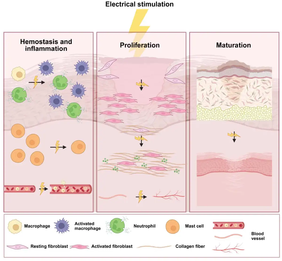
Figure 4.
The role of the electric field in stages of tissue healing. In hemostasis and inflammation phase, the electrical stimulation modulates immune cell activity and enhances vascular responsiveness; in proliferation phase, the electrical stimulation promotes fibroblast proliferation, migration, and secretion, and enhances angiogenesis; in maturation phase, the electrical stimulation modulates collagen deposition by regulating the secretion of matrix metalloproteinases (MMPs). Created with BioRender.com.
4.2. Piezoelectric generator
Piezoelectric materials can convert mechanical energy into electrical energy. When subjected to an external force, these materials undergo internal polarization, resulting in the generation of opposite charges on their specific surfaces. Researchers have explored various methods to generate voltage or current through piezoelectric materials for wound healing, including external stimuli (e.g., ultrasound) and harnessing mechanical energy from bodily movements. Traditionally, piezoelectric materials are categorized into three groups: inorganic piezoelectric materials, organic piezoelectric materials (including natural and synthetic biopolymers), and piezoelectric composites. Inorganic piezoelectric materials typically exhibit stronger piezoelectric effects, superior long-term stability, and high rigidity. In contrast, organic piezoelectric materials, while demonstrating weaker piezoelectric effects, offer advantages in flexibility and biocompatibility. Piezoelectric composites, designed by combining inorganic and/or organic materials with other components, achieve a balance of flexibility and piezoelectric performance. However, their preparation is more complex, and their performance stability may be compromised.
Piezoelectric ceramics are the most widely utilized inorganic piezoelectric materials[76]. Their individual grains can be uniformly polarized, generating a potential difference. Its high stiffness restricts its application in wound healing. In contrast, polyvinylidene fluoride (PVDF) stands out as a representative organic piezoelectric material, offering excellent piezoelectric properties, workability, and mechanical strength. The piezoelectric performance of PVDF is significantly influenced by its β-phase content, which can be enhanced by optimizing parameters such as spinning distance, applied voltage, speed, and annealing processes[77]. Sun et al. developed a PVDF-based self-powered wound dressing integrated with antibiotic release. This dressing not only facilitates wound healing through piezoelectric voltage generation but also enables precise control of hydrophilic drug release by modulating the electrostatic balance between the drug carrier and the drug (Figure 5A, B)[78]. Fu et al. also combined Poly(vinylidene fluoride-co-trifluoroethylene) (P(VDF-TrFE)), TrFE-based nanogenerator and drug delivery system[79]. This nanogenerator generated a relatively low EF at rest, and an EF exceeding 625 mV/mm promoting the rapid release of PTEN inhibitor in motion. This on-demand amplified cell electric responsiveness and avoided overinhibition of PTEN (Figure 5C, D). In a distinct approach, Ren et al. utilized cell adhesion tension to create a dynamic force–electrical closed-loop feedback system[80]. They engineered a piezoelectric dual-network nanofiber dressing incorporating copper/epigallocatechin 3-gallate and PVDF. This design circumvents the temporal limitations of magnetic field- or ultrasound-induced electrical stimulation, synchronizes with cellular physiological states, and mimics the extracellular matrix’s piezoelectric effect to enhance intercellular communication. Experimental results demonstrated that the electrical stimulation generated by this dressing activates calcium-related ion channels and significantly accelerates wound healing (Figure 5E, F). On this dressing surface of, cells further modulated the adaptive electrical stimulation generated by the piezoelectric nanofiber network through their inherent adhesiveness, producing a dynamic mechanoelectric closed-loop feedback signal that mimics the electrical microenvironment of natural physiological conditions. Moreover, it exhibited strong antibacterial and anti-inflammatory capabilities, promoting skin repair through multiple biological pathways.
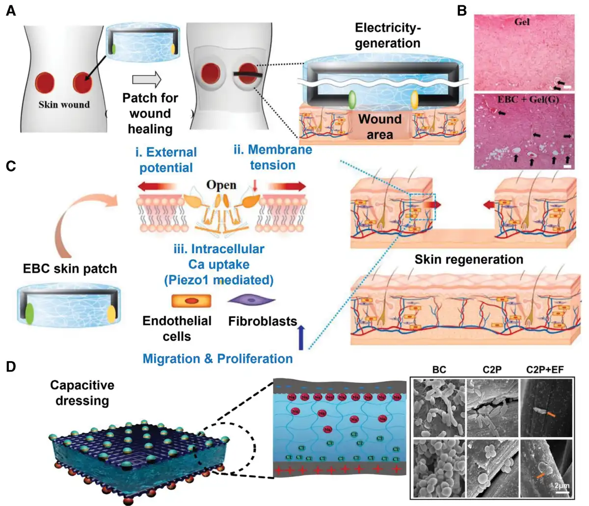
Figure 5.
Battery for application to soft tissue wounds. (A) The skin patch battery over the soft tissue wound[74]. Copyright © 2021, Elsevier. (B) The effect of skin patch battery to help wound healing. (C) The process of wound healing activated via skin patch battery[74]. Copyright 2021, Elsevier. (D) Structural schematic of charged capacitive dressing. (E) The morphology of charged capacitive dressing[75]. Copyright 2024, The Authors.
Composite piezoelectric materials are currently a focal point of research[81]. Xu et al. developed a repetitive mechanical impact-electrical stimulation system by combining barium titanate with polydimethylsiloxane (PDMS)[82]. PDMS, known for its excellent biocompatibility and flexibility, ensures continuous and stable contact with damaged skin, overcoming the limitations of barium titanate nanoparticles. Studies indicated that this electrical stimulation reduced inflammation, enhanced angiogenesis, and significantly accelerated wound healing. Wang et al. designed 4-octyl itaconate-coated Li-doped ZnO/Ply(L-lactide) piezoelectric composite microfibers[83]. The incorporation of Li atoms induces lattice distortion in ZnO, enhancing its piezoelectric properties, while aligned Ply(L-lactide) further amplifies the piezoelectric effect. Electrical stimulation, combined with reactive oxygen species generated by microfibers, restores mitochondrial function. Additionally, the acoustic dynamic effect of Li-doped ZnO achieves a 94.2% bactericidal rate against Staphylococcus aureus. These microfibers facilitate infected wound healing through multiple mechanisms, offering a promising approach for managing infected wounds.
4.3. Triboelectric nanogenerators
Triboelectric nanogenerators (TENGs) operate based on the coupling effect of triboelectricity and electrostatic induction. The triboelectric effect induces the accumulation of positive and negative charges on 2 friction layers, respectively. Subsequently, electrical energy is generated between the electrodes through electrostatic induction. The fundamental structure of TENGs consists of 2 dielectric films with distinct electron gain/loss capabilities, connected by back electrodes. As a self-powered device for wearable electronics, TENG has attracted significant attention due to its high efficiency and wide range of material choices[84].
The output capacity of TENGs is primarily influenced by the surface potential properties of the material, as well as external factors such as ambient temperature and humidity. Venkatesan et al. developed a single-electrode TENG, whose surface charge density and electrical output performance were significantly enhanced through incorporating a TiO₂–MXene–polystyrene nanofiber membrane as a charge-trapping interlayer[85]. When applied to rat skin, the TENG generated an open-circuit voltage of 1 to 10 V through the rat’s daily activities, delivering electrical stimulation to the wound. In vivo studies demonstrated that this electrical stimulation enhances cell proliferation, promotes angiogenesis, and accelerates wound healing. For TENGs to be effective in wound healing, they must operate in a fully biocompatible environment, such as body fluids present at the wound site. Additionally, the materials used in TENGs must be sufficiently flexible to conform to the wound without causing further damage. Hydrogels, known for their softness and water-absorbing properties, are ideal candidates for wound dressings, as they promote wound closure[86]. Combining friction materials with hydrogels represents a promising approach. Qin et al. designed a triboelectric stimulator that integrates a TENG with a triboelectric-responsive drug delivery hydrogel (TR-DDH), using the TR-DDH as an electrode in direct contact with the wound[87]. The TENG generated pulsed currents and triggered volume changes in the polypyrrole within the TR-DDH, releasing curcumin nanoparticles. In vitro and in vivo experiments confirmed that this system enables efficient and controllable drug delivery, significantly promoting the healing of infected wounds (Figure 6A–C). Similarly, Tang et al. fabricated a TENG using electrospun polymer tribo-layers and a chemical vapor-deposited polypyrrole electrode, resulting in a device with excellent flexibility, air permeability, and moisture retention[88]. In vitro and in vivo experiments demonstrated that the TENG upregulates the expression of growth factor-related genes, enabling diabetic rat skin wounds to heal within 2 weeks (Figure 6D–F).
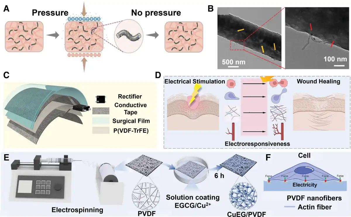
Figure 6.
Piezoelectric generator for application to soft tissue wounds. (A) Schematic diagram of self-powered electric field and drug release. (B) TEM image of PCVE0.03. The pore structure inside the fiber is indicated with yellow arrows. A carboxylated carbon nanotube multi-walled composite with vancomycin hydrochloride embedded within the PCVE0.03 porous piezoelectric fiber is pointed with red arrows[78]. Copyright 2024, Wiley-VCH GmbH. (C) The structure of P(VDF-TrFE)-based nanogenerator. (D) P(VDF-TrFE)-Based nanogenerator realizes self-Powered electrical stimulation therapy and promotes wound healing[79]. Copyright 2023, American Chemical Society. (E) The synthesis of hydrophilic piezoelectric PVDF nanofibers. (F) Generation and mechanism of action of electrical stimulation at the cellular scale[80]. Copyright 2024, The Authors.
4.4. Electret
Electret, also known as a permanent electric body, is a dielectric that can store an excess “real” charge or/ and maintain an “oriented” electric dipole for a long time (much longer than the time required to form the polarization). The former means that it is a nonelectric neutral material. The latter is that after the medium containing the intrinsic electric dipole is polarized by the strong external EF, its polarization state does not completely disappear with the removal of the external EF, and the “oriented” electric dipole is maintained “long-term,” forming the “oriented” electric dipole electret.
As a stimulus source of exogenous electrical stimulation, the electret has significant advantages: stability. Electrets can spontaneously generate microcurrent and micro EF in the humoral environment and maintain the charge for a long time in the absence of external energy input. This means that when selecting and designing the electret material, the stable presence of its charge matches the physiological process of healing in time, ΔT especially in deep wounds. Yao et al. combined the electret thin films with a shape memory alloy to construct an electromechanical synergistic dressing[89]. Biocompatible shape memory alloys can draw the epithelial tissue and provide the appropriate contractile force for wound healing. Electret film could enhance the endogenous EF to promote healing while playing an antibacterial role. In vivo studies in rats showed that this dressing could promote the secretion of EGF and VEGF, regulate the microenvironment of the wound surface, and significantly improve the wound closure rate (Figure 7A–E). Kim et al. applied electrets in the other direction[90]. They obtained higher voltages via a multilayer stacked electret (MS-electret) to suppress skin scarring. MS-electret have surface potential of 3400 V to generate direct current EF (Figure 7F).
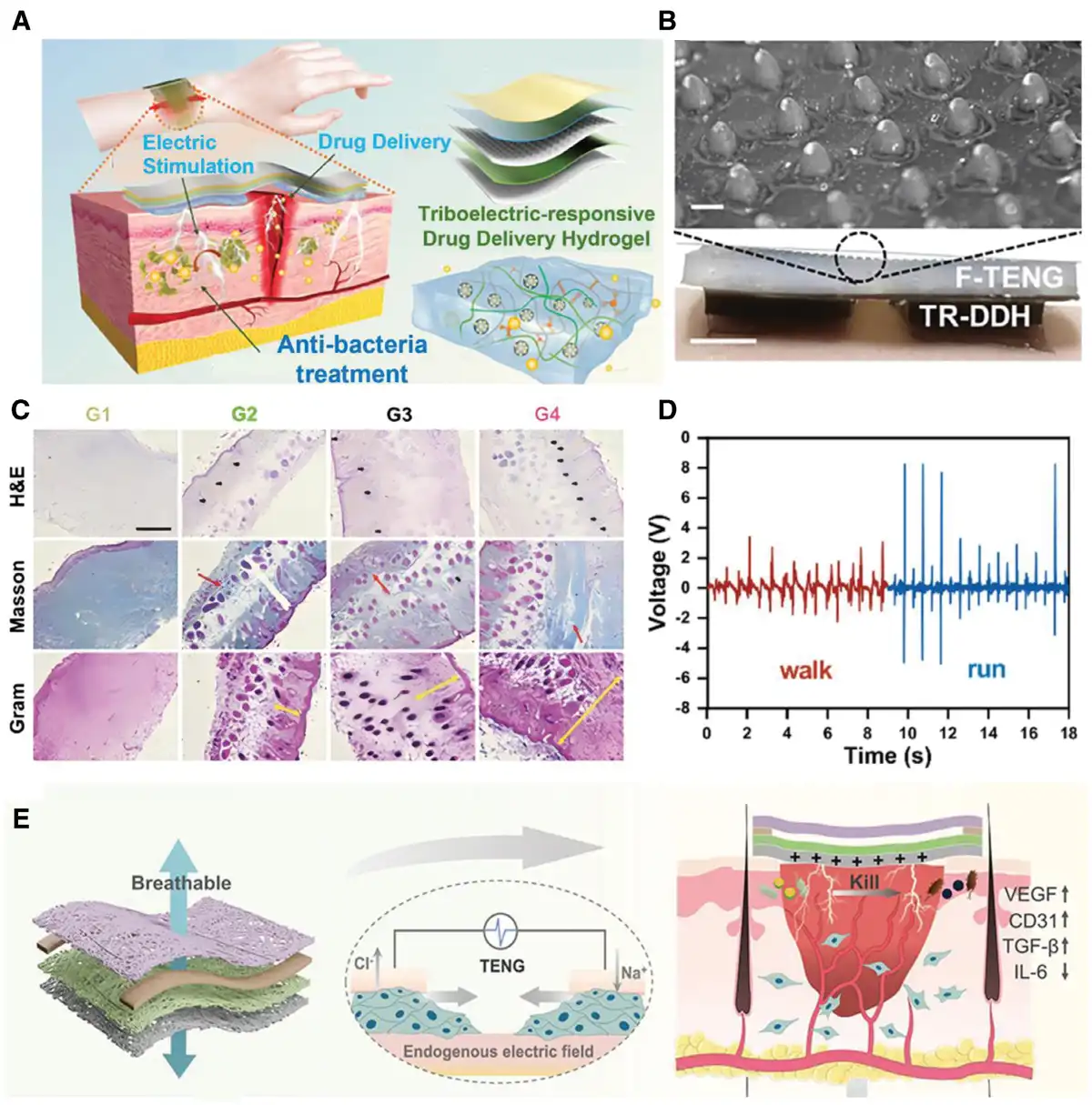
Figure 7.
Triboelectric nanogenerators for application to soft tissue wounds. (A) Design of the wearable triboelectric stimulator (WTS) for bacterially infected wound healing. (B) Optical photo of the microstructure on the surface of the silicone rubber film (scale bar = 300 µm). (C) H&E staining, Masson staining, and Gram staining of the wounds from the experimental mice (scale bar, 100 µm) (hair follicles: black arrows; connective tissue: red arrows; and thicknesses of the epithelium: yellow lines[87]. Copyright 2024, Wiley-VCH GmbH. (D) Output voltage of Teng while human walking and running. (E) Wound-healing-promoting effects[88]. Copyright 2023, American Chemical Society.
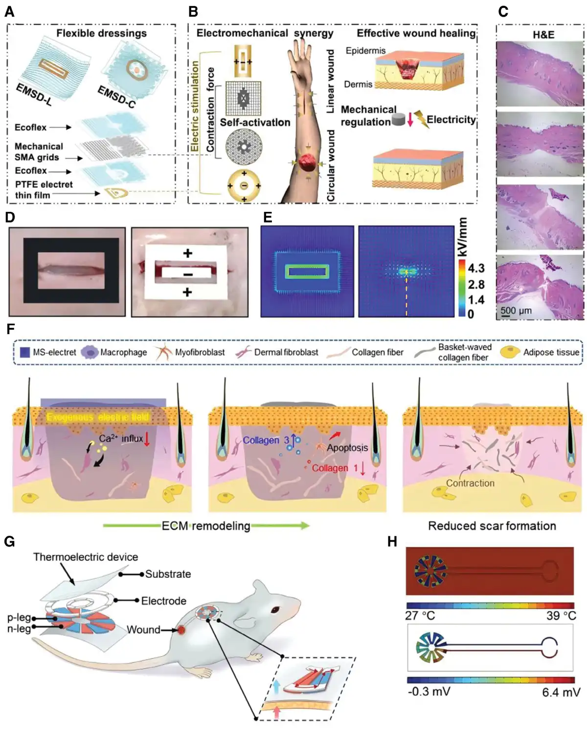
Figure 8.
Electret and thermoelectric generators for application to soft tissue wounds. (A) Exploded illustration of the device components, essential materials, and multilayer structures (bottom). (B) Working principle of wound treatment by programmable and skin temperature-activated electromechanical synergistic dressings. (C) The healing condition of the wound. (D) Optical images of the linear incisional wound and experimental setup for healing. (E) Simulated electric field distribution inside a linear wound[89]. Copyright 2022, The Authors (F) Timeline of inhibition of scar formation from the exogenous electric field, which is created with the multilayer stacked electret[90]. Copyright © 2023, The Authors (G) The left panel depicts the exploded view of the thin-film structured flexible thermoelectric device. (H) The finite element analysis resolves the temperature distribution (up panel) and corresponding electric field (bottom panel) within the flexible thermoelectric device[91]. Copyright 2024, Wiley-VCH GmbH.
4.5. Thermoelectric generators
Thermoelectric nanogenerators utilize the Seebeck effect to convert thermal energy into electrical power. The Seebeck effect manifests as a voltage gradient between 2 dissimilar conductors or semiconductors when subjected to a temperature differential. In pyroelectric materials exhibiting spontaneous polarization, temperature variations modulate polarization strength, generating opposing surface charges through the primary pyroelectric effect. Pyroelectric nanogenerators harness this temperature-dependent charge separation for energy conversion. Unlike piezoelectric counterparts requiring mechanical strain, pyroelectric nanogenerators generate transient currents solely through thermal fluctuations. Secondary pyroelectric effects emerge in wurtzite-structured materials where thermal expansion induces crystallographic strain, subsequently activating piezoelectric charge separation. This synergistic mechanism enables dual energy harvesting through coupled pyroelectric–piezoelectric phenomena.
While thermoelectric and pyroelectric generators find widespread application in electronic component design[92], their direct implementation as exogenous electrical stimulation sources remains limited. Zhang et al. recently demonstrated a breakthrough in flexible biocompatible TE device development[91]. The researchers employed Bi₂Te₃-based compounds, selected for superior near-ambient thermoelectric performance, deposited on flexible polyimide substrates with interdigitated gold electrodes. This configuration achieved exceptional energy conversion efficiency, generating 10 mV output under ΔT = 10 K conditions. When applied topographically to wound sites, the device sustained μV-level potential gradients through dermal thermoelectric coupling. Experimental validation revealed that bioelectrical stimulation derived from physiological (37 °C) to environmental (25 °C) thermal gradients significantly enhanced cellular metabolic activity. Specifically, this is manifested by suppressed secretion of inflammatory cytokines and increased angiogenesis, thereby accelerating wound healing (Figure 7G, H).
Table 2 presents some additional examples.
Table 2
The applications of exogenous electrical stimulation in promoting skin wound healing.
| Charge generation | Key materials | Structure | Type/intensity/frequency of ES | Outcome | References |
|---|---|---|---|---|---|
| Enzymatic biofuel cells | Carbon fiber fabric with redox enzyme (fructose dehydrogenase/ bilirubin oxidase) | Enzymatic electrodes, an elastic conductive resistor, a hydrogel, and medical adhesive tape | About 1 mA/cm2, maintaining for 12 hours | Accelerate wound closure by enhancing cell proliferation and migration | [93] |
| Chemical discharge after charge storage | Perylene polyimide, an aqueous battery material | Hydrazine hydrate-derived polyimide synthesized from perylene-3,4,9,10-tetracarboxylic dianhydride is coated on titanium | Li+-ions diffuse across the solid-liquid interface driven by a concentration gradient | Prevent biofilm formation during the early discharge stage and promote fibroblast proliferation in the later discharge stage | [94] |
| Wound exudate-activated battery | Hydrophilic brushes-grafted cotton yarn | A Mg-wire core, a separator consisting of poly(N,N-(dimethylamino)ethyl methacrylate) brushes-grafted cotton yarn, and an outer layer formed by wrapping of carbon yarn. | Maximum short-circuit current of 2 mA and output voltage of 1.9 V, with good long-term stability | Promote the proliferation and migration of fibroblasts and M2-type polarization of macrophages | [73] |
| Aqueous Zn–MnO2 microbatteries | Zn electrodes and MnO2 electrodes electrodeposited onto the forked fingers | Rechargeable flexible microZn–MnO2 batteries with an annular circuit structure | An operating voltage of 1.5 V | Accelerate cell migration and optimize cytokine distribution | [95] |
| Piezoelectric driven triboelectric nanogenerator | Electrospun fibers of PVDF | A conducting hydrogel component constructed with carbonized polydopamine/polydopamine/polyacrylamide and paired with electroactive electrospun PVDF membrane | Open-circuit output voltage (Voc) and short-circuit current (Isc) of ~42 mV and ~60 nA, respectively, assessed by repetitive human finger tapping on the PVDF membrane | Faster wound closure, re-epithelialization, regeneration of blood vessels and follicles | [96] |
| Single-electrode mode TENG | Polycaprolactone-based polyurethane and poly(acrylic acid) N-hydroxysuccinimide ester–poly(vinyl alcohol) copolymer | The upper membrane a polycaprolactone-based polyurethane as a triboelectric and encapsulation layer, poly(3,4-ethylene dioxythiophene) polystyrene sulfonate as a triboelectric electrode adhere on the a polycaprolactone-based polyurethane film, poly(vinyl alcohol) as a bioadhesive and Mo as electrodes. | Voltage output of about 1.50 V and current output of about 24.20 µA | Accelerate wound healing, attributed to the E-field-promoted cell proliferation and migration. | [97] |
| Vertical contact-separation of triboelectric nano generator | Composite material consisting of thermoplastic polyurethane and Prussian blue | a vertical contact-separation of triboelectric nano generator, a modified Prussian blue layer coated and biodegradable magnesium microneedle patch | The output voltage is 723 V and the output current is 31 μA | Higher collagen density, and enhanced neovascularization | [98] |
| Coin cell battery | – | Conductive hydrogel, a soft chassis made of polydimethylsiloxan, and flexible circuits | Programmable drug release and electrostimulation | Reduce inflammation and enhance neovascularization, thereby improving the healing process of diabetic wounds | [99] |
| Electromagnetic induction | Nano-interlocking structure between Ti3C2Tx MXene and polycaprolactone fibers | Polycaprolactone (PCL)/Ti3C2Tx MXene flexible fibrous membranes and a portable handheld RMF generator | Wireless electrical signals ranging from 4.5 to 10.8 μA | Activate pro-healing pathways while suppressing inflammatory pathways | [100] |
| Thermogalvanic cell | Fe2+/Fe3+ cross-linked alginate hydrogel | A Fe2+/Fe3+ cross-linked sodium alginate hydrogel and dispersed nanofibers | Increase the potential difference by ~53 mV | Facilitate cell migration and promoted the healing of chronic, acute and infected large wounds | [101] |
| TENG | DNA hydrogel with chemically polymerized polypyrrole | A rotatory disc-shaped TENG and DNA/ polymerized polypyrrole hydrogel | The Short-circuit current is measured as 10 μA at 60 rpm and 55 μA at 300 rpm | Exhibit excellent antibacterial and anti-inflammatory properties. Collagen deposition, angiogenesis, and hair follicle regeneration are promoted | [102] |
5. Conclusion and outlook
Endogenous EFs are naturally generated at the site of skin and other tissue injuries, playing a crucial role in promoting wound healing. For chronic wounds, such as those in diabetic patients, pressure ulcers, and chronic infected wounds, exogenous electrical stimulation is being explored as a therapeutic intervention to enhance healing and improve the quality of tissue repair. In cases of extensive wounds, exogenous electrical stimulation is also anticipated to accelerate the healing process, alleviate patient discomfort, and minimize scarring. Additionally, the tissue surrounding implants is often more vulnerable than normal tissue, making it susceptible to damage and infection. Exogenous electrical stimulation holds promise for strengthening the integration of soft tissue around implants and preventing infections. In summary, exogenous electrical stimulation offers broad application prospects as a therapeutic strategy for promoting soft tissue healing.
The integration of bioengineering, materials science, and nanotechnology has facilitated significant advancements in the development of electrical stimulation devices. These devices have become increasingly miniaturized and flexible, with mechanical properties that match those of the surrounding wound tissue. They are designed to exhibit appropriate swelling characteristics and controlled degradation rates, better meeting the requirements of wound dressings. Furthermore, 3D printing and bioprinting technologies have enhanced the biocompatibility of electrical stimulation devices[103]. However, to achieve true clinical applicability, considerations regarding manufacturability and cost-effectiveness in production require further attention. Additionally, the potential long-term adverse effects of various electrical stimulation devices on patients necessitate large-sample, long-term studies. Furthermore, a variety of external electrical devices are now being integrated with antibacterial, drug delivery, and so on, enabling a multifaceted approach to promoting wound healing.
Wound healing is a complex, multistage process involving various cells and intricate interactions among them. The endogenous EF dynamically adjusts in response to changes in the wound state. However, many underlying biological mechanisms remain incompletely understood. Numerous studies have demonstrated that different forms of exogenous electrical stimulation, such as alternating current, pulse stimulation parameters, and applied EFs or currents, can influence wound cell behavior. The lack of standardized protocols for these stimulation methods has resulted in studies that are often incomparable. Moreover, the mechanisms of action vary under different electrical stimulation conditions. Consequently, determining the optimal application of electrical stimulation and exploring the potential for personalized, automated, and intelligent feedback adjustments based on the wound healing status will be critical directions for the future development of electrical stimulation devices. The former requires further in-depth exploration by biomedical researchers, and clearly, there is still a long way to go. For the latter, with the rapid development of artificial intelligence, flexible electronics, and other technologies, a clear direction is emerging. The first milestone is the development of noninvasive biosensors capable of real-time monitoring of the wound’s EF to accurately and continuously assess wound status. Complex algorithms would then compute matching electrical stimulation parameters. Highly controllable electrical stimulation devices, adapted to the wound environment, would subsequently deliver appropriate therapeutic stimuli. Advanced signal acquisition and noise reduction techniques may be necessary for these biosensors. Integrating telemedicine with efficient energy storage and release systems appears to be an area currently underdeveloped in exogenous electrical stimulation devices. Long-term clinical validation of safety and efficacy is also essential. In summary, scientists still have a considerable journey ahead in developing intelligent electrical stimulation systems for promoting wound healing.
Acknowledgements
This work was financially supported by Shaanxi Provincial Health High-level Talent (Team) Cultivation Program and Air Force Military Medical University Interdisciplinary Integration Program (Grant No. 2024JC033).
Conflicts of interests
The authors declare that they have no conflicts of interest.
Data availability
Data sharing not applicable to this article, as no datasets were generated or analyzed during the current study.
Author contributions
Jingwei Yu.: conceived, organized, and wrote the manuscript. Dawei Zhao., Yue Yuan., and Peng Li.: organized and wrote the manuscript. Minghao Zhou. and Peng Li.: conceptualization. Hongbo Wei.: conceptualization, funding acquisition, investigation, project administration, resources, supervision. All authors discussed and approved the final manuscript.
References
- [1] Han G, Ceilley R. Chronic wound healing: a review of current management and treatments. Adv Ther. 2017;34:599–610.
- [2] Sen CK. Human wounds and its burden: an updated compendium of estimates. Adv Wound Care. 2019;8(2):39–48.
- [3] Tiktinsky R, Chen L, Narayan P. Electrotherapy: yesterday, today and tomorrow. Haemophilia. 2010;16(Suppl 5):126–131.
- [4] Parent A. Giovanni Aldini: from animal electricity to human brain stimulation. Can J Neurol Sci. 2004;31(4):576–584.
- [5] Mitcheson J, Stanfield P. Bioelectricity, ionic basis of membrane potentials and propagation of voltage signals. In: Roberts GCK, ed. Encyclopedia of Biophysics. Berlin, Heidelberg: Springer; 2013:189–192.
- [6] Tyler SEB. Nature’s electric potential: a systematic review of the role of bioelectricity in wound healing and regenerative processes in animals, humans, and plants. Front Physiol. 2017;8:627.
- [7] Levin M. Bioelectric mechanisms in regeneration: unique aspects and future perspectives. Semin Cell Dev Biol. 2009;20(5):543–556.
- [8] Sundelacruz S, Levin M, Kaplan DL. Role of membrane potential in the regulation of cell proliferation and differentiation. Stem Cell Rev Rep. 2009;5(3):231–246.
- [9] Foulds IS, Barker AT. Human skin battery potentials and their possible role in wound healing. Br J Dermatol. 1983;109(5):515–522.
- [10] Levin M. Large-scale biophysics: ion flows and regeneration. Trends Cell Biol. 2007;17(6):261–270.
- [11] Nuccitelli R. Endogenous ionic currents and DC electric fields in multicellular animal tissues. Bioelectromagnetics. 1992;13(S1):147–157.
- [12] McCaig CD, Song B, Rajnicek AM. Electrical dimensions in cell science. J Cell Sci. 2009;122(Pt 23):4267–4276.
- [13] Luo R, Dai J, Zhang J, et al. Accelerated skin wound healing by electrical stimulation. Adv Healthc Mater. 2021;10(16).
- [14] Babona-Pilipos R, Droujinine IA, Popovic MR, et al. Adult subependymal neural precursors, but not differentiated cells, undergo rapid cathodal migration in the presence of direct current electric fields. PLoS One. 2011;6(8):e23808.
- [15] Sato MJ, Kuwayama H, van Egmond WN, et al. Switching direction in electric-signal-induced cell migration by cyclic guanosine monophosphate and phosphatidylinositol signaling. Proc Natl Acad Sci USA. 2009;106(16):6667–6672.
- [16] Jiang ZT, You L, Dou WH, et al. Effects of an electric field on the conformational transition of the protein: a molecular dynamics simulation study. Polymers-Basel. 2019;11(2):282.
- [17] Mycielska ME, Djamgoz MB. Cellular mechanisms of direct-current electric field effects: galvanotaxis and metastatic disease. J Cell Sci. 2004;117(Pt 9):1631–1639.
- [18] Jahanshahi A, Schönfeld LM, Lemmens E, et al. In vitro and in vivo neuronal electrotaxis: a potential mechanism for restoration? Mol Neurobiol. 2014;49(2):1005–1016.
- [19] Zhang HL, Peng HB. Mechanism of acetylcholine receptor cluster formation induced by DC electric field. PLoS One. 2011;6(10):e26805.
- [20] McCaig CD, Rajnicek AM, Song B, et al. Controlling cell behavior electrically: current views and future potential. Physiol Rev. 2005;85(3):943–978.
- [21] Li L, El-Hayek YH, Liu BS, et al. Direct-current electrical field guides neuronal stem/progenitor cell migration. Stem Cells. 2008;26(8):2193–2200.
- [22] Lin BJ, Tsao SH, Chen A, et al. Lipid rafts sense and direct electric field-induced migration. Proc Natl Acad Sci USA. 2017;114(32):8568–8573.
- [23] Lin B-j, Tsao S-h, Chen A, et al. Lipid rafts sense and direct electric field-induced migration. Proc Natl Acad Sci USA. 2017;114(32):8568–8573.
- [24] Liu Q, Song B. Electric field regulated signaling pathways. Int J Biochem Cell Biol. 2014;55:264–268.
- [25] Sato MJ, Kuwayama H, van Egmond WN, et al. Switching direction in electric-signal-induced cell migration by cyclic guanosine monophosphate and phosphatidylinositol signaling. Proc Natl Acad Sci USA. 2009;106(16):6667–6672.
- [26] Sun Y, Do H, Gao J, et al. Keratocyte fragments and cells utilize competing pathways to move in opposite directions in an electric field. Curr Biol. 2013;23(7):569–574.
- [27] Huang J, Lu D, Xiao C, et al. Built-in electric field accelerates nanotopography-mediated enhancement of vascularized osseointegration via Cav1.2/Piezo/Ca2+/PI3K signaling. Small Sci. 2500095.
- [28] Dai X, Heng BC, Bai Y, et al. Restoration of electrical microenvironment enhances bone regeneration under diabetic conditions by modulating macrophage polarization. Bioact Mater. 2021;6(7):2029–2038.
- [29] Yan Y, Zhang Y, Li K, et al. Synergistic effects of graphene microgrooves and electrical stimulation on M2 macrophage polarization. Biochem Biophys Res Commun. 2024;711:149911.
- [30] Wu C, Chen X, Huang W, et al. Electric fields reverse the differentiation of keratinocyte monolayer by down-regulating E-cadherin through PI3K/AKT/Snail pathway. Heliyon. 2024;10(12):e33069.
- [31] Liu Q, Telezhkin V, Jiang W, et al. Electric field stimulation boosts neuronal differentiation of neural stem cells for spinal cord injury treatment via PI3K/Akt/GSK-3β/β-catenin activation. Cell Biosci. 2023;13(1):4.
- [32] Kawamura K, Kano Y. Electrical stimulation induces neurite outgrowth in PC12m3 cells via the p38 mitogen-activated protein kinase pathway. Neurosci Lett. 2019;698:81–84.
- [33] Du W, Hu H, Zhang J, et al. The mechanism of MAPK signal transduction pathway involved with electroacupuncture treatment for different diseases. Evid Based Complement Alternat Med. 2019;2019(1):8138017.
- [34] Fusagawa H, Sato T, Yamada T, et al. High-intensity interval training using electrical stimulation ameliorates muscle fatigue in chronic kidney disease-related cachexia by restoring mitochondrial respiratory dysfunction. Front Physiol. 2024;15:1423504.
- [35] Sheikh AQ, Taghian T, Hemingway B, et al. Regulation of endothelial MAPK/ERK signalling and capillary morphogenesis by low-amplitude electric field. J R Soc Interface. 2013;10(78):20120548.
- [36] Fukuda R, Suico MA, Koyama K, et al. Mild electrical stimulation at 0.1-ms pulse width induces p53 protein phosphorylation and G2 arrest in human epithelial cells. J Biol Chem. 2013;288(22):16117–16126.
- [37] Konstantinou E, Zagoriti Z, Pyriochou A, et al. Microcurrent stimulation triggers MAPK signaling and TGF-β1 release in fibroblast and osteoblast-like cell lines. Cells. 2020;9(9):1924.
- [38] Geng K, Wang J, Liu P, et al. Electrical stimulation facilitates the angiogenesis of human umbilical vein endothelial cells through MAPK/ERK signaling pathway by stimulating FGF2 secretion. Am J Physiol Cell Physiol. 2019;317(2):C277–C286.
- [39] Rouabhia M, Park HJ, Abedin-Do A, et al. Electrical stimulation promotes the proliferation of human keratinocytes, increases the production of keratin 5 and 14, and increases the phosphorylation of ERK1/2 and p38 MAP kinases. J Tissue Eng Regen Med. 2020;14(7):909–919.
- [40] Witte MB, Barbul A. General principles of wound healing. Surg Clin North Am. 1997;77(3):509–528.
- [41] Rodrigues M, Kosaric N, Bonham CA, et al. Wound healing: a cellular perspective. Physiol Rev. 2019;99(1):665–706.
- [42] Witte MB, Barbul A. Role of nitric oxide in wound repair. Am J Surg. 2002;183(4):406–412.
- [43] Reich JD, Cazzaniga AL, Mertz PM, et al. The effect of electrical stimulation on the number of mast cells in healing wounds. J Am Acad Dermatol. 1991;25(1 Pt 1):40–46.
- [44] Hoare JI, Rajnicek AM, McCaig CD, et al. Electric fields are novel determinants of human macrophage functions. J Leukoc Biol. 2016;99(6):1141–1151.
- [45] Kong Y, Liu F, Ma BJ, et al. Wireless localized electrical stimulation generated by an ultrasound-driven piezoelectric discharge regulates proinflammatory macrophage polarization. Adv Sci. 2021;8(13).
- [46] Cosgrove KA, Alon G, Bell SF, et al. The electrical effect of two commonly used clinical stimulators on traumatic edema in rats. Phys Ther. 1992;72(3):227–233.
- [47] Choi YD, Lee JH. Edema and pain reduction using transcutaneous electrical nerve stimulation treatment. J Phys Ther Sci. 2016;28(11):3084–3087.
- [48] Lawrence WT, Diegelmann RF. Growth factors in wound healing. Clin Dermatol. 1994;12(1):157–169.
- [49] Wang Y, Rouabhia M, Zhang Z. Pulsed electrical stimulation benefits wound healing by activating skin fibroblasts through the TGFβ1/ERK/NF-κB axis. Biochim Biophys Acta. 2016;1860(7):1551–1559.
- [50] Rouabhia M, Park H, Meng S, et al. Electrical stimulation promotes wound healing by enhancing dermal fibroblast activity and promoting myofibroblast transdifferentiation. PLoS One. 2013;8(8):e71660.
- [51] Nguyen EB, Wishner J, Slowinska K. The effect of pulsed electric field on expression of ecm proteins: collagen, elastin, and MMP1 in human dermal fibroblasts. J Electroanal Chem (Lausanne). 2018;812:265–272.
- [52] Lu B, Ye M, Xia J, et al. Electrical stimulation promotes the vascularization and functionalization of an engineered biomimetic human cardiac tissue. Adv Healthc Mater. 2023;12(19).
- [53] Kurkinen M, Vaheri A, Roberts PJ, et al. Sequential appearance of fibronectin and collagen in experimental granulation tissue. Lab Invest. 1980;43(1):47–51.
- [54] Reed MJ, Puolakkainen P, Lane TF, et al. Differential expression of SPARC and thrombospondin 1 in wound repair: immunolocalization and in situ hybridization. J Histochem Cytochem. 1993;41(10):1467–1477.
- [55] Thawer HA, Houghton PE. Effects of electrical stimulation on the histological properties of wounds in diabetic mice. Wound Repair Regen 2001;9(2):107–115.
- [56] Gouarderes S, Ober C, Doumard L, et al. Pulsed electric fields induce extracellular matrix remodeling through matrix metalloproteinases activation and decreased collagen production. J Invest Dermatol. 2022;142(5):1326–1337.e9.
- [57] Wang CR, Jiang X, Kim HJ, et al. Flexible patch with printable and antibacterial conductive hydrogel electrodes for accelerated wound healing. Biomaterials. 2022;285:121479.
- [58] Zhong S, Lu CH, Liu HY, et al. Electrical and immune stimulation-based hydrogels synergistically realize scarless wound healing via amplifying endogenous electrophysiological function and promoting macrophage phenotype-switching. Chem Eng J. 2024;491:152048.
- [59] Zhu YY, Zhang XW, Li R, et al. Multifunctional polyvinyl alcohol/poly-dopamine hydrogels loaded with bio-nano composites promote wound healing by repairing endogenous electric fields. Int J Biol Macromol. 2024;272:132763.
- [60] Qin W, Li L, Niu W, et al. Effects of electric field-modulated conductive hydrogel on osseoperception and osseointegration of dental implants. Adv Funct Mater. 2024;34(28):2400256.
- [61] Liang Y, Tian H, Liu J, et al. Application of stable continuous external electric field promotes wound healing in pig wound model. Bioelectrochemistry. 2020;135:107578.
- [62] Zheng MH, Wang XC, Yue O, et al. Skin-inspired gelatin-based flexible bio-electronic hydrogel for wound healing promotion and motion sensing. Biomaterials. 2021;276:121026.
- [63] Nguyen N, Lin ZH, Barman SR, et al. Engineering an integrated electroactive dressing to accelerate wound healing and monitor noninvasively progress of healing. Nano Energy. 2022;99:107393.
- [64] Yang SB, Yuan ZD, Wang TT, et al. Rapid and scar free wound repair by using a biologically flexible and conductive dressing under electrical stimulation. Adv Funct Mater. 2024;34(40).
- [65] Yan B, Zhao Y, Peng H. Tissue-matchable and implantable batteries toward biomedical applications. Small Methods. 2023;7(10):2300501.
- [66] Wang Y, Hong M, Venezuela J, et al. Expedient secondary functions of flexible piezoelectrics for biomedical energy harvesting. Bioact Mater. 2023;22:291–311.
- [67] Kim WG, Kim DW, Tcho IW, et al. Triboelectric nanogenerator: structure, mechanism, and applications. ACS Nano. 2021;15(1):258–287.
- [68] Wang WJ, Sun WT, Du YF, et al. Triboelectric nanogenerators-based therapeutic electrical stimulation on skin: from fundamentals to advanced applications. ACS Nano. 2023;17(11):9793–9825.
- [69] Zhu J, Yang Y, Zhang H, et al. More than energy harvesting in electret electronics-moving toward next-generation functional system. Adv Funct Mater. 2023;33(17):2214859.
- [70] Fan Z, Zhang YY, Pan LJ, et al. Recent developments in flexible thermoelectrics: from materials to devices. Renew Sust Energ Rev. 2021:137.
- [71] Li J, Ruan P, Chen X, et al. Aqueous batteries for human body electronic devices. ACS Energy Lett. 2023;8(7):2904–2918.
- [72] Wu Z, Xu W, Zhang H, et al. Body-fluid-driven magnesium–molybdenum battery for wound healing. ACS Appl Energy Mater. 2024;7(11):4768–4778.
- [73] Wu HJ, Xiao G, He XD, et al. A wound exudate-activated yarn battery for antimicrobial electrical fabric dressing. Adv Funct Mater. 2024;34(45).
- [74] Kim TH, Jeon WY, Ji Y, et al. Electricity auto-generating skin patch promotes wound healing process by activation of mechanosensitive ion channels. Biomaterials. 2021;275:120948.
- [75] Wang H, Zheng RZ, He PY, et al. A capacitive polypyrrole-wrapped carbon cloth/bacterial cellulose antibacterial dressing with electrical stimulation for infected wound healing. Adv Compos Hybrid Ma. 2024;7(1).
- [76] Hao J, Li W, Zhai J, et al. Progress in high-strain perovskite piezoelectric ceramics. Mater Sci Eng R Rep. 2019;135:1–57.
- [77] Wu Y, Zou J, Tang K, et al. From electricity to vitality: the emerging use of piezoelectric materials in tissue regeneration. Burns Trauma. 2024;12:tkae013.
- [78] Sun Y, Tang Y, He Y, et al. A Self-powered wound dressing based on “Lock-ON/OFF” drug release combined electric stimulus therapy for accelerated infected wound healing. 2024;34(23):2315086.
- [79] Fu SB, Yi SQ, Ke QF, et al. A self-powered hydrogel/nanogenerator system accelerates wound healing by electricity-triggered on-demand phosphatase and tensin homologue (PTEN) inhibition. ACS Nano. 2023;17(20):19652–19666.
- [80] Ren J, Wang XR, Bao TJ, et al. Piezoelectric dual network dressing with adaptive electrical stimulation for diabetic infected wound repair via antibacterial, antioxidant, anti-inflammation, and angiogenesis. Chem Eng J. 2024;491:151801.
- [81] Wu J, Zhang S, Gu Q, et al. Recent progress in covalent organic frameworks for flexible electronic devices. FlexMat. 2024;1(2):160–172.
- [82] Xu Q, Dai W, Li P, et al. Piezoelectric film promotes skin wound healing with enhanced collagen deposition and vessels regeneration via upregulation of PI3K/AKT. Nano Res. 2024;17(8):7461–7478.
- [83] Wang X, Sun K, Wang C, et al. Ultrasound-responsive microfibers promoted infected wound healing with neuro-vascularization by segmented sonodynamic therapy and electrical stimulation. Biomaterials. 2025;313:122803.
- [84] Kwak W, Yin J, Wang S, et al. Advances in triboelectric nanogenerators for self-powered wearable respiratory monitoring. FlexMat. 2024;1(1):5–22.
- [85] Venkatesan M, Chandrasekar J, Hsu Y-C, et al. Rationally improved surface charge density of triboelectric nanogenerator with TiO2-MXene/polystyrene nanofiber charge trapping layer for biomechanical sensing and wound healing application. Adv Sci. 2024;11(34):e2404019.
- [86] Liu N, Ma H, Li M, et al. Electroconductive hydrogels for bioelectronics: challenges and opportunities. FlexMat. 2024;1(3):269–301.
- [87] Qin X, Shi H, Wen Z, et al. Triboelectric-responsive drug delivery hydrogel for accelerating infected wound healing. 2024;13(16):2303474.
- [88] Tang Q, Ke Q, Chen Q, et al. Flexible, breathable, and self-powered patch assembled of electrospun polymer triboelectric layers and polypyrrole-coated electrode for infected chronic wound healing. ACS Appl Mater Interfaces. 2023;15(14):17641–17652.
- [89] Yao G, Mo X, Yin C, et al. A programmable and skin temperature–activated electromechanical synergistic dressing for effective wound healing. 2022;8(4):eabl8379.
- [90] Kim SW, Cho S, Lee D, et al. Inhibiting scar formation via wearable multilayer stacked electret patch: self-creation of persistent and customizable DC electric field for fibrogenic activity restriction. Infomat. 2024;6(3).
- [91] Zhang YW, Ge BZ, Feng JH, et al. High-performance self-powered flexible thermoelectric device for accelerated wound healing. Adv Funct Mater. 2024;34(40).
- [92] Xu Y, Wu B, Hou C, et al. Reconfigurable flexible thermoelectric generators based on all-inorganic MXene/Bi2Te3 composite films. FlexMat. 2024;1(3):248–257.
- [93] Kai H, Yamauchi T, Ogawa Y, et al. Accelerated wound healing on skin by electrical stimulation with a bioelectric plaster. Adv Healthc Mater. 2017;6(22).
- [94] Wang F, Guan SW, Xing M, et al. A rechargeable coating with temporal-sequence antibacterial activity and soft tissue sealing. Bioact Mater. 2024;39:224–238.
- [95] Sun XT, Yang Y, Liu QW, et al. Accelerating wound healing with flexible zinc ion microbatteries coupled with endogenous electrical fields. Nano Energy. 2024;123:109425.
- [96] Sharma A, Panwar V, Mondal B, et al. Electrical stimulation induced by a piezo-driven triboelectric nanogenerator and electroactive hydrogel composite, accelerate wound repair. Nano Energy. 2022;99:107419.
- [97] Meng XC, Xiao X, Jeon S, et al. An ultrasound-driven bioadhesive triboelectric nanogenerator for instant wound sealing and electrically accelerated healing in emergencies. Adv Mater. 2023;35(12).
- [98] Hwang HJ, Choi Y, Kim S, et al. Synergistic integration of electrical stimulation, reactive oxygen species regulation, and pro-angiogenic for accelerated wound healing. Nano Energy. 2024;131:110200.
- [99] Du NJ, Fan YL, Zhang YT, et al. Wireless, programmable, and refillable hydrogel bioelectronics for enhanced diabetic wound healing. Adv Sci. 2024;11(45).
- [100] Zhou FK, Wei LQ, Tang LQ, et al. Nano-interlocking enhanced electroactive dressing: electromagnetic induction for accelerated diabetic wound healing and wound microenvironment monitoring. Adv Funct Mater. 2025;35.
- [101] Xin J, Gao L, Zhang WJ, et al. A thermogalvanic cell dressing for smart wound monitoring and accelerated healing. Nat Biomed Eng. 2025.
- [102] Guo JR, Wei YH, Guo ZZ, et al. Triboelectro-responsive DNA hydrogels for accelerated healing of bacterium-infected diabetic wounds. Nano Energy. 2025;142:111217.
- [103] Baig MM, Khan SA, Ahmad H, et al. 3D printing of hydrogels for flexible micro-supercapacitors. FlexMat. 2024;1(1):79–99.
This article is available under Open Access.
© 2026 The Authors