Biomedical applications of polymer porous materials: design, properties, and future perspectives
Authors
Yuhan Wang, Ruiling Wang, Qing Zhao, Yi-Le Tian, Kai Li, Li-Yuan Liu, Nian Zhao, Shuxin Jiang, Yuchun Zhu*, Niannian Li*, Hao Wu*, Xudong Deng*, Xue-Ting Wang*
- aSchool of Clinical Medicine, Shandong Second Medical University, Weifang, g Shandong Province, China
- bWeifang People’s Hospital, Shandong Second Medical University, Weifang, Shandong, China
- cDepartment of Biotechnology, School of Life Science and Technology, Northwestern Polytechnical University, Xi’an, Shaanxi, China
- dMax Planck Institute for Polymer Research, Mainz, Germany.
* Correspondence: Address: Yuchun Zhu, School of Clinical Medicine, Shandong Second Medical University, Weifang 261000, Shandong, China. Email: zyc15065658122@163.com (Y. Zhu); Niannian Li, School of Clinical Medicine, Shandong Second Medical University, Weifang 261000, Shandong, China. Email: liniannian123liu@163.com (N. Li); Hao Wu, Max Planck Institute for Polymer Research, Ackermannweg 10, Mainz 55128, Germany. Email: wuh@mpip-mainz.mpg.de (H. Wu); Xudong Deng, Key Laboratory for Space Bioscience and Biotechnology, School of Life Sciences, Northwestern Polytechnical University, Xi’an 710129, Shaanxi, China. Email: dengxd@nwpu.edu.cn (X. Deng); Xue-Ting Wang, School of Clinical Medicine, Shandong Second Medical University, Weifang 261000, Shandong, China. Email: wxting@sdsmu.edu.cn (X.-T. Wang).
MedMat · 2026 · Vol. 3 · No. 2 · pp. 132-146
Abstract
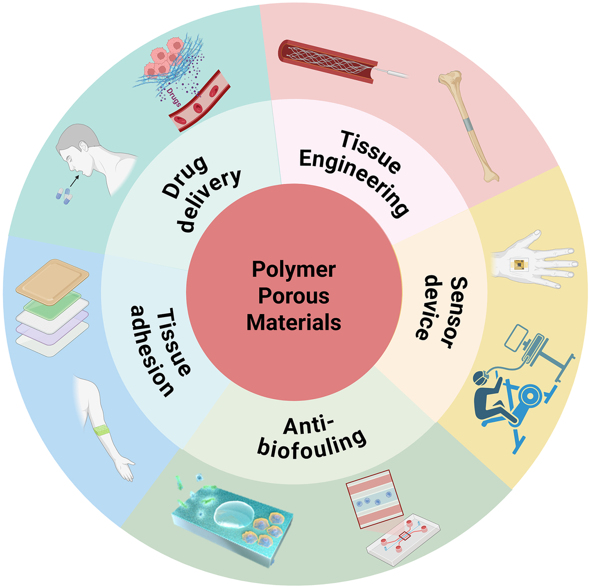
Polymer porous materials (PPMs) are an emerging class of functional biomaterials characterized by tunable pore structures, high specific surface areas, and favorable physicochemical properties. This review highlights the classification of PPMs into inorganic, organic, and organic–inorganic hybrids, with a focus on how structural features influence their biomedical performance. Due to their unique architecture and biocompatibility, PPMs have shown significant potential in various biomedical applications, including drug delivery systems, tissue engineering scaffolds and implantable devices, biosensors, and membranes for functional coating and separation systems. For drug delivery, biodegradable and stimuli-responsive PPMs offer controlled and targeted drug release while minimizing adverse effects. In tissue engineering, PPM-based scaffolds support cell adhesion, proliferation, and extracellular matrix deposition, which would subsequently promote functional tissue regeneration. For biosensing, the high surface-to-volume ratio and selective permeability of PPMs enhance detection sensitivity and specificity. Furthermore, recent advances in responsive hydrogels, antifouling filtration membranes, and bioactive coatings underscore their clinical translation potential. Despite rapid development, challenges such as precise control over pore size, mechanical–biological trade-offs, and long-term safety still remain. Addressing these limitations is critical for advancing PPMs toward clinical application in regenerative medicine, smart therapeutics, and diagnostic technologies.
Translations
Long abstracts in additional languages. The English article is the version of record.
中文zh-Hans
聚合物多孔材料(PPMs)作为一类新兴的功能性生物材料,凭借其可调控的孔隙结构、高比表面积以及优越的物理化学性质,在生物医学领域展现出巨大的应用潜力。然而,尽管其发展迅速,目前仍面临精确控制孔径大小、机械性能与生物学功能之间的权衡矛盾,以及长期安全性评估不足等关键挑战。本文旨在系统综述聚合物多孔材料的分类体系及其结构设计原则,深入探讨材料微观结构特征如何决定其在药物递送、组织工程支架及植入式器件中的生物医学表现,并分析其从实验室研究向临床转化过程中所面临的核心问题与未来发展方向。
本综述首先对聚合物多孔材料进行了系统的分类梳理,将其划分为无机类、有机类以及有机–无机杂化材料三大体系,重点阐述了不同类别材料的结构特征与其功能属性之间的构效关系。文章详细分析了生物可降解和刺激响应型PPMs在药物递送系统中的应用机制,探讨了其如何通过独特的孔隙架构实现药物的可控释放与靶向输送,从而最大限度地减少不良反应。同时,文中还深入剖析了基于PPMs的组织工程支架如何促进细胞粘附、增殖及胞外基质沉积,以及高比表面积材料在生物传感器中提升检测灵敏度与特异性的物理化学基础。
研究指出,聚合物多孔材料的独特架构和优异的生物相容性使其成为多种生物医学应用的理想候选者。在药物递送领域,刺激响应型PPMs能够根据体内环境变化实现药物的精准释放;在组织工程中,其支架结构有效支持了细胞行为并促进了功能性组织的再生。此外,针对生物传感应用,材料的高比表面积和选择性渗透能力显著增强了检测的灵敏度与特异性。近期关于响应性水凝胶、抗污过滤膜及生物活性涂层的最新进展进一步证实了其临床转化的巨大潜力,表明通过合理设计可显著提升材料的整体性能以满足复杂医疗需求。
尽管聚合物多孔材料在再生医学、智能治疗及诊断技术等领域展现出广阔前景,但其大规模临床应用仍受限于若干关键瓶颈。当前主要挑战包括难以实现对孔径尺寸的精确调控、机械强度与生物活性之间的固有矛盾,以及长期体内安全性数据的缺乏。解决这些局限性对于推动该领域发展至关重要。未来的工作应聚焦于开发更先进的制备工艺以优化孔隙结构,平衡材料的力学性能与生物学功能,并开展系统的长期毒理学评估。只有克服上述障碍,聚合物多孔材料才能真正实现从实验室研究到临床应用的跨越,为生物医学工程带来革命性的突破。
Françaisfr
Les matériaux polymères poreux (MPP) constituent une classe émergente de biomatériaux fonctionnels caractérisés par des structures poreuses modulables, de grandes surfaces spécifiques et d'excellentes propriétés physico-chimiques. Malgré leur potentiel considérable dans divers domaines biomédicaux tels que les systèmes de délivrance de médicaments, l'ingénierie tissulaire et la biosensibilisation, le développement rapide de ces matériaux se heurte à des défis majeurs : le contrôle précis de la taille des pores, les compromis entre propriétés mécaniques et biologiques, ainsi que les incertitudes concernant leur sécurité à long terme. L'objectif de cette revue est d'examiner systématiquement la classification des MPP en catégories inorganiques, organiques et hybrides organo-inorganiques, tout en analysant comment leurs caractéristiques structurelles influencent directement leurs performances biomédicales et les perspectives futures pour leur traduction clinique.
Cette synthèse présente une analyse approfondie de l'architecture et du design des matériaux polymères poreux, en mettant l'accent sur la relation entre la microstructure et la fonctionnalité. Les auteurs détaillent comment les MPP biodégradables et à réponse stimulée sont conçus pour permettre un relargage contrôlé et ciblé des médicaments tout en minimisant les effets indésirables dans le cadre de systèmes de délivrance avancés. De plus, l'étude explore la conception d'échafaudages basés sur ces matériaux qui favorisent l'adhésion cellulaire, la prolifération et le dépôt de matrice extracellulaire pour soutenir la régénération tissulaire fonctionnelle. Le texte examine également les mécanismes par lesquels la grande surface spécifique et la perméabilité sélective des MPP améliorent la sensibilité et la spécificité dans les applications de biosensing, soulignant l'importance du design structural.
Les résultats principaux démontrent que grâce à leur architecture unique et leur biocompatibilité, les matériaux polymères poreux offrent des solutions prometteuses pour une gamme étendue d'applications biomédicales. Ils se sont révélés particulièrement efficaces dans la délivrance de médicaments ciblée, le soutien à l'ingénierie tissulaire par la promotion de la régénération fonctionnelle, et l'amélioration des performances des capteurs biologiques grâce à leur rapport surface/volume élevé. Les avancées récentes en matière d'hydrogels intelligents, de membranes de filtration anti-salissures et de revêtements bioactifs soulignent le potentiel croissant pour une transition clinique réussie. Ces matériaux permettent non seulement un relargage contrôlé mais aussi des interactions biologiques optimisées qui sont essentielles pour l'intégration avec les tissus vivants.
En dépit du développement rapide, la traduction clinique de ces matériaux reste entravée par des limitations critiques qu'il est impératif d'aborder. Les défis persistants incluent le contrôle précis de la taille des pores, les compromis inhérents entre les propriétés mécaniques et biologiques, ainsi que l'évaluation à long terme de leur sécurité in vivo. Surmonter ces obstacles est fondamental pour faire progresser les matériaux polymères poreux vers une application clinique réelle dans les domaines de la médecine régénérative, des thérapies intelligentes et des technologies diagnostiques. Les travaux futurs doivent se concentrer sur le développement de stratégies innovantes pour résoudre ces problèmes techniques et biologiques afin d'assurer que ces biomatériaux puissent atteindre leur plein potentiel thérapeutique et sécuritaire dans les environnements cliniques complexes.
Españoles
Los materiales poliméricos porosos (PPMs) constituyen una clase emergente de biomateriales funcionales caracterizados por estructuras porosas sintonizables, altas áreas superficiales específicas y propiedades fisicoquímicas favorables. A pesar de su potencial significativo en diversas aplicaciones biomédicas como sistemas de liberación de fármacos e ingeniería tisular, el desarrollo rápido se enfrenta a desafíos críticos que incluyen el control preciso del tamaño de poro, los compromisos entre las propiedades mecánicas y biológicas, así como la seguridad a largo plazo. Esta revisión tiene como objetivo destacar la clasificación de PPMs en inorgánicos, orgánicos e híbridos orgánico-inorgánicos, analizando cómo las características estructurales influyen en su rendimiento biomédico y explorando sus perspectivas futuras para la traducción clínica en medicina regenerativa y tecnologías diagnósticas.
El enfoque de este trabajo consiste en una síntesis exhaustiva del diseño y clasificación de los materiales poliméricos porosos, con un énfasis especial en cómo las características estructurales determinan su funcionalidad. Se examinan detalladamente los sistemas de liberación de fármacos que utilizan PPMs biodegradables y sensibles a estímulos para lograr una liberación controlada y dirigida mientras se minimizan los efectos adversos. Asimismo, el texto analiza cómo los andamios basados en PPMs apoyan la adhesión celular, la proliferación y la deposición de matriz extracelular para promover la regeneración tisular funcional. Además, se interpreta científicamente cómo la alta relación superficie-volumen y la permeabilidad selectiva de estos materiales mejoran la sensibilidad y especificidad en aplicaciones de biosensado.
Los hallazgos principales indican que debido a su arquitectura única y biocompatibilidad, los PPMs han demostrado un potencial significativo en una amplia gama de aplicaciones biomédicas. En el campo del sensado biológico, las propiedades físicas permiten detectar con mayor precisión, mientras que en la ingeniería tisular facilitan procesos regenerativos complejos. Los avances recientes en hidrogeles responsivos, membranas de filtración antifouling y recubrimientos bioactivos subrayan su potencial para la traducción clínica. Estos materiales no solo ofrecen un control preciso sobre la liberación de agentes terapéuticos sino que también establecen interacciones biológicas óptimas necesarias para la integración exitosa con tejidos vivos, demostrando versatilidad en entornos médicos complejos.
A pesar del desarrollo acelerado, persisten limitaciones importantes como el control preciso del tamaño de poro y los compromisos mecánico-biológicos que deben abordarse para avanzar hacia aplicaciones clínicas. La seguridad a largo plazo sigue siendo una preocupación crítica que requiere más investigación antes de su implementación generalizada en medicina regenerativa o terapias inteligentes. Abordar estas limitaciones es fundamental para el futuro del campo, ya que solo superando estos obstáculos se podrá aprovechar plenamente la capacidad de los PPMs para revolucionar las tecnologías diagnósticas y terapéuticas. El trabajo futuro debe centrarse en desarrollar estrategias innovadoras para optimizar la estructura porosa y garantizar la seguridad a largo plazo dentro del cuerpo humano.
日本語ja
ポリマー多孔質材料(PPMs)は、調整可能な孔隙構造、高い比表面積および優れた物理化学的特性を特徴とする新興の機能性バイオマテリアル群です。しかしながら、その急速な発展にもかかわらず、孔径サイズの精密制御や機械的・生物学的特性間のトレードオフ、ならびに長期安全性に関する課題が依然として残されています。本レビューは、PPMs の無機質、有機質および有機–無機ハイブリッドへの分類を強調し、構造的特徴がいかにして薬物送達システム、組織工学用スキャフォールド、インプラントデバイス、バイオセンサーなどの生体医学的応用に影響を与えるかを体系的に概説することを目的としています。また、これらの材料が臨床転換において直面する核心的な問題と将来の展望についても言及します。
本稿では、PPMs の設計原理および構造的特徴と機能性との関連性を詳細に分析しています。特に生分解性及び刺激応答型PPMsを用いた薬物送達システムにおいては、その独特なアーキテクチャを利用して薬剤を制御・標的化放出し、有害副作用を最小化するメカニズムが解説されています。さらに、組織工学分野では、PPMベースのスキャフォールドが細胞の接着、増殖および細胞外基質沈着をどのように支援し、機能的な組織再生を促進するかについて論じられています。また、高表面体積比と選択的透過性を有するPPMsがバイオセンシングにおいて検出感度と特異性をいかに向上させるかという物理化学的な基礎も明確に示されています。
主要な知見として、独特の構造と生体適合性により、ポリマー多孔質材料は薬物送達から組織再生まで多岐にわたる生体医学応用において顕著な可能性を示すことが確認されました。刺激応答型PPMsは体内環境の変化に応じて薬剤を放出し、スキャフォールドは細胞挙動をサポートして機能性組織の再生を促します。さらに、バイオセンサーにおける高表面積と選択的透過性は検出性能を劇的に向上させます。最近では、反応性ハイドロゲル、抗汚染ろ過膜および生体活性コーティングに関する進展が臨床転換の可能性を裏付けており、これらの材料設計の最適化により複雑な医療ニーズに応える能力が高まることが示されています。
急速な発展にもかかわらず、再生医学、スマート療法、診断技術への応用に向けた臨床実装には依然として克服すべき課題が存在します。孔径サイズの精密制御の難しさや機械的強度と生物活性とのバランスの問題、そして長期安全性データの不足が主要なボトルネックとなっています。これらの限界に対処することは、PPMs の発展にとって不可欠です。今後の研究は、より高度な製造プロセスの開発による孔隙構造の最適化、材料特性間のトレードオフの解決、および体系的な長期的毒性評価に焦点を当てるべきです。これら障壁を取り除くことで初めて、ポリマー多孔質材料は実験室から臨床現場への飛躍を果たし、生体医学工学における革命的な進歩をもたらすことができるでしょう。
العربيةar
تُعد المواد البوليمرية المسامية (PPMs) فئة ناشئة من المواد الحيوية الوظيفية تتميز ببنية مسامية قابلة للتعديل، ومساحات سطحية نوعية عالية، وخصائص فيزيوكيميائية مواتية. وعلى الرغم من إمكاناتها الكبيرة في مجالات مثل أنظمة توصيل الأدوية وهندسة الأنسجة والأجهزة القابلة للزرع، إلا أن تطورها السريع يواجه تحديات جوهرية تشمل التحكم الدقيق في حجم المسام، والمفاضلات بين الخصائص الميكانيكية والبيولوجية، بالإضافة إلى مخاوف تتعلق بالسلامة على المدى الطويل. يهدف هذا الاستعراض إلى تسليط الضوء على تصنيف المواد البوليمرية المسامية إلى فئات غير عضوية وعضوية وهجينة عضوية-غير عضوية، مع التركيز على كيفية تأثير الخصائص الهيكلية على أدائها في التطبيقات الطبية الحيوية ومناقشة آفاقها المستقبلية للانتقال السريري.
يركز هذا العمل على تحليل منهجي لتصميم وتصنيف المواد البوليمرية المسامية، حيث يتم استعراض العلاقة بين الميزات الهيكلية والأداء الوظيفي. يوضح الاستعراض كيف توفر المواد القابلة للتحلل الحيوي والاستجابة للمحفزات إمكانية إطلاق الأدوية بشكل مضبوط ومستهدف مع تقليل الآثار الجانبية في أنظمة توصيل الدواء. كما يستكشف كيفية دعم السقالات القائمة على هذه المواد لالتصاق الخلايا وتكاثرها وترسب المصفوفة خارج الخلية، مما يعزز تجديد الأنسجة الوظيفي. بالإضافة إلى ذلك، يفسر كيف تعزز النسبة العالية بين المساحة والحجم والنفاذية الانتقائية حساسية وكشف المستشعرات الحيوية.
تظهر النتائج الرئيسية أن البنية الفريدة والتوافق الحيوي للمواد البوليمرية المسامية يمنحها إمكانات كبيرة في تطبيقات طبية حيوية متنوعة، بما في ذلك توصيل الأدوية الموجه وهندسة الأنسجة. لقد أثبتت هذه المواد قدرتها على دعم سلوك الخلايا وتعزيز التجديد الوظيفي للأنسجة من خلال سقالات مصممة بعناية. كما أن الخصائص الفيزيائية لهذه المواد تعزز بشكل كبير حساسية وكشف المستشعرات الحيوية، مما يجعلها مثالية للتطبيقات التشخيصية الدقيقة. وتؤكد التطورات الحديثة في الهيدروجيلات الاستجابة والأغشية المرشحة المضادة للتلوث والطلاءات النشطة بيولوجيًا على إمكاناتها الكبيرة للانتقال السريري بنجاح.
على الرغم من التطور السريع، لا تزال هناك تحديات رئيسية يجب معالجتها لتسريع الانتقال إلى التطبيقات السريرية في الطب التجديدي والعلاجات الذكية. تشمل هذه التحديات صعوبة التحكم الدقيق في حجم المسام، والمفاضلات بين الخصائص الميكانيكية والبيولوجية، ونقص البيانات حول السلامة على المدى الطويل. إن معالجة هذه القيود أمر بالغ الأهمية لتطوير هذا المجال؛ حيث يجب أن تركز الأعمال المستقبلية على تطوير تقنيات تصنيع متقدمة لتحسين البنية المسامية وموازنة خصائص المواد وإجراء تقييمات سمية طويلة الأمد شاملة. فقط من خلال التغلب على هذه العقبات يمكن للمواد البوليمرية المسامية تحقيق إمكاناتها الكاملة في revolutionizing التكنولوجيا الطبية الحيوية.
Keywords
Full Text
1. Introduction
Polymer porous materials (PPMs) have emerged as a significant focus in contemporary materials research, particularly in biomedical applications where their porous architecture enables precise control over mass transport, mechanical compliance, and surface interactions. As their name implies, the defining feature of these materials is their porous structure—underscoring the principle that structure determines function. The unique pore architecture of PPMs imparts them with a range of distinctive properties, including tunable mechanical properties, selective adsorption, and enhanced surface activity, which are critical for applications such as drug delivery systems (e.g., controlled release scaffolds[1]), tissue engineering (e.g., cell-instructive scaffolds with biomimetic porosity[2]), and biosensing (e.g., porous electrodes for enhanced analyte capture[3]).
Historically, activated carbon was one of the earliest porous materials recognized for its exceptional adsorption capacity, enabling effective water purification and air filtration—representing an early and widely adopted use of porous materials. Compared with bulk continuum materials, porous materials generally exhibit 4 notable characteristics:
Inorganic porous materials (e.g., activated carbon, zeolite[4], Li materials[5], TiO2[6], porous glass, and Al2O3[7]) are widely used in sensors, catalysis, tissue engineering, and environmental remediation. While these materials offer excellent mechanical strength, their limited structural tunability restricts their adaptability in dynamic biomedical environments.
Organic porous materials, such as hydrogels and metal-organic frameworks (MOFs), include covalent organic frameworks (COFs)[8], conjugated microporous polymers (CMPs)[9], and polymers of intrinsic microporosity (PIMs)[10]. Their high structural tunability and ease of functionalization make them particularly attractive for biomedical applications, including drug-loaded hydrogels and porous scaffolds for cell encapsulation. However, their mechanical limitations remain a challenge for load-bearing applications.
Organic/inorganic composites synergize the strengths of both classes, offering enhanced functional versatility for biomedical applications such as bone tissue engineering (e.g., bioactive glass-polymer composites) and targeted drug delivery(e.g., mesoporous silica nanoparticles coated with polymers).
This article reviews the major application areas of PPMs, with a focus on their mechanisms and practical value in biomedicine, including drug delivery, tissue engineering, and biosensing.
2. Classification and definition of PPMs
PPMs are a class of polymeric materials with tunable pore structures, widely used in drug delivery, tissue engineering, membrane separation, and other fields. Based on material sources, pore size, pore structure, and functional characteristics, they can be systematically classified as follows:
2.1 Classification by source
Based on their origin, PPMs can be classified into 2 main categories:
Natural polymer-based porous materials: for example, collagen, chitosan, and alginate, which exhibit good biocompatibility and biodegradability, making them particularly suitable for biomedical applications such as tissue engineering and drug delivery[11].
Synthetic polymer-based porous materials: for example, polylactic acid (PLA), polycaprolactone (PCL), and polyethylene glycol (PEG), which offer excellent structural tunability and programmable degradation behavior, enabling precise control over material properties for various engineering applications[12].
Natural polymers are typically derived from biological sources and show excellent biofunctionality, and synthetic polymers provide superior processability and property customization. Hybrid systems combining both types are increasingly developed to leverage complementary advantages.
2.2 Classification by pore size
According to the International Union of Pure and Applied Chemistry classification[13], porous materials are categorized by pore size into:
microporous materials (pore diameter < 2 nm);
mesoporous materials (2–50 nm);
macroporous materials (>50 nm).
The pore size distribution fundamentally governs the material’s interfacial properties, mass transport efficiency, and host–guest interactions[14], which collectively dictate their functional performance. These characteristics are particularly pivotal in biomedical applications, where precise control over molecular loading, release kinetics, and surface reactivity is essential. Among porous materials, PPMs have emerged as versatile platforms due to their tunable pore architectures, biocompatibility, and chemical flexibility.
2.3 Classification by pore structure
According to their structural configuration, porous materials can be categorized into 2 fundamental types[15]:
Open-cell structure, which features interconnected pore networks that facilitate fluid transport and mass transfer. For example, filtration membranes, tissue engineering scaffolds, and catalytic supports.
Closed-cell structure, which comprises isolated, sealed pores that optimize physical barrier properties. For example, thermal insulation foams, shock-absorbing materials, and buoyancy components.
Open-cell structures exhibit high permeability and surface accessibility, and closed-cell structures demonstrate superior thermal/acoustic insulation. Hybrid configurations can be engineered for multifunctional applications.
2.4 Classification by responsiveness
According to their interactive properties with external stimuli, porous materials can be functionally categorized as:
Static porous materials: Maintain stable pore architecture under ambient conditions. Typical examples: conventional porous plastics and sintered metal foams.
Stimuli-responsive porous materials: Exhibit dynamic pore structure modulation in response to environmental triggers:
Static types ensure structural reliability for long-term application, and smart types enable on-demand regulation of permeability/adsorption. Hybrid systems combine stability with environmental adaptability.
3. Drug delivery systems
Although traditional modes of drug delivery, such as oral and parenteral administration, are widely utilized, their therapeutic effects are often constrained by various physiological and physicochemical factors. For instance, gastric acid may degrade acid-labile drugs[18]. Additionally, drugs with low water solubility result in decreased bioavailability, with a significant portion of the administered dose potentially being metabolically cleared[19]. Furthermore, the lack of targeted drug delivery can further diminish effective drug concentrations at the lesion site. More critically, many patients with chronic diseases require long-term or even high-dose medication, which can inflict serious damage to human organs, particularly the liver and kidneys. Consequently, there is a pressing need to develop drug delivery systems that address these challenges by utilizing various materials and chemical techniques. Such a carrier must be capable of stably transporting the drug, regulating the rate of drug release, and delivering the drug precisely to the target site[20]. Simultaneously, the carrier itself must exhibit biocompatibility and avoid causing toxic side effects in the target organism. Natural products often have excellent biocompatibility, but are often difficult to use directly and need to be properly modified to meet the needs of drug delivery (Figure 1A).
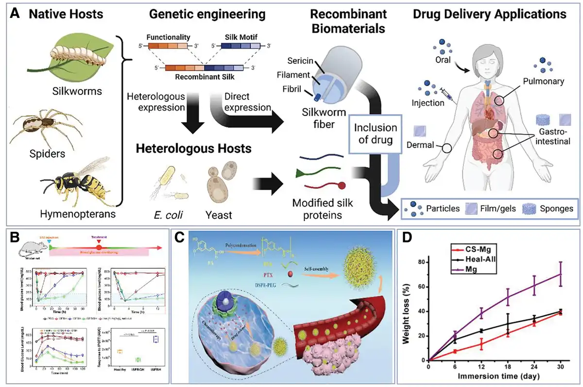
Figure 1.
(A) Principle of drug release[21]. Copyright 2023, Elsevier Ltd. (B) Glucose-responsive self-adjusting injectable hydrogel[22]. Copyright 2023, American Chemical Society. (C) Synthesis of PFA@PTX NPs and CT26 cancer treatment[23]. Copyright 2019, John Wiley & Sons, Inc. (D) Slower degradation rate of porous magnesium scaffolds covered with chitosan coating[24]. Copyright 2019, Elsevier Ltd.
In addition to the high requirements for material biocompatibility, precise regulation of drug release rates is crucial for such applications, as it directly impacts therapeutic efficacy and drug safety. To address this challenge, researchers have developed various rate control strategies based on drug delivery materials. Among these, hydrogels have garnered significant attention and are widely utilized in biomedical engineering due to their exceptional biocompatibility, high permeability, and hydrophilicity[25]. During the formation of these materials, a 3-dimensional network structure is established between monomers through cross-linking[26]. This topology, characterized by its porous features, provides an ideal microenvironment for drug loading and slow release, making hydrogels a highly promising drug carrier[27]. However, due to their poor mechanical properties, hydrogels are often challenging to apply independently. Consequently, they are typically compounded with inorganic porous materials to enhance mechanical strength while retaining their drug-carrying advantages[28]. Furthermore, by designing hydrogels with specific functional monomers or through chemical modification, these materials can be endowed with the ability to respond to external stimuli such as pH[29], temperature[30], light[31], enzymes[32], and glucose concentration[22,33,34] (Figure 1B). This allows the physically cross-linked hydrogels to be reversible and exhibit stimulus responsiveness, with the intensity of the stimulus being used to modulate the rate of drug release.
3.1 Biodegradable drug sustained-release system
Biodegradable materials are defined as substances that can break down into nontoxic products, such as water and carbon dioxide, and re-enter the natural ecological cycle after fulfilling their intended function. In the context of sustained drug release, an ideal drug carrier is one that degrades naturally within the body without requiring external intervention and does not cause secondary tissue damage. Currently, biodegradable materials used in such systems primarily include biodegradable ceramics and degradable polymers, with the latter being more commonly applied.
PLA and poly(lactic-co-glycolic acid) (PLGA) are widely used degradable polymers that undergo hydrolysis in vivo to form lactic acid and glycolic acid, which are subsequently metabolized via the tricarboxylic acid cycle and excreted through urine, sweat, and other routes. However, the degradation of PLA can lead to localized tissue acidification, resulting in inflammation. The incorporation of magnesium has been shown to partially alleviate this inflammatory response by buffering the acidic microenvironment[35]. In vitro, human fetal venous endothelial cells degraded mesoporous silica nanoparticles (MSNs) via cytoplasmic/lysosomal pathways within 48 h, with faster initial degradation (first 2 days). In vivo, intravenously injected MSNs degraded rapidly (2 h), primarily in liver/spleen, with fragments excreted in urine or recirculated to lungs and back to liver/spleen (peaking at 6 h). Bone accumulation occurred after 24 h (increasing by 48 h). Orally administered MSNs showed shape-dependent degradation, where rod-shaped particles reduced degradation, absorption, and liver distribution. These findings inform MSNs’ in vivo behavior and formulation design[36].
In addition to commonly used synthetic polymers such as PLA, PCL, and polyvinyl alcohol, several natural polymers derived from plants and animals—such as chitosan and ferulic acid (FA)—have attracted attention due to their excellent biocompatibility and biodegradability. Studies have demonstrated that the combination of FA with glycol chitosan exhibits potential neuroprotective effects. Zheng et al.[23] were the first to synthesize polymers using the repeating units of FA and employed paclitaxel (PTX) as a model drug to fabricate PTX-loaded poly ferulic acid (PFA) nanoparticles (PFA@PTX NPs) via self-assembly and nanoprecipitation. These nanoparticles had an average diameter of approximately 100 nm. A 50 wt% concentration of PEG with a molecular weight of 3000 (DSPE-PEG3000) was used as a stabilizer during nanoparticle formation. The results indicated that the synthesized polymer exhibited efficient drug delivery capabilities and conferred additional anticancer effects (Figure 1C).
Phenylboronic acid (PBA) exhibits a high affinity for glucose and specifically binds to 1,2- and 1,3-diols through the formation of reversible boronic esters[37]. This interaction induces a glucose-dependent volume change in the PBA-based material, subsequently accelerating the release of physically encapsulated insulin[38]. In comparison to traditional glucose-sensing molecules such as glucose oxidase and lectin, PBA demonstrates greater stability and offers distinct advantages for biomedical applications[39]. Odent et al.[40] successfully synthesized PBA-modified poly(hydroxyethyl methacrylate) (PHEMA)-based hydrogels utilizing high-resolution stereolithography. The light-cured PBA-glucose–responsive derivatives were produced via a two-step photochemical reaction and incorporated into a bioinert PHEMA matrix at varying loadings to create centimeter-scale 3-dimensional drug-eluting implants. These smart implants can be remotely activated by diol molecules, such as glucose, to modulate the on-demand release of drugs, including insulin. The underlying mechanism involves the transformation of the 2:1 binding state of PBA and glucose (PBA-glucose) into two 1:1 complexes as glucose concentration increases. This transformation triggers the glucose concentration-dependent reversible swelling of the hydrogel network, leading to a reduction in the cross-link density of the polymer. Consequently, the swelling facilitates the release of physically encapsulated drugs by decreasing the expansion of the pore space and diffusion resistance.
Biodegradable (e.g., PLA/PLGA, chitosan) polymers exhibit excellent biocompatibility and controllable degradability, metabolizing into nontoxic byproducts (e.g., CO2/H2O). Natural polymers (e.g., FA composites) can provide additional therapeutic benefits (e.g., neuroprotection). Structural modifications (e.g., PBA-modified hydrogels) enable smart stimulus-responsive drug release (e.g., glucose-dependent insulin release). However, their degradation may cause localized microenvironment acidification (requiring magnesium ion buffering), and their poor mechanical properties often necessitate composite reinforcement materials. Some synthesis processes (e.g., photocured PBA hydrogels) are also relatively complex. Overall, these materials demonstrate significant advantages in targeting and biosafety, but further optimization of degradation kinetics and mechanical stability is needed to meet clinical requirements.
3.2 Nonbiodegradable drug sustained-release system
Compared with biodegradable drug sustained-release systems, there have been relatively few reports of nonbiodegradable drug sustained-release systems in recent years, but they still have unique advantages. In addition to good biocompatibility, these materials often have good mechanical properties, good chemical and physical stability, and processability. Porous silicon has emerged as one of the most promising inorganic drug carrier materials due to its excellent biocompatibility, biodegradability[41,42], and tunable pore size distribution[43]. Leveraging these advantageous properties, researchers frequently construct composite drug delivery systems by integrating porous silica with hydrogels to achieve precise control over drug release. For instance, Perelman et al.[16] synthesized a pH/temperature dual-responsive hydrogel-poly(N-isopropylacrylamide-co-acrylic acid) nanohybrid system within the mesoporous channels of an oxidized porous silicon template through an in situ polymerization reaction. This system can modulate the thermal responsiveness of the hybrids and eliminate the lower critical solution temperature at pH 7.
Nonbiodegradable (e.g., porous silicon) systems offer distinct advantages, including superior mechanical strength, excellent physicochemical stability, and enhanced processability compared with biodegradable systems. Their tunable pore structure (e.g., mesoporous silica) enables precise drug loading and controlled release, particularly when combined with stimuli-responsive components (e.g., pH/temperature-sensitive hydrogels) for advanced composite drug delivery systems. However, their nondegradable nature raises potential long-term safety concerns, requiring careful evaluation for clinical applications.
4. Tissue engineering implants
Tissue engineering is an emerging interdisciplinary field that was first systematically proposed and defined by Robert Langer and Joseph Vacanti in 1993[44]. As a significant branch of regenerative medicine, it constructs tissue-like structures through the synergistic action of biochemical and physical factors, such as living cells, biocompatible materials, and growth factors, including cyclic mechanical loading. The bioengineered constructs derived from this field provide implantable solutions for tissue repair and organ function replacement[45,46], offering substantial clinical value and paving new avenues for the treatment of many challenging diseases. The essence of tissue engineering lies in the integration of 3 key components: porous scaffolds, cells, and biomolecules[47]. Among these, scaffolds serve as crucial support structures for cell growth and tissue regeneration and are composed of a diverse range of materials, including natural polymers, synthetic polymers, and ceramic materials. Scaffolds must provide a structural foundation for cell adhesion, spreading, migration, proliferation, and extracellular matrix (ECM) synthesis[48]. Biocompatibility is essential for their application; they must be nonthrombogenic, nonimmunogenic, and resistant to infection to prevent adverse immune reactions or infections postimplantation. Furthermore, scaffolds must possess adequate mechanical stability to endure surgical manipulation, be rupture-resistant, accurately conform to the shape of the target tissue, and avoid mechanical damage to the organism[49]. These characteristics are crucial to ensure the safety and effectiveness of scaffolds.
Tissue engineering aims to promote endogenous tissue repair, with implants designed for temporary residence in the body. Therefore, the implants need good biocompatibility not only, when implanted in the human body, to ensure that it does not adversely affect the body but also to ensure that it does not harm the body during its functioning, and the rate of scaffold degradation needs to match the rate of neoplastic tissue formation[50]. That is, entering the human body requires good biocompatibility and leaving the human body requires good biodegradability.
In general, mechanical properties, biocompatibility, and biodegradability are the 3 most fundamental and essential requirements for tissue engineering applications[51–52–53]. Of course, if such materials are applied to the surface of the skin, they may not have the ability to biodegrade, for example, polypropylene (PP) and polyethylene terephthalate (PET), which are often used for epidermal healing of the inguinal hernia[54–55–56].
Before this, when the tissues and organs in the body were damaged or lack of function, most of them relied on treatment or transplantation methods. These methods can play a certain role, but there are also many defects. If the injury is more serious and requires transplantation, the price is very expensive, the success rate of surgery is not very high, and after transplantation, a series of immune rejections will occur, which is very painful for the patient, and even threatens the life of the patient after the transplantation[51].
Tissue-engineered scaffolds have been extensively studied and applied across various fields, including bone[57], skin[58], nerve[59], blood vessels[60], and heart valves[61]. The rapid development of stent research can be attributed to the shortage of donor tissues and the limitations associated with donor area damage in both autologous and allogeneic grafts[62]. Commonly utilized materials encompass polymeric synthetic substances and modified metal stents. Cheng et al.[63] found that biodegradable polymer/hydroxyapatite composites could serve as substitutes for bone grafts, with nanohydroxyapatite/polyamide 6 (n-hydroxyapatite/PA6) emerging as a preferred synthetic bone material due to its composition and structure closely resembling that of natural bone mineral. Kong et al.[64] demonstrated that apatite-coated chitosan/n-hydroxyapatite composite scaffolds exhibit superior cell proliferation capabilities compared with apatite-coated chitosan scaffolds. The introduction of n-hydroxyapatite enhances bioactivity, offering new insights into the development of bone tissue engineering and the coating technology for bone biomaterials.
Porous biometallic materials have gained significant traction in biomedical applications due to their excellent biocompatibility, low in vivo corrosion rates, and high mechanical strength[48]. In tendon tissue engineering, Deepthi et al.[65] utilized a layered approach by loading porous chitosan-collagen composite hydrogels onto nanofibrous membranes, effectively mimicking the arrangement of collagen fiber bundles found in natural tendons. Their findings demonstrated that cells exhibited favorable attachment and oriented growth within the aligned fibers, as well as good attachment and infiltration within the hydrogel portion. This strategy was shown to significantly enhance the repair of ligament scar tissue. Beyond organic polymers, various inorganic porous materials have been extensively investigated to improve implant biocompatibility. Notably, magnesium alloys, in comparison to traditional nondegradable metal implants, exhibit remarkable physico-mechanical properties, with their specific strength and modulus of elasticity closely resembling those of natural bone. Additionally, magnesium is biodegradable in body fluids, allowing it to be metabolized and gradually resorbed alongside the repair and regeneration of bone tissues, thereby mitigating the risk of secondary surgeries[66]. However, magnesium scaffolds are prone to corrosion in body fluids, which can lead to localized alkaline pH changes. This issue necessitates enhancements through surface treatments or coatings to establish protective ceramic, polymer, or composite layers[67]. Guo et al.[24] pretreated porous magnesium scaffolds with a silane coupling agent in a phosphoric acid solution, subsequently preparing chitosan-coated magnesium alloy membranes (CS-Mg) through chitosan dip-coating. These membranes demonstrated favorable degradation characteristics, excellent in vitro biocompatibility, and significant in vivo bone regeneration promotion. As illustrated in Figure 1D, the degradation curve of CS-Mg exhibited a gradual decline during the initial 12 days, followed by an increased slope, indicating that the degradation occurred via hydrolysis. This slow degradation rate in the early stage effectively mitigated the degradation rate of the porous magnesium scaffolds.
Polymer porous scaffolds are essential in tissue engineering, offering excellent biocompatibility, tunable biodegradability (matching tissue regeneration rates), and adequate mechanical strength for surgical handling. Natural/synthetic polymers (e.g., chitosan and PLGA) and composites (e.g., nHA/PA6) mimic native ECM, supporting cell adhesion and tissue growth. However, challenges include balancing degradation kinetics (e.g., rapid Mg alloy corrosion requiring coatings) and mechanical stability. While nondegradable polymers (e.g., PP and PET) suit epidermal applications, most scaffolds require precise degradation-to-regeneration synchronization to avoid secondary surgeries or immune responses.
5. Sensor
As signal transduction devices, sensors transform external stimuli (including optical, electrical, and chemical signals) into quantifiable responses, analogous to but technologically distinct from biological sensory systems, similar to biological sensory systems in humans (e.g., vision, olfaction, audition, and tactition). As early as 1997, Saffo[68] published an article entitled “Sensors: The next wave of innovation,” in which he believed that sensors would flourish in the next few decades and may even become a new technology revolution. The sensor is mainly composed of 2 parts: the molecular recognition component and the signal conversion component (Figure 2A). The former is used for the acceptance of external stimuli, and the latter is the information that can be utilized by converting the received external signal into an electrical signal. There are also many kinds of materials for making sensors. At present, most of the inorganic materials on the market are semiconductor materials such as silicon[73], germanium[74], and cadmium[75]; ceramic materials such as iron oxide[75], zinc oxide[76], and aluminum oxide[77], and metal materials such as gold[78], silver[79], and copper[80]. Polymer organic materials have been studied as sensors. However, due to its poor mechanical properties and easy degradation, most of them are still in the experimental research stage or used to modify existing inorganic sensor materials. At present, there are many kinds of sensors on the market, which are filled in every corner of life.
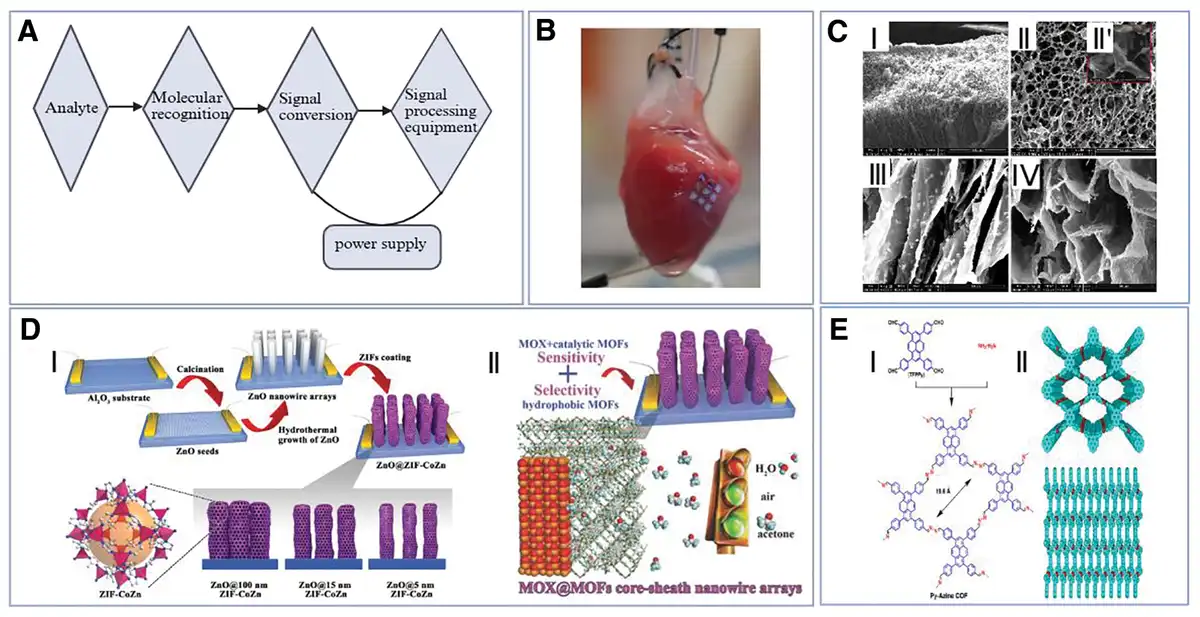
Figure 2.
(A) Sensor working process flow chart. (B) Conduction of nanoporous materials in the heart[69]. Copyright 2022, Springer Nature. (C) Hydrogel sensor for wound dressing moisture monitoring by immobilizing acting substances on a xyloglucan-polyvinyl alcohol hydrogel substrate[70]. Copyright 2018, Elsevier Ltd. (D) (I) Schematic diagram of the synthesis process of MOX@MOFs and the schematic diagram of the microstructure of ZIF@CoZn; (II) Schematic diagram of the overall structure of MOX@MOFs and the selectivity of the sensing part to gases[71]. Copyright 2016, WILEY-VCH Verlag GmbH & Co. KGaA, Weinheim. (E) (I) Azine-linked COF (Py-Azine COF) schematic diagram of synthesis; (II) top view and side view of Py-Azine COF[72]. Copyright 2013, American Chemical Society.
Sensors, as critical signal transduction devices, have revolutionized biomedical applications by converting biochemical, physical, and optical stimuli into quantifiable electrical signals. Unlike their industrial counterparts, biomedical sensors require stringent performance characteristics, including exceptional biocompatibility, high sensitivity in complex biological matrices, and long-term stability under physiological conditions. This review systematically examines cutting-edge sensor technologies specifically developed for healthcare applications, ranging from implantable diagnostic devices to wearable health monitoring systems. We highlight fundamental working principles, material innovations, and translational challenges while providing insights into future directions for clinical implementation.
5.1 Biosensor
In the 1960s, Clark, an American scholar, invented the enzyme electrode, marking the birth of biosensors by converting enzyme signals into electrical signals[81]. With advancements in technology, biomolecules and materials such as antibodies[82], cells[83], nucleic acids[84], and bacteria[85] have been employed as molecular recognition elements for biosensors. Porous materials present significant opportunities for interdisciplinary research in the field of sensing. Their high specific surface area, combined with interconnected channels and high porosity, promotes mass diffusion, enhancing sensitivity, accelerating response times, and shortening reaction and recovery periods. Furthermore, selective adsorption based on pore size or solubility facilitates the detection and differentiation of specific analytes[86]. Notably, nanoporous materials outperform traditional bulk material sensors due to their high surface-to-volume ratio, which increases the active sites for interaction with target analytes. For instance, nanoporous silicon has been applied to flexible biosensing platforms, such as those used for heart monitoring (Figure 2B)[69].
There are also many hydrogel sensors that are implemented by immobilizing a substance on the hydrogel substrate that can interact with the analyte of interest. A hydrogel membrane (HM) based on xyloglucan-polyvinyl alcohol was developed to construct an ultrahigh frequency radio frequency identification sensor. This sensor integrates the functionality of a smart wound dressing with monitoring capabilities, allowing it to absorb simulated body fluids from the affected area and to monitor the moisture content of the dressing contact area in real time. Scanning electron microscopy characterization revealed that the pore distribution on the surface of the HM and within the cross-section was homogeneous, although the pore sizes were heterogeneous. After 24 hours of immersion in water, the pore size increased, confirming the swelling behavior (Figure 2C)[70].
The enzyme sensor is the world’s first biosensor. It was developed by the American scientist Clark in 1962. In 1967, Updike successfully made the enzyme electrode and realized the real enzyme sensor[87]. However, these are based on inorganic materials. With the deepening of research by scientists, various enzyme sensors have been produced, which are intended to improve the sensitivity, detection accuracy, and widening of the types of detection. Niculescu et al.[88] used a poly(ethylene glycol) diglycidyl ether to graft a quinoprotein alcohol dehydrogenase with an Os-complex-modified poly(vinylimidazole) redox polymer for ethanol content determination during wine fermentation. Enzyme sensors also have great application prospects in food safety testing. Meats such as fish and meat are highly susceptible to deterioration. When a fish dies, it will be accompanied by a series of enzyme reactions, such as xanthine (XA). It will make the bitterness of the unfresh fish. In other words, if the fish is stored for a longer time, the more XA is produced, and then the XA can be used to determine whether the fish is fresh[89,90]. Yazdanparast et al.[91] used a glassy carbon electrode (GCE) as a substrate to bind a multiwalled carbon nanotube to poly(L-aspartic acid) on GCE as a composite membrane for immobilized xanthine oxidase (XO), and finally chemically coupled using 1-ethyl-3-(3 dimethylaminopropyl) carbodiimide/N-hydroxysuccinimide (EDC/NHS) to graft XO onto the composite membrane. The sensor has a simple synthesis process and a short response time, and poly(L-Asp)/multi-walled carbon nanotube (MWCNT)/GCE can also immobilize other enzymes as enzyme biosensors.
Immunosensors are biosensors that integrate highly sensitive sensing technology with specific immune responses to monitor antigen–antibody interactions, making them widely utilized in biomedical research. PEG, a hydrophilic polymer, can form a hydrated layer that protects drug molecules from enzymatic degradation and is extensively used in biopharmaceuticals due to its excellent biocompatibility[92]. However, the antibody isoforms IgM and IgG can accelerate the blood clearance of PEGylated drugs, thereby diminishing their therapeutic efficacy and safety[93]. Surface modification of PEG reduces undesirable interactions between the drug and its environment, facilitating targeted delivery. To address this issue, Zhang et al.[94] modified the sensor chip with methoxypolyethylene glycol (mPEG) and synthesized poly[poly(ethylene glycol) methyl ether methacrylate] (PEGMA) brushes on the surface of a gold chip using surface-initiated atom transfer radical polymerization to create a homogeneous polymer coating. This coating offers high sensitivity, rapid quantitative detection, resistance to nonspecific adsorption, and ease of operation, enabling the rapid and quantitative detection of anti-PEG antibodies from diluted serum. Overall, the coating presents several advantages, including high sensitivity, quick quantitative detection, resistance to nonspecific adsorption, and operational simplicity.
5.2 Physical sensor
The physical sensor converts various physical signals (force[95,96], light[97], gas[98], etc.) into electrical signals and further analyzes and processes the device. Electronic skin (e-skin) is a kind of flexible electronic material. At present, most e-skins are mainly synthesized based on. The e-skin is applied to the surface of the human body as a strain sensor, which can monitor the physiological changes of the human body in real time, which is good for maintaining health.
Tong et al.[99] synthesized cellulose ionic hydrogels (CIHs) by free radical polymerization in a NaOH/urea aqueous solution. The hydrogels exhibited good compression and tensile properties while also having high transparency (UV–vis), spectrum at 550 nm (~89%), and the ionic conductivity of 0.16 mS cm−1. In addition, CIHs are able to operate in a wide range of temperatures without losing their transparency (−20 °C), with the potential to be a stable skin strain sensor. Shu et al.[100] developed a ZnCl2/CaCl2 binary electrolyte-incorporated hydrogel system exhibiting remarkable ionic conductivity (5.48 S m−1). Mechanistically, applied mechanical deformations induce dynamic reconfiguration of the ionic conduction pathways, resulting in measurable resistance fluctuations. This intrinsic property enables effective mechanoelectrical signal conversion, demonstrating significant potential for high-performance strain/pressure sensing applications. Ionic conductive hydrogels have emerged as a promising class of materials for developing multifunctional sensors with significant potential in healthcare monitoring and flexible electronic applications. Hou et al.[101] presents a wearable fingerprint electronic skin (WFES) sensor that integrates a graphene oxide/polydimethylsiloxane (GO/PDMS) hybrid dielectric layer with a biomimetic fingerprint-structured PDMS electrode, achieving high sensitivity (0.21 kPa−1, 0–1 kPa) and rapid response (15 ms, ΔC/C = 17%), while resolving microgrooves spaced only 30 µm apart. By systematically analyzing how scanning speed, surface roughness, and fingerprint pitch modulate the vibrational spectrum, the work provides new evidence for the subcutaneous perceptual mechanism proposed in psychophysics. For the first time, historical fingerprints were employed to fabricate sensors, revealing how different ridge patterns govern vibrational signals. When attached to a fingertip, the device enables artificial recognition of fabrics and microgrooves, underscoring its potential as electronic skin. Further integration with hydrogel technology is expected to accelerate its development.
5.3 Chemical sensor
A chemical sensor is a kind of sensor that converts chemical signals into electrical signals through chemical reactions, including various metal ion detection, gas detection, pH detection, alcohol detection, etc. Chemical sensors are one of the most common sensors in life. For example, as early as 1927, Bogen designed a breathing alcohol detector[102,103]. And now, the traffic police use an alcohol detector to determine whether a driver is drunk driving or not. When the alcohol molecules pass the sensor, they will be converted into acetaldehyde. The released electrons can cause the current to change, and then it can judge whether the driver is drunk or not. And the carbon monoxide detector in the kitchen is also like this. Lee et al.[104] designed PAF-1-SMe, a 3-dimensional porous aromatic framework (PAF) densely functionalized with thioether groups for highly selective capture and concentration of copper ions from biofluids and aqueous samples. PAF-1-SMe exhibits remarkable selectivity for copper over other biologically relevant metals, with a saturation capacity exceeding 600 mg/g. Furthermore, the combination of PAF-1-SMe as a copper-capturing material from biological samples with 8-hydroxyquinoline as a colorimetric indicator establishes an effective method for detecting abnormal copper elevation in urine samples from Wilson disease model mice and tracing exogenously added copper in serum. Dalapati et al.[72,105] showed that 1,3,6,8-tetrakis(4-formylphenyl)pyrene and diazabutadiene synthesize covalent organic skeletons (COFs) with permanent porosity and high specific surface area, which can be used to sensitively detect the presence of 4,6-trinitrophenol in many different solvents (Figure 2E).
Demonstrate unique advantages for sensor applications due to their high surface area, tunable porosity, and excellent biocompatibility, enabling enhanced sensitivity, selective detection, and flexible designs for wearable and implantable devices. These materials have proven particularly valuable in developing smart wound dressings, health monitoring systems, and food quality detectors. However, challenges remain regarding their mechanical stability in physiological environments, potential degradation issues, and nonspecific binding interference. Current research focuses on composite material strategies and surface modifications to address these limitations while maintaining the materials’ inherent benefits. The future development of these sensors will require balancing key trade-offs between sensitivity and robustness, biocompatibility and durability, and design flexibility and manufacturing consistency, with advanced material hybrids and nanoscale engineering approaches showing particular promise for overcoming current limitations.
6. Oil/water separation
Porous materials, in addition to the aforementioned extensive applications in the field of biology, have great application value in water pollution control, such as porous membranes for oil/water separation. Among them, water pollution of emulsified oil is very common, and this kind of pollution is easily spread and difficult to treat. In response to this problem, porous membranes for oil/water separation have emerged. These membranes are mainly divided into 2 major categories (Figure 3A), namely superhydrophobic porous membranes[106] and superhydrophilic porous membranes[111]. While oil/water separation technology has been primarily applied in environmental engineering, its potential value in medical applications—such as surgical wastewater treatment[112] and medical instrument cleaning—has been attracting increasing attention[113]. Fan et al.[114] developed an innovative hydrophilic poly(vinylidene fluoride) (PVDF) membrane fabrication method through in situ assembly of phytic acid (PA)-polyethylenimine (PEI) polyelectrolyte complexes with PA-Fe3+ dual-cross-linked structures, successfully creating PVDF/PA-PEI/Fe3+ membranes with exceptional oil/water separation performance. The membranes demonstrated superhydrophilicity (contact angle [CA] 19°), high water flux (3200 L m−2 h−1), and outstanding retention efficiency for herbal volatile oils (>90%), along with superior antifouling properties (86.5% flux recovery after 5 cycles) and long-term stability (30-day performance maintenance). Using response surface methodology to optimize the preparation process, this environmentally friendly membrane material effectively addressed the corrosion issues of conventional PVDF membranes in herbal volatile oil separation, providing an efficient solution for active pharmaceutical ingredient extraction in traditional Chinese medicine and industrial oil/water separation applications.
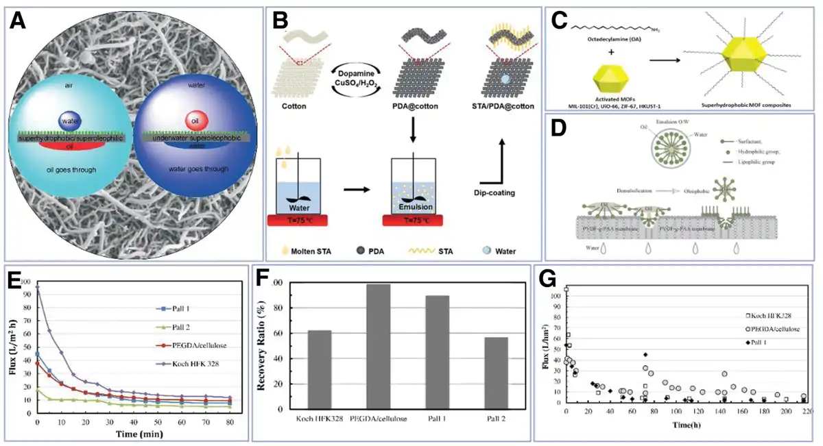
Figure 3.
(A) Superhydrophobic (left) and superoleophobic (right) porous membranes[106]. Copyright 2015, WILEY-VCH Verlag GmbH & Co. KGaA, Weinheim. (B) STA/PDA@cotton synthesis process[107]. Copyright 2018, Royal Society of Chemistry. (C) Formation process of surface-modified superhydrophobic MOF materials[108]. Copyright 2019, American Chemical Society. (D) Possible mechanism of O/W emulsion separation by PVDF-g-PAA membrane[109]. Coryright 2016, High Education Press. (E) Rate of decline of 4 filter membrane fluxes in short-term fouling experiments. (F) Recovery after 80-min fouling test. (G) Changes in filtration membrane flux and recovery rate in long-term fouling experiments[110]. Copyright 2013, Elsevier Ltd.
6.1 Superhydrophobic porous materials
Superhydrophobic materials are a new type of material. The surface static water CA is greater than 150°, and the rolling angle is less than 10°[115]. Generally, these materials have strong self-cleaning ability. For example, if the superhydrophobic property is applied to porous materials, the material can be cleaned against external objects, and it can prevent itself from being polluted. An important indicator for measuring the performance of superhydrophobic materials is the solid–liquid contact time. The shorter the solid–liquid contact time, the faster the mass, momentum, and energy exchange between the solid–liquid surface interface, and the better the performance of the material interface. Anti-icing, self-cleaning, and antifouling antibacterial on the surface. Liu et al.[116] have prepared a submillimeter superhydrophobic system called “pancake rebound mechanism,” which shortens the theoretical limit of solid–liquid contact time by 80% and successfully challenges the Guinness World Record. Some of St. Pauli has installed a superhydrophobic coating on some of the blocks to punish those who urinate anywhere, and this material will splash their urine back onto them.
In the process of applying hydrophobic porous materials to oil/water separation, it is often the case that the material is degraded after it has been used, and this degradation product will further pollute the environment. In response to this problem, Dong et al. developed a superhydrophobic coating, STA/PDA@cotton, which is simple to manufacture, low in cost, environmentally friendly, and has efficient oil/water separation performance. This process eliminates the need for additional organic solvents and uses CuSO4 as a trigger to form a stable, environmentally friendly superhydrophobic coating (Figure 3B) with a CA of approximately 162° and a sliding angle of approximately 7.8°. And due to the presence of dopamine, it can achieve self-healing under daylight conditions, broadening its practical application range[107].
MOF is an emerging porous material with a higher porosity than traditional materials such as activated carbon and zeolite, so it has a wide range of applications. Gao et al. modified a MOF by grafting octadecylamine—an agent with low surface energy—onto its surface via coordination with unsaturated metal sites. This modification yielded a superhydrophobic porous material (Figure 3C) with excellent hydrophobic oil absorption properties and strong adsorption capacities for chemical solvents such as chloroform and toluene[108]. Xue et al. prepared a superhydrophilic and underwater superoleophobic PAM hydrogel-coated stainless steel mesh was designed in an oil/water/solid 3-phase system. It demonstrated selective and effective water/oil separation, and it is also a new attempt to use special wettability to design next-generation materials for oil/water separation[117].
6.2 Hydrophilic porous materials
Research on the application of hydrophilic porous materials in oil/water separation has been reported in recent years. Gao et al.[109] grafted PAA on the surface of the PVDF membrane and formed a superhydrophilic porous material using the organic porogen PVP. The principle of oil/water separation of the material is shown in Figure 3D. For the oil-in-water (O/W) emulsion, the process is divided into 2 processes: demulsification and oleophobic: the demulsification process is the adsorption of the porous material to the emulsion surfactant. Destruction of its O/W structure; the oleophobic process uses its hydrophilic and oleophobic principles to separate water from oil.
Hydrogel-tethered polysulfone (PSF) membranes synthesized by grafting propargyl-poly(ethylene glycol) (pro-PEG) onto azide-functionalized PSF membrane surfaces via copper (I) catalyzed azide-alkyne cycloaddition (CuAAC) reaction showed better antifouling performance and permeance[118]. Chen et al.[119] prepared a porous nickel foam coated with polyacrylamide (PAM)-hydrogel showed superhydrophilicity and underwater superoleophobicity, and achieved oil/water separation by allowing water to permeate through.
The application of porous materials to oil/water separation is now a hot topic in porous materials research. However, porous materials currently used in oil/water separation often have many problems: insufficient mechanical strength, difficulty in recycling, secondary pollution, and the like. At present, most of the porous materials used in oil/water separation are only in the laboratory stage, and it is difficult to achieve industrial production. How to apply them to real life is an urgent problem to be solved.
Polymer porous membranes demonstrate excellent performance in oil/water separation, with superhydrophobic types (CA >150°) enabling efficient oil absorption and self-cleaning properties, while superhydrophilic variants (CA <10°) achieve selective water permeation and antifouling capabilities. These materials show particular promise for treating emulsified oil pollution and have potential medical applications in surgical wastewater treatment. However, key challenges include insufficient mechanical strength, recycling difficulties, and risks of secondary pollution from degradation products. Current research focuses on developing environmentally friendly, self-healing coatings and MOF-based composites to address these limitations, though scaling up from laboratory to industrial production remains a significant hurdle.
Polymer porous membranes (e.g., PVDF and polysulfone) demonstrate excellent filtration performance with high rejection rates (>98%) and improved antifouling properties when modified with hydrophilic groups or nanoparticles. These membranes achieve enhanced water flux and contamination resistance through various modification strategies, including hydroxyl group grafting and nanoparticle incorporation. However, significant challenges persist, particularly regarding long-term stability, as modified surfaces often degrade over time, leading to reduced antifouling performance. Additionally, issues with coating durability, recycling difficulties, and the need for secondary cleaning processes hinder industrial scalability. While laboratory results are promising, transitioning these advanced filtration membranes to industrial applications remains a critical challenge that requires solutions for coating stability and sustainable operation.
7. Filtration membrane
7.1 In situ synthetic filter membrane
At present, porous materials are widely used in water purification. This is also known as membrane separation, and materials currently used in this aspect include polysulfone[120], polyvinyl alcohol[121], polyacrylonitrile[122], PEG[123], PVDF[124], and the like. Zhang et al.[125] prepared a calcium alginate/polyacrylamide hydrogel nanofiltration membrane with good antipollution ability and detected it with bovine serum albumin solution (BSA) and yeast suspension, which is for BSA and yeast. The retention rate is as high as 98.53% and 99.64%. Wang et al.[110] used PEG to cross-link with ultrafine cellulose nanofibers (CN) to form an ultrafiltration membrane. The ultrafiltration membrane not only has good hydrophilicity but also has good antifouling properties. In the short-term fouling test (80 min), the antipollution ability of PEG-CN was detected by means of pollution-cleaning, and the recovery rate after pollution was significantly higher than that of other commercial films (Koch HFK 328, pall 1, pall 2; Figure 3E). In the long-term pollution test (220 h), PEG-CN showed good antistaining performance. After 72 h of filtration, it could recover 88% of the flux, which was higher than the other 3 commercial films (Figure 3F, G).
7.2 Filter membrane antifouling modification
An important problem is that porous materials used as filter membranes during application are highly contaminated, especially for filtering protein solutions, although in some filter membranes, the pore size is larger than the protein, but due to the accumulation of protein, after a long period of use, there will still be pollution, resulting in a decrease in water purification efficiency, and the water flux will be greatly affected.
Jang et al.[126] combine polyvinyl alcohol with chloroacetic acid under alkaline conditions (Figure 4A) for modification of PVDF membrane, which is higher than the pure PVDF membrane. The purified water flux and the lower rate of purified water flux are both highly hydrophilic.
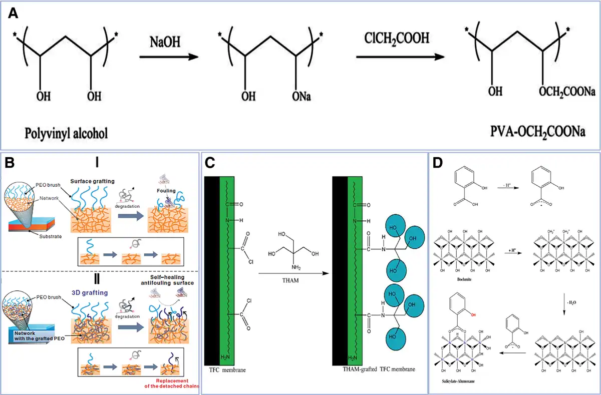
Figure 4.
(A) Etherification of polyvinyl alcohol and chloroacetic acid under alkaline conditions[126]. Copyright 2014, Wiley Periodicals, Inc. (B) Surface grafting and 3D grafting difference: (I) surface grafting: when the grafted polymer chain is degraded, the material’s antifouling ability will be lost; (II) 3D grafting: PEO is grafted onto the surface and inside of P2VP. When the antifouling layer on the surface is degraded, the internally grafted polymer will be rearranged to the interface due to the new gradient in the chemical potential[127]. Copyright 2013, WILEY-VCH Verlag GmbH & Co. KGaA, Weinheim. (C) THAM is bound to the TFC membrane by covalent attachment (interfacial polymerization via an amino group with an acid chloride group)[128]. Copyright 2017, Wiley Periodicals, Inc. (D) The process of deprotonation of salicylic acid and the process of binding to boehmite nanoparticle[129]. Copyright 2016, Elsevier B.V.
The purified water flux and the lower rate of purified water flux are both highly hydrophilic. Grafting new materials on the surface of existing filter membranes can improve their antifouling ability to a certain extent, but as the use time prolongs, the material grafted on the surface degrades or falls off, and its antifouling performance is lost. In response to this problem, Chen et al.[127] proceeded from the structure and adopted the 3D grafting method to prolong the antipollution performance of the host material (Figure 4B).
The hydrophilicity of the hydroxyl group is excellent. If a large amount of hydroxyl groups is grafted on the surface of the existing porous filtration membrane, the hydrophilicity of the filtration membrane is improved, so that the dirt is difficult to deposit on the surface of the membrane, and when the membrane is contaminated, physical rinsing also makes it easier for dirt to leave the surface of the membrane. Based on this principle, Chen et al. grafted Tris(hydroxymethyl)aminomethane onto a thin film composite RO membrane (Figure 4C) to obtain a composite membrane with better antifouling properties than the original membrane. The increase in the content of (hydroxymethyl) aminomethane increases the antifouling properties of the film, further demonstrating that increasing the hydrophilicity of the film can improve the antifouling properties of the film to some extent[128].
In addition to the aforementioned methods for polymer-modified filter membranes, many scholars have devoted themselves to research the use of nanoparticle modification to improve the antifouling ability of porous filter membranes. Yao et al.[71] developed a MOF-coated ZnO nanowire core–sheath structure, achieving significantly enhanced acetone sensing performance with improved selectivity, higher sensitivity, lower detection limit, and reduced operating temperature (Figure 2D). Mokhtari et al.[129] modified the polysulfone (PS) membrane by using salicylate-aluminoxane (SA) nanoparticles and prepared SA nanoparticles by the reaction between boehmite nanoparticles and salicylic acid. This is mainly due to the deprotonation of salicylic acid and the combination of 2 processes with boehmite nanoparticle (Figure 4D). Then SA/PS nanocomposite membranes were synthesized by the reversed phase method. The results show that the composite porous membrane not only has improved flux but also has a significant decrease in irreversible pollution rate. Ma et al.[130] deposited an iron/aluminum hydrolyzed precipitate onto a porous ultrafiltration membrane to improve the antipollution performance of the membrane. The experimental results show that the aluminum hydrolyzed precipitation layer improves the antipollution performance of the membrane, using HA and bovine serum. When the protein is characterized by antipollution performance, the author analyzes a large part of the reason because the aluminum sulfate precipitate layer is positively charged, and both HA and BSA are negatively charged. Because of the positive and negative electric attraction, the pollutants are more easily adsorbed by the sediment layer. Subsequently, Ma et al. modified the polyvinylidene fluoride porous filter membrane with nanoscale zerovalent iron ions (NZVI). The results showed that the NZVI layer has obvious scale-inhibition ability, especially for macromolecules and medium molecules of humic acid (HA)[131].
It is also convenient to apply the filter membrane to solve the water pollution problem. Therefore, the research on the porous membrane material of the filter membrane is also one of the hot topics, including the research on the in situ synthetic membrane and the target. Existing research on filtration membrane modification. However, in addition to the existing industrial membranes, most of them only stay in the laboratory stage, and it is difficult to carry out industrial production. For example, for the modification of the polymer porous membrane, there are still a series of problems: the coating layer is easy to fall off, and it is difficult to recycle, the second cleaning consumes extra resources, and so on.
Polymer porous filtration membranes demonstrate excellent separation efficiency (98%–99% rejection rates) and improved antifouling properties through hydrophilic modifications (e.g., hydroxyl group grafting) and nanoparticle incorporation, yet face challenges including surface coating degradation, limited long-term stability, and difficulties in industrial-scale production.
8. Other applications
8.1 Adhesive/bandage
There are many applications for, involving agriculture, industry, biomedicine, and environmental fields. For example, the newly developed “dry double-sided tape” by Zhao Xuanhe team combines PAA with N-hydroxysuccinimide and gelatin (or chitosan) by UV light, and then fully dried[132]. Due to the interfacial water often being between the organ and the adhesive material, the adhesive material has difficulty adhering to the tissue organ, or the adhesion is not strong[133,134]. The traditional method is to make the 2 bonds through the diffusion of the interface water, but this tape is to remove the interface water by a dry cross-linking mechanism and then bond (Figure 5A).
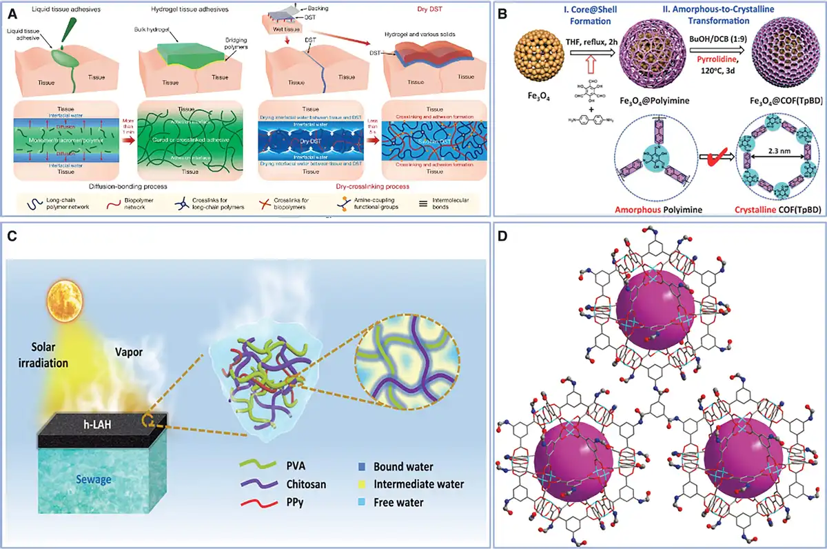
Figure 5.
(A) Schematic diagram of the action of the traditional binder and its mechanism of action[132]. Copyright 2019, Springer Nature Limited. (B) The synthesis process of Fe3O4@polyimine microspheres, the surface of citrate-treated Fe3O4 has a large amount of –COOH, and the benzidine adheres to the surface of the nucleus through hydrogen bonding, and then forms an amorphous polyaniline shell through Schiff base reaction. The classical solvothermal process forms a crystalline network structure[17]. Copyright 2016, Wiley-VCH Verlag GmbH & Co. KGaA, Weinheim.
8.2 Electroconductive PPM
Electroconductive PPMs have excellent properties and have broad application prospects, but most PPMs are not electrically conductive and limit their application under certain conditions, such as biosensors[135,136], ion-responsive drug delivery systems[137], artificial muscles[138]., and skin tissue[139]. However, if the electrochemical properties of PPM are imparted, the field of application will be greatly expanded. The hydrogel has good biocompatibility and a large specific surface area. It is easy to combine with metal particles (Fe[140], Cu[141]), conductive polymers (polyaniline[142], poly(3,4-ethylenedioxythiophene)-poly(styrenesulfonate) [PEDOT:PSS][143], polypyrrole[139]), and other conductive materials (carbon nanofibers[144], graphene[145]). Since the first report of conductive hydrogels by Gilmore et al. in 1994[146], it has received widespread attention. In recent years, conductive hydrogels have flourished, and more and more applications have been discovered (e.g., PEDOT:PSS hydrogel). PEDOT was first produced by Bayer, Germany. With PSS, it can greatly improve its solubility and make it highly conductive[147]. Lu et al. prepared a pure PEDOT:PSS hydrogel film by adding dimethyl sulfoxide to PEDOT:PSS solution and then drying and annealing several times. The purification process is simple, and the product is in pure water. With conductivity up to 40 S·cm−1 and adjustable swelling behavior, it opens up new avenues for hydrogel electronics[148]. Naficy et al.[149] added PEDOT to poly(ethylene glycol) methyl ether methacrylate and PAA double-network hydrogel. The product has both pH response and good electrical conductivity, and its maximum conductivity is 4.3S·cm−1 and is among the best in high swelling materials.
8.3 Reactor
A bioreactor is a device that performs biochemical reactions in vitro, such as enzymes and microorganisms. Due to its special porous structure, good biocompatibility, high specific surface area, and adjustable pore size, it has unique application advantages in the field of bioreactors. Zhang et al.[150] used a simple method to prepare MOF MIL-101(Cr)-NH2. The MOF material has good moisture resistance, acid resistance, and thermal stability and also has a high specific surface area and a large amount of -–NH2. Extremely strong N-chain glycopeptide enrichment advantage (N-linkage). Tan et al.[17] synthesized Fe3O4@polyimine microspheres (Figure 5B) by controlling the amorphous to crystalline transition. The microspheres are COFs with typical core-shell structures. It proves that the microsphere has good photothermal conversion ability, even comparable to some mature photosensitizers, which has broad prospects in the field of phototherapy.
PPMs demonstrate versatile applications with distinct advantages and limitations across different fields. As adhesives/bandages (e.g., dry double-sided tape), they enable strong tissue adhesion through innovative dry cross-linking mechanisms, overcoming interfacial water challenges, though long-term biocompatibility requires further validation. Conductive PPMs, particularly hydrogels integrated with materials like PEDOT:PSS, exhibit tunable conductivity (up to 40 s cm−1) and stimuli-responsiveness, expanding their use in biosensors and artificial muscles, yet face scalability and stability hurdles. In bioreactors, their high surface area and pore tunability (e.g., MOF-based systems) enhance biocatalytic efficiency and photothermal performance, but cost and complex synthesis limit widespread adoption.
8. Conclusion and future perspectives
This article mainly introduces the accumulation of the most popular and discusses its application, including the drug delivery systems, tissue engineering implants, biosensors, filtration membrane antifouling, and other popular applications. Of course, there are many other types of PPMs that are not described in detail herein, such as covalent triazine-based frameworks, CMPs, and PIMs[105]. And the application of porous materials is not limited to this; there are many aspects, such as molecular sieves[151–152–153], insulation[154,155], sound insulation[156], and cushion[157]. PPMs demonstrate promising biomedical applications, yet critical challenges remain in precise pore structure control and targeted drug release. Future research should focus on enhancing clinical translation potential through optimization of degradation safety and mechanical–biological performance balance. In addition, drug delivery systems are also a popular application of PPM (as mentioned earlier), but in such a complex environment, how to control the precise targeted release of drugs and the precise control of drug delivery is also a problem. After the release of the drug, what is the remaining drug carrier? In the complex environment of living organisms, PPM will inevitably degrade, and where is the degradation product? Taking PAA hydrogel as an example, when PAA is combined with chitosan, gelatin and the like, the biocompatibility is good, but the PAA itself is not biocompatible, and it has a large amount of the negative charge, and it is also a carcinogen, if degradation in the body will inevitably have a negative effect, these are all problems that need further solution. In addition, PPM has great application prospects, but this often requires modification of PPM to make it better applied to specific environments, but often a series of problems occur in the modification process, through graft modification, etc. Means give PPM specific performance while often accompanied by a loss of other performance. When the hydrogel is given certain properties, its mechanical properties often fail to meet the requirements, but when the mechanical properties are improved, other properties are lost. 2APBA-modified hyaluronic acid/poly-(vinyl alcohol) and alginate/calcium (Interpenetrating Polymer Network) hydrogel, for example, with the increase of the content of alginate/calcium, its mechanical properties can be improved, but its self-adhesive performance is getting worse[158]. Therefore, how to make PPMs get better performance, raw materials are cheaper and more environmentally friendly, which is an urgent problem to be solved.
There are many more in terms of the type of application or materials. This article covers only a small part of it. It is undeniable that PPMs are a complex of many disciplines, and their future must have broad prospects for development.
Acknowledgements
This study was financially supported by Shandong Provincial Natural Science Foundation Youth Project (Grant No. ZR2024QC085) and the National Natural Science Foundation of China International (Regional) Cooperation Project (Grant No. 32261160571).
Conflicts of interests
The authors declare that they have no conflicts of interest.
Author contributions
Yuhan Wang contributed to conceptualization and writing—original draft; Ruiling Wang contributed to data curation; Qing Zhao and Yi-Le Tian contributed to investigation; Kai Li, Li-Yuan Liu, and Nian Zhao contributed to resources; Shuxin Jiang contributed to the formal analysis; Yuchun Zhu and Niannian Li contributed to supervision; Xudong Deng contributed to supervision and writing—review and editing. Xue-Ting Wang contributed to supervision, writing—original draft, and writing—review and editing.
References
- [1] Zhang F, Feng Q, Zhan J, et al. NIR-activatable antibacterial 3D-printed hydrogel scaffold with controllable drug release for enhanced vascularized bone regeneration. ACS Appl Mater Interfaces. 2025;17(28):40035–40051.
- [2] Robb KP, Shridhar A, Flynn LE. Decellularized matrices as cell-instructive scaffolds to guide tissue-specific regeneration. ACS Biomater Sci Eng. 2018;4(11):3627–3643.
- [3] Fiori S, Bruckschlegel C, Weiss K, et al. Laser-induced carbon nanofibers as permeable nonenzymatic sensor for biomarker detection in breath aerosol. Anal Chem. 2025;97(8):4293–4298.
- [4] DeCastro C, Sauvage E, Valkenberg MH, et al. Immobilised ionic liquids as Lewis acid catalysts for the alkylation of aromatic compounds with dodecene. J Catal. 2000;196(1):86–94.
- [5] Yu L, Wu HB, Lou XW. Mesoporous Li4Ti5O12 hollow spheres with enhanced lithium storage capability. Adv Mater. 2013;25(16):2296–2300.
- [6] Chen X, Mao SS. Titanium dioxide nanomaterials: synthesis, properties, modifications, and applications. Chem Rev. 2007;107(7):2891–2959.
- [7] Masuda H, Fukuda K. Ordered metal nanohole arrays made by a 2-step replication of honeycomb structures of anodic alumina. Science. 1995;268(5216):1466–1468.
- [8] Ding SY, Wang W. Covalent organic frameworks (COFs): from design to applications. Chem Soc Rev. 2013;42(2):548–568.
- [9] Jiang JX, Su F, Trewin A, et al. Conjugated microporous poly (aryleneethynylene) networks. Angew Chem Int Ed. 20078578;46(45):8574.
- [10] Carta M, Malpass-Evans R, Croad M, et al. An efficient polymer molecular sieve for membrane gas separations. Science. 2013;339(6117):303–307.
- [11] Cui J, Dai Y, Xu S, et al. Ultralight carbon aerogels with highly hierarchical porous structures synthesized from sodium alginate-nanocellulose composites for high-performance supercapacitors. Polymers. 2025;17(11):1544.
- [12] Lee S, Kim A, Koo J, et al. Injectable PEG-PCL-PEG copolymers for skin rejuvenation: in vitro cell studies to in vivo collagen induction. Polymers. 2025;17(14):1892.
- [13] Rouquerol J, Avnir D, Fairbridge CW, et al. Recommendations for the characterization of porous solids. Pure Appl Chem. 1994;66(8):1739–1758.
- [14] Armatas GS. Determination of the effects of the pore size distribution and pore connectivity distribution on the pore tortuosity and diffusive transport in model porous networks. Chem Eng Sci. 2006;61(14):4662–4675.
- [15] Liu Q, Xiong J, Lin W, et al. Porous polymers: structure, fabrication and application. Mater Horiz. 2025;12(8):2436–2466.
- [16] Perelman LA, Moore T, Singelyn J, et al. Preparation and characterization of a pH- and thermally responsive poly(N-isopropylacrylamide-co-acrylic acid)/porous SiO(2) hybrid. Adv Funct Mater. 2010;20(5):826–833.
- [17] Tan J, Namuangruk S, Kong W, et al. Manipulation of amorphous-to-crystalline transformation: towards the construction of covalent organic framework hybrid microspheres with NIR photothermal conversion ability. Angew Chem Int Ed Engl. 2016; 55(45):13979–13984.
- [18] Aun R. Solid nanoparticles for oral antimicrobial drug delivery: a review. Drug Discov Today. 2019;24(3):858–866.
- [19] Aun R. Oral meropenem for superbugs: challenges and opportunities. Drug Discov Today. 2021;26(2):551–560.
- [20] Yu L, Liu S, Jia S, et al. Emerging frontiers in drug delivery with special focus on novel techniques for targeted therapies. Biomed Pharmacother. 2023;165:115049.
- [21] Gonzalez-Obeso C, Jane Hartzell E, Albert Scheel R, et al. Delivering on the promise of recombinant silk-inspired proteins for drug delivery. Adv Drug Deliv Rev. 2023;192:114622.
- [22] Maity B, Moorthy H, Govindaraju T. Glucose-responsive self-regulated injectable silk fibroin hydrogel for controlled insulin delivery. ACS Appl Mater Interfaces. 2023;15(43):49953–49963.
- [23] Zheng YH, You X, Guan S, et al. Poly(ferulic acid) with an anticancer effect as a drug nanocarrier for enhanced colon cancer therapy. Adv Funct Mater. 2019;29(15).
- [24] Guo Y, Yu Y, Han L, et al. Biocompatibility and osteogenic activity of guided bone regeneration membrane based on chitosan-coated magnesium alloy. Mater Sci Eng C Mater Biol Appl. 2019;100:226–235.
- [25] Dimatteo R, Darling NJ, Segura T. In situ forming injectable hydrogels for drug delivery and wound repair. Adv Drug Deliv Rev. 2018;127:167–184.
- [26] Calo E, Khutoryanskiy VV. Biomedical applications of hydrogels: a review of patents and commercial products. Eur Polym J. 2015;65:252–267.
- [27] Ahmed EM. Hydrogel: preparation, characterization, and applications: a review. J Adv Res. 2015;6(2):105–121.
- [28] Hoffman AS. Hydrogels for biomedical applications. Adv Drug Deliv Rev. 2002;54(1):3–12.
- [29] Tang Q, Zhao D, Yang H, et al. A pH-responsive self-healing hydrogel based on multivalent coordination of Ni2+ with polyhistidine-terminated PEG and IDA-modified oligochitosan. J Mater Chem B. 2019;7(1):30–42.
- [30] Sapino S, Chirio D, Peira E, et al. Ocular drug delivery: a special focus on the thermosensitive approach. Nanomaterials (Basel). 2019;9(6):884.
- [31] Iwaso K, Takashima Y, Harada A. Fast response dry-type artificial molecular muscles with [c2] daisy chains. Nat Chem. 2016;8(6):625–632.
- [32] Skardal A, Murphy SV, Crowell K, et al. A tunable hydrogel system for long-term release of cell-secreted cytokines and bioprinted in situ wound cell delivery. J Biomed Mater Res B Appl Biomater. 2017;105(7):1986–2000.
- [33] Chen S, Matsumoto H, Moro-Oka Y, et al. Smart microneedle fabricated with silk fibroin combined semi-interpenetrating network hydrogel for glucose-responsive insulin delivery. ACS Biomater Sci Eng. 2019;5(11):5781–5789.
- [34] Thambi T, Phan VHG, Lee DS. Stimuli-sensitive injectable hydrogels based on polysaccharides and their biomedical applications. Macromol Rapid Commun. 2016;37(23):1881–1896.
- [35] Kum CH, Cho Y, Seo SH, et al. A poly(lactide) stereocomplex structure with modified magnesium oxide and its effects in enhancing the mechanical properties and suppressing inflammation. Small. 2014;10(18):3783–3794.
- [36] Sarkar S, Ekbal Kabir M, Kalita J, et al. Mesoporous silica nanoparticles: drug delivery vehicles for antidiabetic molecules. Chembiochem. 2023;24(7):e202200672.
- [37] Meng H, Zheng J, Wen X, et al. pH- and sugar-induced shape memory hydrogel based on reversible phenylboronic acid-diol ester bonds. Macromol Rapid Commun. 2015;36(6):533–537.
- [38] Alexeev VL, Sharma AC, Goponenko AV, et al. High ionic strength glucose-sensing photonic crystal. Anal Chem. 2003;75(10):2316–2323.
- [39] Wu Q, Wang L, Yu H, et al. Organization of glucose-responsive systems and their properties. Chem Rev. 2011;111(12):7855–7875.
- [40] Odent J, Baleine N, Torcasio SM, et al. 3D-printed phenylboronic acid-bearing hydrogels for glucose-triggered drug release. Polymers. 2024;16(17):2502.
- [41] Low SP, Williams KA, Canham LT, et al. Evaluation of mammalian cell adhesion on surface-modified porous silicon. Biomaterials. 2006;27(26):4538–4546.
- [42] Low SP, Voelcker NH, Canham LT, et al. The biocompatibility of porous silicon in tissues of the eye. Biomaterials. 2009;30(15):2873–2880.
- [43] Jarvis KL, Barnes TJ, Prestidge CA. Surface chemistry of porous silicon and implications for drug encapsulation and delivery applications. Adv Colloid Interface Sci. 2012;175:25–38.
- [44] Langer R. Tissue engineering: perspectives, challenges, and future directions. Tissue Eng. 2007;13(1):1–2.
- [45] Berthiaume F, Maguire TJ, Yarmush ML. Tissue engineering and regenerative medicine: history, progress, and challenges. Annu Rev Chem Biomol Eng. 2011;2:403–430.
- [46] Ventura-Jiménez G, Meléndez-Ortiz HI, Toriz G, et al. Preparation of thermo-responsive antimicrobial hydrogel from agave bagasse xylan-net-oxazoline and phosphate mesoporous silica SBA-15. Int J Biol Macromol. 2025;320(Pt 2):145883.
- [47] Zhang S, Lin A, Tao Z, et al. Microsphere-containing hydrogel scaffolds for tissue engineering. Chem Asian J. 2022;17(20):e202200630.
- [48] Chan BP, Leong KW. Scaffolding in tissue engineering: general approaches and tissue-specific considerations. Eur Spine J. 2008;17(Suppl 4):467–479.
- [49] Mao Z, Fan B, Wang X, et al. A systematic review of tissue engineering scaffold in tendon bone healing in vivo. Front Bioeng Biotechnol. 2021;9.
- [50] Velasco MA, Narvaez-Tovar CA, Garzon-Alvarado DA. Design, materials, and mechanobiology of biodegradable scaffolds for bone tissue engineering. Biomed Res Int. 2015;2015:1–21.
- [51] O’Brien FJ. Biomaterials & scaffolds for tissue engineering. Mater Today. 2011;14(3):88–95.
- [52] Lyons FG, Al-Munajjed AA, Kieran SM, et al. The healing of bony defects by cell-free collagen-based scaffolds compared to stem cell-seeded tissue engineered constructs. Biomaterials. 2010;31(35):9232–9243.
- [53] Brown BN, Valentin JE, Stewart-Akers AM, et al. Macrophage phenotype and remodeling outcomes in response to biologic scaffolds with and without a cellular component. Biomaterials. 2009;30(8):1482–1491.
- [54] Faulk DM, Londono R, Wolf MT, et al. ECM hydrogel coating mitigates the chronic inflammatory response to polypropylene mesh. Biomaterials. 2014;35(30):8585–8595.
- [55] Shankaran V, Weber DJ, Reed RL, et al. A review of available prosthetics for ventral hernia repair. Ann Surg. 2011;253(1):16–26.
- [56] Cevasco M, Itani KMF. Ventral hernia repair with synthetic, composite, and biologic mesh: characteristics, indications, and infection profile. Surg Infect. 2012;13(4):209–215.
- [57] Moreno Madrid AP, Vrech SM, Sanchez MA, et al. Advances in additive manufacturing for bone tissue engineering scaffolds. Mater Sci Eng C Mater Biol Appl. 2019;100:631–644.
- [58] Chaudhari AA, Vig K, Baganizi DR, et al. Future prospects for scaffolding methods and biomaterials in skin tissue engineering: a review. Int J Mol Sci. 2016;17(12):1974.
- [59] Cheng X, Xu B, Lei B, et al. Opposite mechanical preference of bone/nerve regeneration in 3D-printed bioelastomeric scaffolds/conduits consistently correlated with YAP-mediated stem cell osteo/neuro-genesis. Adv Healthcare Mater. 2024;13(7):e2301158.
- [60] Zhang WJ, Liu W, Cui L, et al. Tissue engineering of blood vessel. J Cell Mol Med. 2007;11(5):945–957.
- [61] Snyder Y, Jana S. Strategies for development of decellularized heart valve scaffolds for tissue engineering. Biomaterials. 2022;288:121675.
- [62] Fitzpatrick V, Martín-Moldes Z, Deck A, et al. Functionalized 3D-printed silk-hydroxyapatite scaffolds for enhanced bone regeneration with innervation and vascularization. Biomaterials. 2021;276:120995.
- [63] Cheng L, Li Y, Zuo Y, et al. Nano-hydroxyapatite/polyamide 6 scaffold as potential tissue engineered bone substitutes. Mater Res Innov. 2008;12(4):192–199.
- [64] Kong L, Gao Y, Lu G, et al. A study on the bioactivity of chitosan/nano-hydroxyapatite composite scaffolds for bone tissue engineering. Eur Polym J. 2006;42(12):3171–3179.
- [65] Deepthi S, Nivedhitha Sundaram M, Deepti Kadavan J, et al. Layered chitosan-collagen hydrogel/aligned PLLA nanofiber construct for flexor tendon regeneration. Carbohydr Polym. 2016;153:492–500.
- [66] Hornberger H, Virtanen S, Boccaccini AR. Biomedical coatings on magnesium alloys – a review. Acta Biomater. 2012;8(7):2442–2455.
- [67] Witte F, Hort N, Vogt C, et al. Degradable biomaterials based on magnesium corrosion. Curr Opin Solid State Mater Sci. 2008;12(5-6):63–72.
- [68] Saffo P. Sensors: the next wave of innovation. Commun ACM. 1997;40(2):93–97.
- [69] Prominski A, Shi J, Li P, et al. Porosity-based heterojunctions enable leadless optoelectronic modulation of tissues. Nat Mater. 2022;21(6):647–655.
- [70] Ajovalasita A. Development and characterization of xyloglucan-poly(vinyl alcohol) hydrogel membrane for wireless smart wound dressings. Eur Polym J. 2018;106:214–222.
- [71] Yao MS, Tang W-X, Wang G-E, et al. MOF thin film-coated metal oxide nanowire array: significantly improved chemiresistor sensor performance. Adv Mater. 2016;28(26):5229–5234.
- [72] Dalapati S, Jin S, Gao J, et al. An azine-linked covalent organic framework. J Am Chem Soc. 2013;135(46):17310–17313.
- [73] Xu YZ, Hu X, Kundu S, et al. Silicon-based sensors for biomedical applications: a review. Sensors. 2019;19(13).
- [74] Boone DW, Harp EE, Whatley MM. Germanium imaging sensor. IEEE J Solid-State Circuits. 1970;5(5):266–272.
- [75] Adithya GT, Rangabhashiyam S, Sivasankari C. Lanthanum-iron binary oxide nanoparticles: as cost-effective fluoride adsorbent and oxygen gas sensor. Microchem J. 2019;148:364–373.
- [76] Govindasamy M, Wang S-F, Subramanian B, et al. A novel electrochemical sensor for determination of DNA damage biomarker (8-hydroxy-2’-deoxyguanosine) in urine using sonochemically derived graphene oxide sheets covered zinc oxide flower modified electrode. Ultrason Sonochem. 2019;58:104622.
- [77] Kumar S, Islam T, Raina KK. Study of long term drift of aluminum oxide thin film capacitive moisture sensor. IEEE Trans Device Mater Reliab. 2018;18(2):180–188.
- [78] Tang J, Huang R, Zheng S, et al. A sensitive and selective electrochemical sensor based on graphene quantum dots/gold nanoparticles nanocomposite modified electrode for the determination of luteolin in peanut hulls. Microchem J. 2019;145:899–907.
- [79] Xu DQ, Hou B, Qian L, et al. Non-enzymatic electrochemical sensor based on sliver nanoparticle-decorated carbon nanotubes. Molecules. 2019;24(18).
- [80] Dat PV, Viet NX. Facile synthesis of novel areca flower like Cu2O nanowire on copper foil for a highly sensitive enzyme-free glucose sensor. Mater Sci Eng C Mater Biol Appl. 2019;103:109758.
- [81] Clark LC, Lyons C. Electrode systems for continuous monitoring in cardiovascular surgery. Ann N Y Acad Sci. 1962;102:29–45.
- [82] Kim YS, Raston NHA, Gu MB. Aptamer-based nanobiosensors. Biosens Bioelectron. 2016;76:2–19.
- [83] Wallace GG, Smyth M, Zhao H. Conducting electroactive polymer-based biosensors. TRAC Trends Anal Chem. 1999;18(4):245–251.
- [84] Liu J, Lu Y. Fast colorimetric sensing of adenosine and cocaine based on a general sensor design involving aptamers and nanoparticles. Angew Chem Int Ed Engl. 2005;45(1):90–94.
- [85] Shao S, Gao S, Li Y, et al. Rapid screening and synthesis of abiotic synthetic receptors for selective bacterial recognition. ACS Appl Mater Interfaces. 2023;15(13):16408–16419.
- [86] Vasanth A, Ashok A, Do TN, et al. Advancements in flexible porous nanoarchitectonic materials for biosensing applications. Adv Colloid Interface Sci. 2025;339:103439.
- [87] Updike SJ, Hicks GP. Enzyme electrode. Nature. 1967;214(5092):986–988.
- [88] Niculescu M, Erichsen T, Sukharev V, et al. Quinohemoprotein alcohol dehydrogenase-based reagentless amperometric biosensor for ethanol monitoring during wine fermentation. Anal Chim Acta. 2002;463(1):39–51.
- [89] Torres AC, Ghica ME, Brett CMA. Design of a new hypoxanthine biosensor: xanthine oxidase modified carbon film and multi-walled carbon nanotube/carbon film electrodes. Anal Bioanal Chem. 2013;405(11):3813–3822.
- [90] Hong H, Regenstein JM, Luo YK. The importance of ATP-related compounds for the freshness and flavor of post-mortem fish and shellfish muscle: a review. Crit Rev Food Sci Nutr. 2017;57(9):1787–1798.
- [91] Yazdanparast S, Benvidi A, Abbasi S, et al. Enzyme-based ultrasensitive electrochemical biosensor using poly(L-aspartic acid)/MWCNT bio-nanocomposite for xanthine detection: a meat freshness marker. Microchem J. 2019;149:104000.
- [92] Wang XT, Deng X, Zhang TD, et al. A versatile hydrophilic and antifouling coating based on dopamine modified four-arm polyethylene glycol by one-step synthesis method. ACS Macro Lett. 2022;11(6):805–812.
- [93] Sanchez Armengol E, Unterweger A, Laffleur F. PEGylated drug delivery systems in the pharmaceutical field: past, present and future perspective. Drug Dev Ind Pharm. 2022;48(4):129–139.
- [94] Zhang P, Sun F, Hung H-C, et al. Sensitive and quantitative detection of anti-poly(ethylene glycol) (PEG) antibodies by methoxy-PEG-coated surface plasmon resonance sensors. Anal Chem. 2017;89(16):8217–8222.
- [95] Pan H, Chen Z, Zhu S, et al. Photoelastic colloidal gel for a high-sensitivity strain sensor. Nanotechnology. 2018;29(17):175502.
- [96] Xia S, Song S, Li Y, et al. Highly sensitive and wearable gel-based sensors with a dynamic physically cross-linked structure for strain-stimulus detection over a wide temperature range. J Mater Chem C. 2019;7(36):11303–11314.
- [97] Rieter WJ, Taylor KML, Lin WB. Surface modification and functionalization of nanoscale metal-organic frameworks for controlled release and luminescence sensing. J Am Chem Soc. 2007;129(32):9852.
- [98] Yang ZM, Zhang DZ, Chen HN. MOF-derived indium oxide hollow microtubes/MoS2 nanoparticles for NO2 gas sensing. Sens Actuators B. 2019;300.
- [99] Tong RP, Chen G, Pan D, et al. Highly stretchable and compressible cellulose ionic hydrogels for flexible strain sensors. Biomacromolecules. 2019;20(5):2096–2104.
- [100] Shu L, Wang Z, Zhang XF, et al. Highly conductive and anti-freezing cellulose hydrogel for flexible sensors. Int J Biol Macromol. 2023;230:123425.
- [101] Hou S, Huang Q, Zhang H, et al. Biometric-tuned E-skin sensor with real fingerprints provides insights on tactile perception: Rosa Parks had better surface vibrational sensation than Richard Nixon. Adv Sci (Weinh). 2024;11(34):e2400234.
- [102] Bogen E. Drunkenness - a quantitative study of acute alcoholic intoxication. J Am Med Assoc. 1927;89:1508–1511.
- [103] Kitagawa T, Wright BM. A quantitative detector-tube method for breath-alcohol estimation. Br Med J. 1962;2(5305):652–653.
- [104] Lee S, Barin G, Ackerman CM, et al. Copper capture in a thioether-functionalized porous polymer applied to the detection of Wilson’s disease. J Am Chem Soc. 2016;138(24):7603–7609.
- [105] Wu JL, Xu F, Li S, et al. Porous polymers as multifunctional material platforms toward task-specific applications. Adv Mater. 2019;31(4).
- [106] Chu ZL, Feng YJ, Seeger S. Oil/water separation with selective superantiwetting/superwetting surface materials. Angew Chem Int Ed. 2015;54(8):2328–2338.
- [107] Dong XL, Gao S, Huang J, et al. A self-roughened and biodegradable superhydrophobic coating with UV shielding, solar-induced self-healing and versatile oil-water separation ability. J Mater Chem A. 2019;7(5):2122–2128.
- [108] Gao ML, Zhao S-Y, Chen Z-Y, et al. Superhydrophobic/superoleophilic MOF composites for oil-water separation. Inorg Chem. 2019;58(4):2261–2264.
- [109] Gao H, Duan YQ, Yuan ZH. Preparation of superhydrophilic and underwater superoleophobic PVDF-g-PAA porous membranes and their oil-water separation performance. Chem J Chin Universities-Chinese. 2016;37(6):1208–1215.
- [110] Wang Z, Ma H, Hsiao BS, et al. Nanofibrous ultrafiltration membranes containing cross-linked poly(ethylene glycol) and cellulose nanofiber composite barrier layer. Polymer. 2014;55(1):366–372.
- [111] He K, Duan H, Chen GY, et al. Cleaning of oil fouling with water enabled by zwitterionic polyelectrolyte coatings: overcoming the imperative challenge of oil-water separation membranes. ACS Nano. 2015;9(9):9188–9198.
- [112] Baig N, Salhi B, Sajid M, et al. Recent progress in microfiltration/ultrafiltration membranes for separation of oil and water emulsions. Chem Rec 2022;22(7):e202100320.
- [113] Wang Y, Zhang Y, Jin X, et al. An electro-responsive copper foam with self-cleaning capability for continuous high-flux demulsification of surfactant-stabilized oil-in-water emulsions. Small. 2025;21:e07597.
- [114] Fan W, Wu J, Sun L, et al. Preparation of hydrophilic PVDF membranes through in situ assembly of phytate-polyethyleneimine-Fe3+ for efficient separation of herbal volatile oil from oily water. Environ Sci Pollut Res Int. 2024;31(55):63800–63818.
- [115] Feng L, Li S, Li Y, et al. Super-hydrophobic surfaces: from natural to artificial. Adv Mater. 2002;14(24):1857–1860.
- [116] Liu YH, Moevius L, Xu X, et al. Pancake bouncing on superhydrophobic surfaces. Nat Phys. 2014;10(7):515–519.
- [117] Xue ZX, Wang S, Lin L, et al. A novel superhydrophilic and underwater superoleophobic hydrogel-coated mesh for oil/water separation. Adv Mater. 2011;23(37):4270–4273.
- [118] Yuan T, Meng J, Hao T, et al. Polysulfone membranes clicked with poly (ethylene glycol) of high density and uniformity for oil/water emulsion purification: effects of tethered hydrogel microstructure. J Membr Sci. 2014;470:112–124.
- [119] Chen BY, Ju G, Sakai E, et al. Underwater low adhesive hydrogel-coated functionally integrated device by a one-step solution-immersion method for oil-water separation. RSC Adv. 2015;5(106):87055–87060.
- [120] Gao HW, Sun XH, Gao CL. Antifouling polysulfone ultrafiltration membranes with sulfobetaine polyimides as novel additive for the enhancement of both water flux and protein rejection. J Membr Sci. 2017;542:81–90.
- [121] Liu LF, Xiao L, Yang FL. Terylene membrane modification with polyrotaxanes, TiO2 and polyvinyl alcohol for better antifouling and adsorption property. J Membr Sci. 2009;333(1-2):110–117.
- [122] Melbiah JSB, Joseph P, Rana D, et al. Customized antifouling polyacrylonitrile ultrafiltration membranes for effective removal of organic contaminants from aqueous stream. J Chem Technol Biotechnol. 2019;94(3):859–868.
- [123] Akbari A, Yegani R, Pourabbas B, et al. Analysis of antifouling behavior of high dispersible hydrophilic poly (ethylene glycol)/vinyl functionalized SiO2 nanoparticles embedded polyethylene membrane. Desalin Water Treat. 2017;76:83–97.
- [124] Wahab MY, Muchtar S, Jeon S, et al. Synergistic effects of organic and inorganic additives in preparation of composite poly(vinylidene fluoride) antifouling ultrafiltration membranes. J Appl Polym Sci. 2019;136(27).
- [125] Zhang XX, Lin B, Zhao K, et al. A free-standing calcium alginate/polyacrylamide hydrogel nanofiltration membrane with high anti-fouling performance: preparation and characterization. Desalination. 2015;365:234–241.
- [126] Jang H, Song D-H, Kim I-C, et al. Fouling control through the hydrophilic surface modification of poly(vinylidene fluoride) membranes. J Appl Polym Sci. 2015;132(21).
- [127] Kuroki H, Tokarev I, Nykypanchuk D, et al. Stimuli-responsive materials with self-healing antifouling surface via 3D polymer grafting. Adv Funct Mater. 2013;23(36):4390–4390.
- [128] Chen LY, Wu L, Zhang H, et al. Tris(hydroxymethyl) aminomethane polyamide thin-film-composite antifouling reverse osmosis membrane. J Appl Polym Sci. 2018;135(8).
- [129] Mokhtari S, Rahimpour A, Shamsabadi AA, et al. Enhancing performance and surface antifouling properties of polysulfone ultrafiltration membranes with salicylate-alumoxane nanoparticles. Appl Surf Sci. 2017;393:93–102.
- [130] Ma BW, Yu W, Liu H, et al. Effect of iron/aluminum hydrolyzed precipitate layer on ultrafiltration membrane. Desalination. 2013;330:16–21.
- [131] Ma BW, Yu W, Jefferson WA, et al. Modification of ultrafiltration membrane with nanoscale zerovalent iron layers for humic acid fouling reduction. Water Res. 2015;71:140–149.
- [132] Yuk H, Varela CE, Nabzdyk CS, et al. Dry double-sided tape for adhesion of wet tissues and devices. Nature. 2019;575(7781):169–174.
- [133] Dumville JC, Coulthard P, Worthington HV, et al. Tissue adhesives for closure of surgical incisions. Cochrane Database Syst Rev. 2014;2014(11).
- [134] Rose S, Prevoteau A, Elzière P, et al. Nanoparticle solutions as adhesives for gels and biological tissues. Nature. 2014;505(7483):382–385.
- [135] Yin MJ, Yao M, Gao S, et al. Rapid 3D patterning of poly(acrylic acid) ionic hydrogel for miniature pH sensors. Adv Mater. 2016;28(7):1394–1399.
- [136] Ye FM, Li M, Ke D, et al. Ultrafast self-healing and injectable conductive hydrogel for strain and pressure sensors. Adv Mater Tech. 2019;4(9).
- [137] Nuthanakanti A, Srivatsan SG. Surface-tuned and metal-ion-responsive supramolecular gels based on nucleolipids. ACS Appl Mater Interfaces. 2017;9(27):22864–22874.
- [138] Sun ZZ, Li F, Zhang D, et al. High-performance all-gel-state nano-biopolymer artificial muscles enabled by macromolecularly interconnected conductive microporous chitosan and graphene loaded carbon nanosheet based ionic electrolyte membrane. J Electrochem Soc. 2018;165(13):H820–H830.
- [139] Han L, Yan L, Wang M, et al. Transparent, adhesive, and conductive hydrogel for soft bioelectronics based on light-transmitting polydopamine-doped polypyrrole nanofibrils. Chem Mater. 2018;30(16):5561–5572.
- [140] Jia ZR, Zeng Y, Tang P, et al. Conductive, tough, transparent, and self-healing hydrogels based on catechol-metal ion dual self-catalysis. Chem Mater. 2019;31(15):5625–5632.
- [141] Lin JM, Tang Q, Wu J, et al. Two steps synthesis and conductivity of polyacrylamide/Cu conducting hydrogel. Polym Compos. 2009;30(8):1132–1137.
- [142] de Oliveira ACS, Ugucioni JC, da Rocha RA, et al. Chitosan/polyaniline conductive blends for developing packaging: electrical, morphological, structural and thermal properties. J Polym Environ. 2019;27(10):2250–2258.
- [143] Khan S, Ul-Islam M, Khattak WA, et al. Bacterial cellulose-poly(3,4-ethylenedioxythiophene)-poly(styrenesulfonate) composites for optoelectronic applications. Carbohydr Polym. 2015;127:86–93.
- [144] Wu FX, Gao A, Liu J, et al. High modulus conductive hydrogels enhance in vitro maturation and contractile function of primary cardiomyocytes for uses in drug screening. Adv Healthcare Mater. 2018;7(24).
- [145] Han L, Lu X, Wang M, et al. A mussel-inspired conductive, self-adhesive, and self-healable tough hydrogel as cell stimulators and implantable bioelectronics. Small. 2017;13(2).
- [146] Gilmore K, Hodgson AJ, Luan B, et al. Preparation of hydrogel conducting polymer composites. Polym Gels Networks. 1994;2(2):135–143.
- [147] Kirchmeyer S, Reuter K. Scientific importance, properties and growing applications of poly(3,4-ethylenedioxythiophene). J Mater Chem. 2005;15(23):2077–2338.
- [148] Lu BY, Yuk H, Lin S, et al. Pure PEDOT:PSS hydrogels. Nat Commun. 2019;10.
- [149] Naficy S, Razal JM, Spinks GM, et al. Electrically conductive, tough hydrogels with pH sensitivity. Chem Mater. 2012;24(17):3425–3433.
- [150] Zhang YW, Li Z, Zhao Q, et al. A facilely synthesized amino-functionalized metal-organic framework for highly specific and efficient enrichment of glycopeptides. Chem Commun. 2014;50(78):11504–11506.
- [151] Zhou XY, Zhao F, Guo Y, et al. Architecting highly hydratable polymer networks to tune the water state for solar water purification. Sci Adv. 2019;5(6).
- [152] Zheng BS, Bai J, Duan J, et al. Enhanced CO2 binding affinity of a high-uptake rht-type metal-organic framework decorated with acylamide groups. J Am Chem Soc. 2011;133(4):748–751.
- [153] Zendehboudi A, Angrisani G, Li XT. Parametric studies of silica gel and molecular sieve desiccant wheels: experimental and modeling approaches. Int Commun Heat Mass Transfer. 2018;91:176–186.
- [154] Morikawa A, Suzuki K, Asano K. Enameled wire having polyimide-silica hybrid insulation layer prepared by sol-gel process. J Photopolym Sci Technol. 2015;28(2):151–155.
- [155] Xu Y, Zhang S, Peng X, et al. Fabrication and mechanism of poly(butylene succinate) urethane ionomer microcellular foams with high thermal insulation and compressive feature. Eur Polym J. 2018;99:250–258.
- [156] Liu JF, et al. Polymerizable nonconventional gel emulsions and their utilization in the template preparation of low-density, high-strength polymeric monoliths and 3D printing. Macromolecules. 2019;52(6):2456–2463.
- [157] Wu WC, Xu C, Zheng Z, et al. Strengthened, recyclable shape memory rubber films with a rigid filler nano-capillary network. J Mater Chem A. 2019;7(12):6901–6910.
- [158] Deng XD, Attalla R, Sadowski LP, et al. Autonomously self-adhesive hydrogels as building blocks for additive manufacturing. Biomacromolecules. 2018;19(1):62–70.
This article is available under Open Access.
© 2026 The Authors