Can optogenetics decode human-specific hyperexcitability circuits?
Authors
Yuchun Wang, Narasimha M. Beeraka, Yinghao Zhu, Minyan Ge, Vladimir N. Nikolenko*, Shumao Xu*
- aDepartment of Rehabilitation Medicine, Huashan Hospital, Institute of Science and Technology for Brain-inspired Intelligence (ISTBI), Fudan University, Shanghai, a China
- bDepartment of Human Anatomy and Histology, Institute of Clinical p Medicine N.V. Sklifosovsky, FSAEI HE I.M. Sechenov First Moscow State Medical T University (Sechenov University), Moscow, Russia. d
* Correspondence: Address: Shumao Xu, Department of Rehabilitation a Medicine, Huashan Hospital, Institute of Science and Technology for Braine inspired Intelligence (ISTBI), Fudan University, No. 826 Zhangheng Road, Pudong New Area, Shanghai 201203, China. Email: shumaoxu@fudan.edu.cn a (S. Xu); Vladimir N. Nikolenko, Department of Human Anatomy and Histology, r Institute of Clinical Medicine N.V. Sklifosovsky, FSAEI HE I.M. Sechenov First C Moscow State Medical University (Sechenov University), Moscow 125009, r Russia. Email: vn.nikolenko@yandex.ru (V. N. Nikolenko). f
MedMat · 2025 · Vol. 2 · No. 3 · pp. 140-144
Translations
Long abstracts in additional languages. The English article is the version of record.
中文zh-Hans
癫痫影响全球超过5000万人,其中近三分之一患者对现有药物或手术无持久疗效。这一治疗僵局主要源于临床前模型与人类病理生理学之间的脱节。人脑网络动力学对于揭示由谷氨酸能兴奋和GABA能抑制失衡、突触可塑性异常及离子通道失调引发的癫痫性过度兴奋至关重要。尽管啮齿类动物模型阐明了包括发作间期尖波、高频振荡和传播去极化在内的 seizure 机制,但其转化相关性受限于物种特异性差异。人类锥体神经元树突长度是啮齿类的5倍(见表1),这种非线性突触求和能力放大了过度兴奋性;海马癫痫标志性的theta-gamma相位振幅耦合通过人源特有的HCN1通道分布和星形胶质细胞缝隙连接以独特方式组织;超过60%在啮齿类中有效的抗 seizure 药物在临床上失败,这与人脑特异性CYP3A4/CYP2C9代谢及血脑屏障处的P-糖蛋白外排有关。这些微结构差异、受体动力学和神经胶质信号传导的物种变异,创造了独特的时空特征,使人类过度兴奋性难以被啮齿类模型复制。此外,特定皮层层中兴奋性神经元比例较高、星形胶质细胞 - 神经元信号传导差异以及具有不同激活/失活动力学的NMDA受体亚型等人类特异性特征,以啮齿类无法模拟的方式塑造了 seizure 动力学和药物反应。这些差距凸显了对人源模型的需求,以解码驱动癫痫的分子、细胞和网络水平病理相互作用的复杂性,确保治疗策略能应对疾病的临床复杂性。
本文提出了一种闭环光遗传学平台框架,旨在实现对癫痫神经元活动的自适应控制和精确调节。该平台通过实时尖峰检测触发光遗传性沉默,并结合免疫组化染色以标记HcKCR1-eYFP在兴奋性神经元中的表达。传统开环神经刺激因对异质网络应用固定参数而加剧了这种脱节;闭环光遗传学则有望通过结合生物标志物(如theta相干性和波传播)检测与细胞类型特异性抑制来转变这一范式。然而,当前进展面临三大障碍:啮齿类优化的CaMKIIα启动子在人类兴奋性神经元中的特异性低于20%(病毒靶向问题);商业神经刺激器无法解析微域特异性的过度活动(如海马齿状回与CA3区之间的差异),存在生物标志物盲区;广谱抑制会破坏对记忆编码至关重要的中间神经元可塑性,导致网络失稳。既往在人脑组织中的工作多局限于急性切片或单细胞分析,未能捕捉癫痫核心的同步化网络活动,且现有模型常依赖药理学诱导的过度兴奋性,可能无法完全反映人源癫痫网络的内在病理特征。
本视角文章综合分析了人类脑片与啮齿类模型的比较数据,指出人类脑片可保留啮齿类中缺失的人类特异性病理(如硬化、苔藓纤维发芽)。研究强调了物种间在神经元架构和电路行为上的根本差异:人类锥体神经元的树突长度是啮齿类的5倍,支持非线性突触求和;theta-gamma相位振幅耦合通过人源特有的HCN1通道分布进行组织。超过60%的抗 seizure 药物在啮齿类有效但在临床失败,这与CYP3A4/CYP2C9代谢差异及血脑屏障P-糖蛋白外排有关。文章指出,人类皮层特定层级中兴奋性神经元比例较高、星形胶质细胞 - 神经元信号传导的差异以及NMDA受体亚型的独特动力学,共同构成了啮齿类无法复制的癫痫动态特征。这些发现表明,仅靠药理学诱导或啮齿类模型不足以模拟人源网络的内在病理,必须依赖人类来源的组织来解码驱动疾病的复杂相互作用。
本框架强调了开发人源模型的必要性,以解决临床前与临床之间的转化鸿沟,确保治疗策略能应对癫痫的复杂性。尽管闭环光遗传学具有变革潜力,但病毒靶向特异性低、生物标志物检测盲区及网络稳定性风险仍是当前主要局限。未来工作需致力于优化针对人类兴奋性神经元的启动子设计,开发能够解析微域特异性的新型刺激器,并探索更精细的抑制策略以保护记忆编码所需的突触可塑性。通过整合分子、细胞和网络层面的病理特征,人源模型有望揭示物种特异性的高频振荡和传播去极化机制,从而推动针对人类癫痫的新型治疗手段的开发,最终改善这一全球性疾病的预后。
Françaisfr
L'épilepsie touche plus de 50 millions de personnes dans le monde, mais près d'un tiers ne trouve aucun soulagement durable avec les traitements actuels. Cette impasse thérapeutique découle largement des discontinuités entre les modèles précliniques et la physiopathologie humaine. La dynamique des réseaux cérébraux humains est cruciale pour comprendre l'hyperexcitabilité épileptique, caractérisée par un déséquilibre entre excitation glutamatergique et inhibition GABAergique, ainsi que par une plasticité synaptique aberrante et une dysrégulation des canaux ioniques. Bien que les modèles rongeurs aient éclairci les mécanismes de crise incluant les pointes interictales et les oscillations à haute fréquence, leur pertinence translationnelle est limitée par des divergences spécifiques aux espèces. Les neurones pyramidaux humains présentent une longueur dendritique 5 fois supérieure (Tableau 1), permettant une sommation synaptique non linéaire qui amplifie l'hyperexcitabilité ; le couplage phase-amplitude theta-gamma, marqueur hippocampique de crise, est organisé distinctement via des distributions spécifiques aux humains du canal HCN1 et des jonctions communicantes astrocytaires. De plus, plus de 60 % des médicaments antipileptiques efficaces chez les rongeurs échouent cliniquement en raison d'une métabolisation spécifique (CYP3A4/CYP2C9) et d'un efflux par la glycoprotéine P à la barrière hémato-encéphalique. Ces disparités microarchitecturales créent des signatures spatiotemporelles uniques de l'hyperexcitabilité humaine que les modèles rongeurs ne peuvent reproduire.
Cette perspective propose une plateforme d'optogénétique en boucle fermée pour un contrôle adaptatif et une modulation précise de l'activité neuronale dans l'épilepsie. Le système déclenche le silencement optogénétique par détection en temps réel des pics, couplé à un marquage immunohistochimique HcKCR1-eYFP pour identifier l'expression du construct chez les neurones excitateurs. L'approche vise à surmonter la connectivité exacerbée par les neurostimulations ouvertes traditionnelles appliquant des paramètres fixes aux réseaux hétérogènes, en pivotant vers une détection de biomarqueurs (cohérence theta, propagation d'ondes) couplée à une inhibition cellulaire spécifique. Cependant, trois barrières entravent le progrès : la spécificité inférieure à 20 % du promoteur CaMKIIα optimisé pour rongeurs dans les neurones excitateurs humains ; l'incapacité des stimulateurs commerciaux à résoudre l'hyperactivité microdomaine-spécifique (comme entre le gyrus denté et CA3) ; et la perturbation de la plasticité interneuronale essentielle au codage mnésique par une inhibition large spectre. Les travaux antérieurs sur tissu cérébral humain se limitaient souvent à des tranches aiguës ou analyses unicellulaires, ne capturant pas l'activité synchrone centrale à l'épilepsie.
L'analyse comparative met en évidence que les tranches humaines conservent la pathologie spécifique (sclérose, bourgeonnement de fibres moussues) absente chez les rongeurs. Les études soulignent que des caractéristiques spécifiques aux humains, telles qu'une proportion plus élevée de neurones excitateurs dans certaines couches corticales et des sous-types récepteurs NMDA avec des cinétiques d'activation/désactivation distinctes, façonnent la dynamique des crises d'une manière non reproductible chez les rongeurs. Les différences de métabolisme pharmacologique et de perméabilité de la barrière hémato-encéphalique expliquent pourquoi les médicaments efficaces sur les modèles animaux échouent souvent en clinique. La synthèse démontre que l'induction pharmacologique de l'hyperexcitabilité dans les modèles existants ne reflète pas pleinement la pathologie intrinsèque des réseaux épileptiques humains, soulignant le besoin impératif de modèles dérivés d'humains pour décoder l'interplay complexe entre pathologies moléculaires, cellulaires et au niveau du réseau.
La signification de ce cadre réside dans la nécessité urgente de développer des modèles humains pour adresser la complexité clinique de l'épilepsie. Les limitations actuelles incluent le faible ciblage viral, les angles morts biomarqueurs et les risques de déstabilisation du réseau par inhibition large spectre. Le travail futur doit se concentrer sur l'optimisation des promoteurs spécifiques aux neurones excitateurs humains, le développement d'appareils capables de résoudre l'hyperactivité microdomaine-spécifique, et la formulation de stratégies inhibitrices préservant la plasticité synaptique nécessaire à l'encodage mnésique. En intégrant les signatures spatiotemporelles uniques des réseaux humains, ces avancées pourraient permettre de décoder les mécanismes d'hyperexcitabilité spécifiques aux espèces et de concevoir des stratégies thérapeutiques plus précises pour la majorité des patients qui ne répondent pas actuellement aux traitements existants.
Españoles
La epilepsia afecta a más de 50 millones de personas en todo el mundo, y casi un tercio no encuentra alivio duradero con los medicamentos o la cirugía existentes. Este callejón sin salida terapéutico se debe principalmente a las discontinuidades entre los modelos preclínicos y la fisiopatología humana. La dinámica de las redes cerebrales humanas es fundamental para desentrañar la hiperexcitabilidad epiléptica, donde las crisis surgen del desequilibrio entre excitación glutamatérgica e inhibición GABAergética, junto con plasticidad sináptica aberrante y disregulación de canales iónicos. Aunque los modelos roedores han iluminado mecanismos como picos interictales y oscilaciones de alta frecuencia, su relevancia traslacional está limitada por divergencias específicas de especie. Las neuronas piramidales humanas tienen una longitud dendrítica 5 veces mayor que la de los roedores (Tabla 1), lo que permite una suma sináptica no lineal que amplifica la hiperexcitabilidad; el acoplamiento fase-amplitud theta-gamma, un sello distintivo de crisis en el hipocampo, se organiza distintamente a través de distribuciones específicas del canal HCN1 y uniones comunicantes astrocitarias. Además, más del 60% de los fármacos antiepilépticos efectivos en roedores fallan clínicamente, agravado por un metabolismo específico humano (CYP3A4/CYP2C9) y el eflujo de glicoproteína P en la barrera hematoencefálica.
Esta perspectiva propone una plataforma optogenética de bucle cerrado para el control adaptativo y modulación precisa de la actividad neuronal en epilepsia, con detección de picos en tiempo real que activa silenciamiento optogenético, junto a tinción inmunohistoquímica HcKCR1-eYFP para marcar la expresión del constructo en neuronas excitadoras. La estimulación neuromoduladora abierta tradicional exacerba esta desconexión al aplicar parámetros fijos; el bucle cerrado podría pivotar este paradigma mediante detección de biomarcadores (coherencia theta, propagación de ondas) y inhibición específica por tipo celular. Sin embargo, tres barreras impiden el progreso: los promotores CaMKIIα optimizados para roedores muestran menos del 20% de especificidad en neuronas excitadoras humanas; los estimuladores comerciales no resuelven la hiperactividad microdominio-específica (ej., giro dentado vs. CA3); y la inhibición de amplio espectro interrumpe la plasticidad interneuronal esencial para el codificado de memoria, desestabilizando la red. Los trabajos previos en tejido cerebral humano se han limitado a cortes agudos o análisis unicelulares que no capturan la actividad sincronizada central.
El análisis comparativo demuestra que los cortes humanos retienen patología específica (esclerosis, brote de fibras musgosas) ausente en roedores. Las características específicas humanas como una mayor proporción de neuronas excitadoras en ciertas capas corticales y subtipos receptores NMDA con cinéticas distintas moldean la dinámica de crisis de formas que los modelos roedores no replican. Estas disparidades se extienden a las respuestas farmacológicas, donde fármacos efectivos para suprimir crisis en roedores a menudo tienen un rendimiento inferior en humanos debido a variaciones en metabolismo y permeabilidad de barrera hematoencefálica. Estos hallazgos subrayan la necesidad de modelos derivados de humanos para descifrar el entrelazamiento de patologías moleculares, celulares y de red que impulsan la epilepsia.
La importancia radica en la necesidad imperativa de desarrollar modelos humanos para abordar la complejidad clínica del trastorno. Las limitaciones actuales incluyen baja especificidad de targeting viral, ceguera a biomarcadores microdominio-específicos y riesgos de desestabilización por inhibición amplia. El trabajo futuro debe centrarse en optimizar promotores específicos para neuronas excitadoras humanas, desarrollar estimuladores capaces de resolver hiperactividad microdominio específica, y explorar estrategias inhibitorias más finas que preserven la plasticidad sináptica esencial para el codificado mnésico. Al integrar las firmas espacio-temporales únicas de la hiperexcitabilidad humana, estos avances podrían permitir descifrar los mecanismos específicos de especie y asegurar que las estrategias terapéuticas aborden verdaderamente la complejidad clínica del trastorno epiléptico en pacientes que no responden a tratamientos actuales.
日本語ja
世界で5000万人以上が癫痫に苦しんでおり、既存の薬物や手術による永続的な緩和を得られない患者は約3分の1に達します。この治療的行き詰まりは主に、臨床前モデルとヒト病理生理学との間の断絶に起因しています。グルタミン酸作動性興奮とGABA作動性抑制の不均衡、異常なシナプス可塑性およびイオンチャネルの調節不全に伴う発作が起きる癫痫過剰興奮を解明するには、ヒト脳ネットワーク動態が不可欠です。ラットモデルは間欠期スパイクや高周波振動などのメカニズムを明らかにしましたが、種特異的な神経構造と回路行動の違いにより翻訳的関連性は制限されています。ヒトのピラミッドニューロンは樹状突起長さがラットの5倍(表1)であり、非線形シナプス加算によって過剰興奮を増幅します;海馬発作の特徴であるシータ・ガンマ位相振幅結合は、ヒト特異的なHCN1チャネル分布とアストロサイトギャップジャンクションを通じて独自に組織化されます。また、ラットで有効な抗癫痫薬の60%以上が臨床的に失敗しており、これはCYP3A4/CYP2C9代謝や血液脳関門でのP-糖タンパク質排出というヒト特異的要因と相まって生じます。これらの微細構造上の差異は、ラットモデルでは再現できないヒト過剰興奮の独自の時空間シグネチャを生み出します。
本稿では、癫痫における神経活動の適応制御および精密調節のためのクローズドループ光遺伝学プラットフォームを提案しています。このシステムはリアルタイムスパイク検出によって光遺伝的沈黙を開始し、HcKCR1-eYFPによる免疫組織化学染色で興奮性ニューロンへの発現マーカーとして機能します。従来のオープンループ神経刺激が不均一ネットワークに固定パラメータを適用することで断絶を悪化させるのに対し、クローズドループ光遺伝学は生物学的指標(シータコヒーレンス、波伝播)と細胞種特異的抑制を組み合わせてこのパラダイムを変革する可能性があります。しかし、3つの障壁が進展を阻んでいます:ラット最適化CaMKIIαプロモーターのヒト興奮性ニューロンにおける特異性は20%未満(ウイルス標的化)、市販神経刺激器による微ドメイン特異的活動(歯状回対CA3など)の解像度不足、広域抑制が記憶符号に不可欠な中間ニューロンの可塑性を破壊しネットワーク不安定化を引き起こす点です。既存の研究は急性切片や単細胞解析に限られ、癫痫の中核である同期化ネットワーク活動を捉えきれていません。
ヒト切片とラットモデルの比較分析により、ヒト切片にはラットに欠如する硬化症や苔状線維芽生いなどのヒト特異的病理が保持されることが示されています。特定の皮質層における興奮性ニューロンの割合が高いこと、アストロサイト - ニューロンシグナリングの違い、および異なる活性化/不活化動力学的特性を持つNMDA受容体サブタイプなど、ヒト特有の機能はラットモデルでは再現できない発作動態を形成します。薬物代謝や血液脳関門透過性の違いにより、ラットで抑制に成功する薬剤が臨床では期待通りに機能しないことが確認されています。これらの知見は、既存のモデルが薬理学的誘導による過剰興奮に依存しており、ヒト癫痫ネットワークの内在性病理を完全に反映していないことを示唆しています。
本枠組みは、分子・細胞・ネットワークレベルの病態相互作用を解明し、臨床的複雑性に即した治療戦略を確保するためにヒト由来モデルを開発する必要性を強調します。現在の限界にはウイルス標的特異性の低さ、生物学的指標の盲点、および広域抑制によるネットワーク不安定化リスクが含まれます。今後の研究では、ヒト興奮性ニューロンに対するプロモーター設計の最適化、微ドメイン特異的過剰活動を解像できる新型刺激装置の開発、記憶符号に不可欠なシナプス可塑性を保護するより精密な抑制戦略の探求が求められます。これらの進展により、種特異的な高周波振動や伝播脱分極メカニズムを解読し、既存治療に応答しない患者のための新たな治療法開発へと繋げることが期待されます。
العربيةar
تؤثر الصرع على أكثر من 50 مليون شخص حول العالم، حيث يجد ثلث المرضى تقريبًا لا راحة دائمة من الأدوية أو الجراحة الحالية. ينشأ هذا الجمود العلاجي بشكل كبير عن الانقطاعات بين النماذج قبل السريرية والفسيولوجيا المرضية البشرية. تعد ديناميكيات شبكات الدماغ البشري محورية لفك تشابك فرط النشاط الصرعي، حيث تنشأ النوبات من اختلال التوازن بين الإثارة الغلوتاماتيرجية والتثبيط GABAergic، مقترنًا بمرونة مشبكية شاذة واضطراب في قنوات الأيونات. بينما أضاءت نماذج القوارض آليات النوبات بما في ذلك قمم ما قبل الصرع والاهتزازات عالية التردد، فإن ارتباطها الانتقالي محدود باختلافات محددة بين الأنواع. تتميز الخلايا العصبية الهرمية البشرية بطول شجري أكبر بخمس مرات من القوارض (الجدول 1)، مما يتيح جمعًا مشبكيًا غير خطي يعزز فرط النشاط؛ ويتم تنظيم اقتران طور-سعة ثيتا-جامما، وهو سمة مميزة لنوبات الحصين، بشكل مميز عبر توزيعات محددة للإنسان لقنوات HCN1 والاتصالات الفجوية في الخلايا النجمية. علاوة على ذلك، يفشل أكثر من 60% من الأدوية المضادة للنوبات الفعالة لدى القوارض سريريًا، مما يفاقمه استقلاب CYP3A4/CYP2C9 المحدد للإنسان وطرد بروتين P-جليكوبروتين عند حاجز الدم في الدماغ.
تقدم هذه المنظور منصة ضوئية وراثية مغلقة الحلقة للتحكم التكيفي والتعديل الدقيق للنشاط العصبي في الصرع، مع كشف النوبات في الوقت الفعلي لبدء كبت الضوء الوراثي، جنبًا إلى جنب مع صبغة مناعية نسيجية لـ HcKCR1-eYFP لتحديد تعبير البناء الضوئي في الخلايا العصبية المثيرة. يفاقم التحفيز العصبي المفتوح التقليدي هذا الانفصال بتطبيق معاملات ثابتة على شبكات غير متجانسة؛ ويمكن للضوء الوراثي المغلق الحلقة أن يحول هذا النموذج من خلال كشف المؤشرات الحيوية (ترابط ثيتا، انتشار الموجات) مقترنًا بالتثبيط المحدد لنوع الخلية. ومع ذلك، فإن ثلاثة عوائق تعيق التقدم: يظهر محفز CaMKIIα المحسن للقوارض أقل من 20% تحديدًا في الخلايا العصبية المثيرة البشرية (استهداف الفيروس)، ولا تستطيع أجهزة التحفيز العصبي التجارية حل النشاط المفرط المحدد للمناطق الدقيقة (مثل الحافة السنية مقابل CA3، عمى المؤشرات الحيوية)، والتثبيط واسع الطيف يعطل المرونة الداخلية الضرورية لتشفير الذاكرة مما يؤدي إلى عدم استقرار الشبكة.
يوضح التحليل المقارن بين شرائح الدماغ البشرية ونماذج القوارض أن الشرائح البشرية يمكنها الاحتفاظ بمرض محدد للإنسان (تصلب، نمو ألياف موسية) غائب في القوارض. تشير الدراسة إلى أن ميزات محددة للإنسان مثل نسبة أعلى من الخلايا العصبية المثيرة في بعض الطبقات القشرية، واختلافات إشارات الخلية النجمية-العصبية، وأنواع فرعية لمستقبلات NMDA ذات حركيات تنشيط/إيقاف مميزة تشكل ديناميكيات النوبات بطرق تفشل نماذج القوارض في تكرارها. تمتد هذه الفجوات إلى الاستجابات الدوائية، حيث غالبًا ما تكون الأدوية الفعالة في كبت نوبات القوارض أقل أداءً لدى البشر بسبب اختلافات في استقلاب الدواء ونفاذية حاجز الدم في الدماغ وتعبير الهدف.
تؤكد أهمية هذا الإطار على ضرورة النماذج المشتقة من الإنسان لفك تشابك التفاعل المعقد بين الأمراض الجزيئية والخلوية وشبكية التي تقود الصرع، مما يضمن معالجة الاستراتيجيات العلاجية للتعقيد السريري للاضطراب. تشمل القيود الحالية ضعف تحديد الفيروسات في الخلايا البشرية، وعمى المؤشرات الحيوية للمناطق الدقيقة، واضطرابات الشبكة بسبب التثبيط الواسع. يجب أن يركز العمل المستقبلي على تحسين تصميم المحفزات المحددة للخلايا العصبية المثيرة البشرية، وتطوير أجهزة تحفيز جديدة قادرة على حل النشاط المفرط المحدد للمناطق الدقيقة، واستكشاف استراتيجيات تثبيط أكثر دقة للحفاظ على المرونة المشبكية الضرورية لتشفير الذاكرة. من خلال دمج التوقيعات الزمنية والمكانية الفريدة لفرط النشاط البشري، يمكن لهذه التطورات أن تكشف آليات فرط النشاط المحددة للأنواع وتدفع نحو تطوير علاجات جديدة تعالج التعقيد السريري الحقيقي للمرض لدى المرضى الذين لا يستجيبون للعلاجات الحالية.
Full Text
A closed-loop optogenetic platform is developed for adaptive control and precise modulation of neuronal activity in epilepsy, with real-time spike detection triggering optogenetic silencing, alongside immunohistochemistry staining for HcKCR1-eYFP to mark the optogenetic construct’s expression in excitatory neurons.
For over 50 million people with epilepsy globally, the harsh reality is that nearly one-third find no lasting relief from existing drugs or surgery.[1–2–3] This therapeutic impasse stems largely from discontinuities between preclinical models and human pathophysiology. Human brain network dynamics are pivotal for unraveling epileptic hyperexcitability, where seizures arise from imbalanced glutamatergic excitation and GABAergic inhibition, coupled with aberrant synaptic plasticity and ion channel dysregulation.[4] While rodent models have illuminated seizure mechanisms including interictal spikes, high-frequency oscillations, and spreading depolarization,[5] their translational relevance is constrained by species-specific divergences in neuronal architecture and circuit behavior. Human pyramidal neurons exhibit 5× greater dendritic length than rodents (Table 1), enabling nonlinear synaptic summation that amplifies hyperexcitability[6–7–8]; theta-gamma phase-amplitude coupling, a hippocampal seizure hallmark, is organized distinctly through human-specific HCN1 channel distributions and astrocytic gap junctions[9,10]; and over 60% of antiseizure drugs effective in rodents fail clinically, compounded by human-specific CYP3A4/CYP2C9 metabolism and P-glycoprotein efflux at the blood–brain barrier.[11] These microarchitectural divergences, alongside species variations in receptor kinetics and glial signaling, create unique spatiotemporal signatures of human hyperexcitability that evade rodent replication. Additionally, human-specific features like a higher proportion of excitatory neurons in certain cortical layers, differences in astrocyte-neuron signaling, and N-Methyl-d-Aspartate (NMDA) receptor subtypes with distinct activation/deactivation kinetics shape seizure dynamics in ways that rodent models fail to replicate.[12] These disparities extend to pharmacological responses, and drugs effective in suppressing rodent seizures often underperform in humans due to variations in drug metabolism, blood–brain barrier permeability, and target expression.[2,13–14–15–16] These gaps underscore the necessity of human-derived models to decode the interplay of molecular, cellular, and network-level pathologies driving epilepsy, ensuring therapeutic strategies address the clinical complexity of the disorder.
Table 1
Comparative analysis of human slices versus rodent models.
| Parameter | Human slices* | Typical rodent models | Insights |
|---|---|---|---|
| Optogenetic construct | AAV9-CAMK2A-HcKCR1 (human CAMK2A promoter) | AAV-CaMKIIα-ChR2 (rodent CaMKIIα promoter) | Human CAMK2A promoters ensure excitatory neuron specificity in human tissue |
| Network activity mapping | High-density MEA (26,400 electrodes) resolves single-cell and network dynamics | MEAs (typically ≤256 electrodes). | Detects propagating theta waves (4–8 Hz) and microcircuit motifs unique to human epilepsy |
| Hyperexcitability | Endogenous (epileptic tissue) + 0-Mg2+/KA induction | Pharmacologic induction (bicuculline, 4-AP) | Combines intrinsic pathology with provoked activity, modeling clinical TLE heterogeneity |
| Translational biomarkers | Theta-coherent propagating waves, rhythmic bursting | High-frequency oscillations (ripples, fast ripples) | Theta coherence in human tissue correlates with ictogenic networks, unlike rodent biomarkers |
| Closed-loop intervention | Real-time spike detection triggers optogenetic silencing | Open-loop electrical stimulation (RNS devices) | Precision targeting reduces off-target effects; mimics adaptive neuromodulation strategies |
| Control experiments | Nontransduced slices, ChR2 (excitatory opsin) controls | Sham surgeries, saline-injected controls | Validates HcKCR1-specific suppression and rules out light-induced artifacts |
| Immune response | Limited immune reaction in acute ex vivo culture | Chronic immune activation in rodent AAV models | Requires immune-stealth AAVs in future human in vivo applications |
| Long-term stability | Short-term viability (5–8 days postculture) | Chronic expression (weeks–months in rodents) | Human tissue models require improved culture systems for prolonged functional studies |
| Therapeutic specificity | Targets CAMK2A+ excitatory neurons (key drivers of TLE) | Broad-spectrum inhibition (DBS, VNS) | Cell-type specificity minimizes cognitive side effects compared with nonselective methods |
*
Human slices can retain human-specific pathology (sclerosis, mossy fiber sprouting) absent in rodents.
AAV, adeno-associated virus; AAV9, adeno-associated virus serotype 9; CAMK2A (for the human gene/promoter)/CaMKIIα (for the rodent protein), calcium/calmodulin-dependent protein kinase II alpha; ChR2, channelrhodopsin-2; DBS, deep-brain stimulation; HcKCR1, K⁺-selective channelrhodopsin; KA, kainic acid; 0-Mg2+, magnesium-free (solution); RNS, responsive neurostimulation; TLE, temporal lobe epilepsy; VNS, vagus nerve stimulation.
Traditional open-loop neurostimulation exacerbates this disconnect by applying fixed parameters across heterogeneous networks.[17–18–19] Closed-loop optogenetics could pivot this paradigm through real-time biomarker detection (theta coherence, wave propagation) paired with cell-type-specific inhibition,[20–21–22–23–24–25] yet 3 barriers impede progress: Rodent-optimized CaMKIIα promoters show <20% specificity in human excitatory neurons (viral targeting)[26,27]; commercial neurostimulators cannot resolve microdomain-specific hyperactivity (eg, dentate gyrus versus CA3, biomarker blindness)[28,29]; and broad-spectrum inhibition disrupts interneuronal plasticity essential for memory encoding (network destabilization).[30] Prior work in human brain tissue has been limited to acute slices or single-cell analyses, which fail to capture the synchronized network activity central to epilepsy.[31] Moreover, existing models often rely on pharmacological induction of hyperexcitability, which may not fully mirror the intrinsic pathology of human epileptic networks.
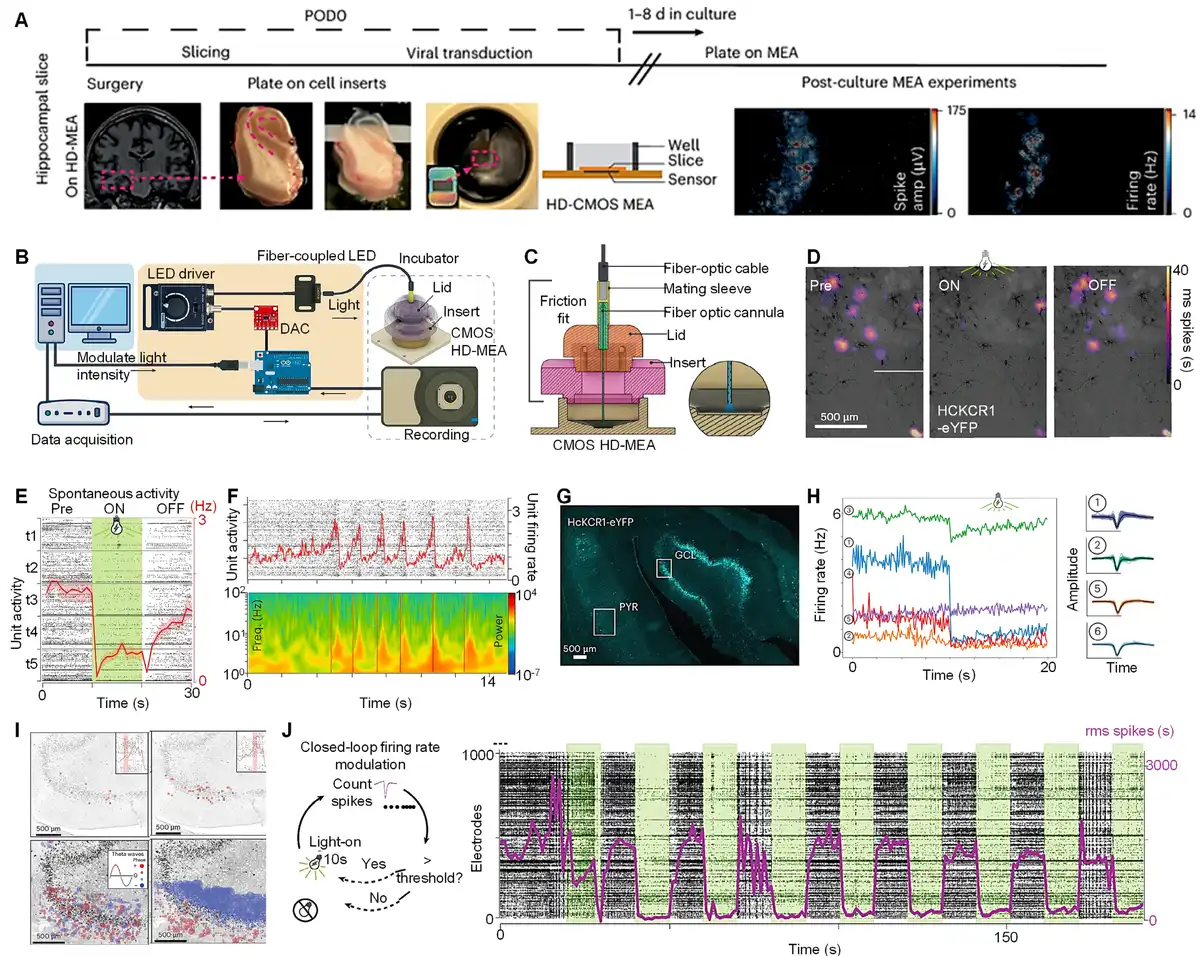
A recent work published in Nature Neuroscience addresses these gaps by establishing a robust ex vivo platform to evaluate optogenetic interventions in surgically resected human hippocampal tissue under clinically relevant epileptic conditions.[30] The organotypic slices (300 µm thick) from hippocampal tissue donated by patients with drug-resistant epilepsy were cultured at the air–liquid interface in serum-free media to model human epileptiform activity (Figure 1A). Slices were transduced with adeno-associated virus serotype 9 (AAV9) carrying the inhibitory potassium-conducting channelrhodopsin, HcKCR1, under the CAMK2A promoter, which restricts expression to excitatory neurons, alongside the enhanced yellow fluorescent protein (eYFP) for visualization. Bicuculline, a GABAa receptor antagonist, was used to pharmacologically elevate neuronal firing by blocking inhibitory signaling, thereby modeling seizure-like network activity.
Figure 1.
Closed-loop optogenetics for human hippocampal circuit interrogation and adaptive seizure control. (A) Workflow for preparing and recording human hippocampal slices. Resected tissue was sliced, cultured, transduced with AAV9 vectors, and recorded after 5–8 days. (B) Optogenetic hardware integration with HD-MEA system. A custom Arduino-controlled LED driver modulates light delivery (530 nm) synchronized with electrophysiological recordings. (C) Cross-sectional schematic of the 3D-printed optogenetic well insert, ensuring reproducible light delivery to tissue on the MEA. (D) Heat maps representing spike activity (5× RMS threshold) across the HD-MEA during 10-second intervals, highlighting regions of hyperactivity. Black denotes HcKCR1-eYFP staining. (E) Stacked raster plot of single-unit activity during optogenetic silencing trials. The average firing rate (red) shows suppression during illumination (green). (F) Rhythmic bursting activity (top) and theta-band LFP increases (bottom) following kainic acid application. (G) eYFP expression co-localizes with neuron-dense regions in the AAV9-HcKCR1-transduced hippocampal slice. (H) (Left) Average firing rate dynamics across response clusters. (Right) Representative extracellular waveforms for each cluster. (I) Spatiotemporal propagation of theta-phase coherence during epileptiform bursts. Dot size reflects phase magnitude; color indicates phase direction. (J) Closed-loop optogenetic control for bicuculline-provoked activity suppression. Reproduced with permission from Andrews et al. [30] Copyright 2024, Springer Nature.
To enable precise interrogation of network dynamics, high-density microelectrode arrays (MEAs; 26,400 electrodes) were integrated with a closed-loop system featuring fiber-coupled 530 nm light emitting diodes (LEDs) for spatiotemporally precise illumination (Figure 1B) and 3D-printed inserts for precise light delivery (Figure 1C). Custom software enabled real-time spike detection and adaptive control of light parameters (intensity, duration, and frequency), triggering optogenetic silencing when firing rates exceeded hyperactivity thresholds. This system achieved rapid suppression, with 15% to 40% of bicuculline-treated slices exhibiting near-complete silencing (≥90% reduction in firing rates). Continuous 530 nm illumination (10-second duration) consistently silenced spontaneous hippocampal activity in CAMK2A+ neurons (Figure 1D). In kainate-treated slices, rhythmic bursting emerged alongside theta-frequency (4–8 Hz) local field potential (LFP) coherence (Figure 1E). While optogenetic inhibition suppressed overall firing rates, coordinated bursts persisted in a subset of network activity, suggesting residual synchronization mechanisms resistant to targeted intervention (Figure 1F). Postexperiment immunohistochemistry confirmed CAMK2A-driven eYFP expression in hippocampal neurons, with enrichment observed in dentate gyrus granule cells and pyramidal neurons (Figure 1G). Waveform clustering revealed 12 neuronal subtypes with distinct anatomical and functional profiles. Neurons localized to nongranule cell layer (GCL) regions (CA1/CA3) consistently exhibited lower baseline firing rates (3–8 Hz) and stronger optogenetic suppression (Figure 1H). Conversely, dentate gyrus GCL-localized neurons showed characteristically high baseline activity (10–25 Hz) with attenuated responses to HcKCR1-mediated silencing. This functional-anatomical divergence suggests dentate granule cells drive network hyperexcitability while displaying relative resistance to inhibition, potentially due to compensatory glutamatergic signaling or incomplete transduction efficacy.
This work establishes a platform for precision neuromodulation in epilepsy by integrating cell-specific optogenetics, human-derived circuit analysis, and adaptive closed-loop control. Central to its innovation is the use of the CAMK2A promoter to restrict optogenetic inhibition (via HcKCR1) to excitatory neurons, key drivers of seizure propagation, while sparing inhibitory interneurons. This specificity is critical, as inhibitory neurons are often compromised in epilepsy, and indiscriminate silencing risks exacerbating network instability. High-density MEAs enabled the identification of human-specific biomarkers, such as theta-frequency coherence (4–8 Hz) and propagating wave dynamics, which are poorly replicated in rodent models. These biomarkers reflect pathological synchronization and spatial spread of hyperactivity, aligning with clinical seizure phenotypes. A modified Hodgkin–Huxley-based network model revealed that theta coherence facilitates long-range communication between epileptic foci while propagating waves mediate localized signal spread. Theta-phase propagation analysis during epileptiform bursts revealed spatiotemporal wave dynamics across the dentate gyrus, partially disrupted by optogenetic intervention (Figure 1I). This underscores that modulating oscillatory patterns can halt seizures without suppressing healthy activity, aligning with clinical goals of preserving cognitive function.
Conventional neurostimulation devices, such as vagus nerve stimulators, lack cellular specificity and temporal precision, often disrupting both pathological and physiological activities.[17–18–19] The closed-loop system’s targeted approach remained effective even with partial transduction of excitatory neurons (10%–54% efficacy), a realistic clinical scenario given the current limitation in viral delivery. This robustness arises from focusing on hyperactive dentate granule cells rather than requiring full neuronal coverage, highlighting the therapeutic potential of precision targeting. This challenges the assumption that high transduction efficiency is mandatory for therapeutic efficacy. The system dynamically modulated epileptiform activity in 2 distinct models: GABAergic blockade (mimicking disinhibition) and kainate-induced glutamate hyperactivity. Optogenetic suppression proved more potent in GABAergic blockade (Figure 1J), suggesting that disinhibited networks rely disproportionately on excitatory neuron synchronization, whereas glutamate-driven hyperactivity involves diffuse mechanisms requiring combinatorial strategies. This differential efficacy highlights the need to tailor interventions to the underlying circuit pathology, a cornerstone of personalized therapy.
1. Immune-optimized gene delivery for chronic stability
However, the translation of optogenetic interventions from ex vivo human hippocampal slices to clinical applications presents critical challenges that remain partially unresolved. While acute optogenetic control in cultured slices has been demonstrated, long-term safety and efficacy in intact human brains are unproven.[32] Chronic AAV-mediated opsin expression risks triggering immune responses or losing potency, particularly within the neuroinflammatory environment and gliosis characteristic of sclerotic hippocampal tissue in drug-resistant epilepsy. The incomplete suppression of rhythmic bursts in models, stemming from limited spatial targeting (10%–54% transduction rates) and spectral specificity (inability to fully block theta-frequency coherence), highlights the potential for off-target effects from the CAMK2A promoter or diffuse light scattering disrupting nonpathological circuits.[33] Moreover, current closed-loop systems, often reliant on simple firing rate thresholds, may miss subtler biomarkers like pre-ictal theta-gamma coupling[34] and fail to adapt effectively to evolving seizure dynamics, such as propagation shifts across hippocampal subfields.
To address these challenges, several potential strategies can be employed. Enhancing tropism for epileptic neurons requires developing engineered AAV capsids through directed evolution in human brain organoids[35,36]; variants like AAV-PHP, exploiting human-specific LY6A receptors, could improve transduction in sclerotic regions. Immune responses might be mitigated by creating stealth capsids with reduced antigenicity and introducing glycosylation sites into opsins like HcKCR1 to mask immunogenic epitopes, paralleling strategies used in therapeutic antibodies.[37] Chronic safety and efficacy should be tested in humanized animal models, “epilepsy-on-a-chip” co-cultures integrating human neurons, glia, and immune cells, or organoids.[38] Improving specificity necessitates dual-promoter systems, combining CAMK2A with activity-dependent promoters (FOS or NPY), to restrict opsin expression to hyperactive neurons, sparing quiescent cells and reducing off-target effects and immune exposure.[39] Spectral limitations and light scattering could be countered by integrating red-shifted opsins like Jaws for deeper, more focused penetration.[40] Meanwhile, overcoming detection limitations involves training machine learning algorithms on human-derived high-density microelectrode array data to recognize patient-specific epileptiform signatures (propagating wave directionality) and optimize stimulation parameters in real time.[24] These refinements engineered capsids, stealth opsins, dual-promoter targeting, red-shifted tools, and adaptive closed-loop systems can extend optogenetic efficacy from acute slices to chronically seizing networks, aligning with the clinical imperative for lifelong disease management.
2. Adaptive closed-loop architectures for predictive control
Despite suppressing seizure-like events by targeting firing rate thresholds, the closed-loop system failed to fully abolish theta-coherent bursts in kainate models, exposing a critical limitation of biomarker blindness. Current systems lack the sensitivity to detect latent precursors like pre-ictal theta-gamma coupling and cannot adapt to dynamically shifting propagation patterns. Overcoming this, multimodal biomarker detection should incorporate LFPs, intracellular Ca2+ imaging via fiber photometry, and astrocytic glutamate sensors into closed-loop algorithms.[24] Crucially, astrocyte-derived glutamate surges precede neuronal hyperactivity by 300 to 500 ms in human tissue, providing a vital predictive trigger for preemptive intervention. Moreover, machine learning-driven adaptation is essential; neural networks should be trained on human-specific seizure signatures using high-density microelectrode array datasets, enabling reinforcement learning to optimize stimulation parameters (wavelength, pulse width) in real time while preserving physiological theta rhythms critical for memory.[23] Meanwhile, deploying red-shifted optoelectronics replacing 530 nm LEDs with far-red light sources paired with opsins like Jaws or ChrimsonR will enable deeper tissue penetration and artifact-free concurrent electrophysiology, which is vital for targeting subcortical foci in intact brains.[22] These strategies would transform closed-loop optogenetics from reactive “firefighters” into adaptive “prevention engines,” capable of decoding the spatiotemporal grammar of human seizures.
The convergence of human-derived models and optogenetics has revealed a fundamental principle: effective epilepsy therapy must align with the biological uniqueness of human neural networks. Complementary evidence from rodent and theoretical models reinforces that pathological oscillations, whether arising from cortical-thalamic loops,[41] hypothalamic excitatory surges,[42] or leader neuron-initiated cortical bursts,[43] can be modulated by precise, scalable neurotechnologies. Emerging multimodal platforms further expand this toolkit: Sono-optogenetic systems enable noninvasive deep-brain targeting,[44] while engineered bacterial phytochromes operating in biliverdin-enriched environments achieve cell-specific control with enhanced spectral precision.[45] These advances, combined with our CAMK2A-guided closed-loop platform, signal a transformative shift toward biologically realistic therapeutic strategies. By pairing immune-stealth gene delivery with intelligent neuromodulation systems, we can extend ex vivo success to in vivo durability, evolving therapies alongside the brain’s dynamic complexity to realize personalized epilepsy management.
Acknowledgments
The authors would like to express their gratitude for the startup support from the Institute of Science and Technology for Brain-Inspired Intelligence (ISTBI) at Fudan University and CFFF platform of Fudan University. This work honors ISTBI’s 10th anniversary, during which its innovations in neuroscience and intelligent technology have advanced interdisciplinary research. Furthermore, this study aligns with the celebration of Fudan University’s 120th anniversary (1905–2025), marking a significant milestone in its commitment to advancing research at the cutting edge of science and technology.
Funding source
This study was funded by the National Natural Science Foundation of China (No. 22205254), the National Yong Talent Project, and Shanghai Pudong Elite Talent.
Conflicts of interests
The authors declare that they have no conflicts of interest.
Author contributions
Conceptualization: V.N.N. and S.X.; Writing – original draft: Y.W., N.M.B., Y.Z., and M.G.; Writing – review and editing: Y.W., N.M.B., V.N.N., and S.X.; Visualization: Y.Z. and M.G.; Supervision: V.N.N. and S.X.; Project administration: S.X.
References
- [1] Tang X, Jaenisch R, Sur M. The role of GABAergic signalling in neurodevelopmental disorders. Nat Rev Neurosci. 2021;22:290–307.
- [2] Wang L, Chen F, Zhang C, et al. Reactive oxygen species and neurodegenerative diseases: insights into nanozyme therapeutics. Med Mat. 2024;1:55–73.
- [3] Cui X, Wu L, Zhang C, et al. Implantable self‐powered systems for electrical stimulation medical devices. Adv Sci (Weinh). 2025;12:e2412044.
- [4] Rho JM, Boison D. The metabolic basis of epilepsy. Nat Rev Neurol. 2022;18:333–347.
- [5] Calia AB, Masvidal-Codina E, Smith TM, et al. Full-bandwidth electrophysiology of seizures and epileptiform activity enabled by flexible graphene microtransistor depth neural probes. Nat Nanotechnol. 2022;17:301–309.
- [6] Spruston N. Pyramidal neurons: dendritic structure and synaptic integration. Nat Rev Neurosci. 2008;9:206–221.
- [7] Mertens EJ, Leibner Y, Pie J, et al. Morpho-electric diversity of human hippocampal CA1 pyramidal neurons. Cell Rep. 2024;43:114100.
- [8] Vanderhaeghen P, Polleux F. Developmental mechanisms underlying the evolution of human cortical circuits. Nat Rev Neurosci. 2023;24:213–232.
- [9] Király B, Domonkos A, Jelitai M, et al. The medial septum controls hippocampal supra-theta oscillations. Nat Commun. 2023;14:6159.
- [10] Fischer P, Lipski WJ, Neumann W-J, et al. Movement-related coupling of human subthalamic nucleus spikes to cortical gamma. eLife. 2020;9:e51956.
- [11] Fromm MF. Importance of P-glycoprotein at blood–tissue barriers. Trends Pharmacol Sci. 2004;25:423–429.
- [12] Vezzani A, Ravizza T, Bedner P, et al. Astrocytes in the initiation and progression of epilepsy. Nat Rev Neurol. 2022;18:707–722.
- [13] Klein P, Kaminski RM, Koepp M, et al. New epilepsy therapies in development. Nat Rev Drug Discov. 2024;23:682–708.
- [14] Wang Y, Yang Z, Wang J, et al. Brain-body interactions in ischemic stroke: VNS reprograms microglia and FNS enhances cerebellar neuroprotection. Stroke. 2025;56:051470.
- [15] Xu S, Manshaii F, Chen J. Is deep brain imaging on the brink of transformation with a bioluminescence molecule? BMEMat. 2024;2:e12115.
- [16] Zhao F, Zhao Z, Gao H, et al. Cuproptosis: an emerging domain for copper-based nanomaterials mediated cancer therapy. Med Mat. 2024;1:74–94.
- [17] Soleimani G, Nitsche MA, Bergmann TO, et al. Closing the loop between brain and electrical stimulation: towards precision neuromodulation treatments. Transl Psychiatry. 2023;13:279.
- [18] Xu S, Manshaii F, Xiao X, et al. Triboelectric nanogenerators for self-powered neurostimulation. Nano Res. 2024;17:8926–8941.
- [19] Bai L, Wu Y, Li J, et al. Organoids in motion: biohybrid robotics futures. Med Mat. 2025;2:79–84.
- [20] Kumari LS, Kouzani AZ. Electrophysiology-based closed loop optogenetic brain stimulation devices: recent developments and future prospects. IEEE Rev Biomed Eng. 2022;16:91–108.
- [21] Zhang Y, Karadas M, Liu J, et al. Interaction of acetylcholine and oxytocin neuromodulation in the hippocampus. Neuron. 2024;112:1862–1875.e5.
- [22] Xu S, Momin M, Ahmed S, et al. Illuminating the brain: advances and perspectives in optoelectronics for neural activity monitoring and modulation. Adv Mater. 2023;35:2303267.
- [23] Xu S, Liu Y, Lee H, et al. Neural interfaces: bridging the brain to the world beyond healthcare. Exploration (Beijing, China). 2024;4:20230146.
- [24] Xu S, Xiao X, Manshaii F, et al. Injectable fluorescent neural interfaces for cell-specific stimulating and imaging. Nano Lett. 2024;24:4703–4716.
- [25] Bansal A, Shikha S, Zhang Y. Towards translational optogenetics. Nat Biomed Eng. 2023;7:349–369.
- [26] Banks E, Gutekunst CA, Vargish GA, et al. An enhancer-AAV approach selectively targeting dentate granule cells of the mouse hippocampus. Cell Rep Methods. 2024;4:100684.
- [27] Lesuis SL, Park S, Hoorn A, et al. Stress disrupts engram ensembles in lateral amygdala to generalize threat memory in mice. Cell. 2025;188:121–140.
- [28] Kahana MJ, Ezzyat Y, Wanda PA, et al. Biomarker-guided neuromodulation aids memory in traumatic brain injury. Brain Stimul. 2023;16:1086–1093.
- [29] Deng Q, Wu C, Parker E, et al. Mystery of gamma wave stimulation in brain disorders. Mol Neurodegener. 2024;19:96.
- [30] Andrews JP, Geng J, Voitiuk K, et al. Multimodal evaluation of network activity and optogenetic interventions in human hippocampal slices. Nat Neurosci. 2024;27:2487–2499.
- [31] Hadjiabadi D, Lovett-Barron M, Raikov IG, et al. Maximally selective single-cell target for circuit control in epilepsy models. Neuron. 2021;109:2556–2572.
- [32] Peters CW, Maguire CA, Hanlon KS. Delivering AAV to the central nervous and sensory systems. Trends Pharmacol Sci. 2021;42:461–474.
- [33] Emiliani V, Entcheva E, Hedrich R, et al. Optogenetics for light control of biological systems. Nat Rev Methods Primers. 2022;2:55.
- [34] Patterson R, Brooks H, Mirjalili M, et al. Theta phase-gamma amplitude coupling during working memory and its relationships with demographic, clinical, genetic, neurochemical, and neurostructural measures in older adults at risk for dementia. Biol Psychiatry. 2021;89:S350–S351.
- [35] Nisanov AM, de Jesús JAR, Schaffer DV. Advances in AAV capsid engineering: integrating rational design, directed evolution and machine learning. Mol Ther. 2025;33:1937–1945.
- [36] Ling Q, Herstine JA, Bradbury A, et al. AAV-based in vivo gene therapy for neurological disorders. Nat Rev Drug Discov. 2023;22:789–806.
- [37] Freeman KG, Robotham AC, Parks OB, et al. Virion glycosylation influences mycobacteriophage immune recognition. Cell Host Microbe. 2023;31:1216–1231.
- [38] Liu J, Sternberg AR, Ghiasvand S, et al. Epilepsy-on-a-chip system for antiepileptic drug discovery. IEEE Trans Biomed Eng. 2018;66:1231–1241.
- [39] Vivot K, Meszaros G, Pangou E, et al. CaMK1D signalling in AgRP neurons promotes ghrelin-mediated food intake. Nat Metab. 2023;5:1045–1058.
- [40] Chuong AS, Miri ML, Busskamp V, et al. Noninvasive optical inhibition with a red-shifted microbial rhodopsin. Nat Neurosci. 2014;17:1123–1129.
- [41] Paz J, Bryant A, Peng K, et al. A new mode of corticothalamic transmission revealed in the Gria4−/− model of absence epilepsy. Nat Neurosci. 2011;14:1167–1173.
- [42] Li H, Viskaitis P, Bracey E, et al. Transient targeting of hypothalamic orexin neurons alleviates seizures in a mouse model of epilepsy. Nat Commun. 2024;15:1249.
- [43] Kobayashi T, Shimba K, Narumi T, et al. Revealing single-neuron and network-activity interaction by combining high-density microelectrode array and optogenetics. Nat Commun. 2024;15:9547.
- [44] Roy S, Pyari G, Bansal H. Theoretical analysis of low-power deep synergistic sono-optogenetic excitation of neurons by co-expressing light-sensitive and mechano-sensitive ion-channels. Commun Biol. 2025;8:379.
- [45] Kasatkina L, Ma C, Sheng H, et al. Deep-tissue high-sensitivity multimodal imaging and optogenetic manipulation enabled by biliverdin reductase knockout. Nat Commun. 2025;16:6469.
This article is available under Open Access.
© 2025 The Authors