Application of microenvironment biomimetic materials in spinal cord injury and future prospects
Authors
Wenzhao Wang, Mingjie Sun, Zhengdong Zhang, Sen Guo, Haijian Sun*, Zhijian Wei*, Bin Ning*
- aDepartment of Orthopedics, Jinan Central Hospital, Shandong University, Jinan, Shandong, China
- bDepartment of Orthopedics, Centre for Orthopedics, Qilu Hospital, Advanced Medical Research Institute, Shandong University, Jinan, China
- cDepartment of Orthopedic Surgery, Center for Joint Surgery, The Second Affiliated Hospital of Chongqing Medical University, Chongqing, China
- dDepartment of Orthopedics, The First Affiliated Hospital of Chengdu Medical College, Chengdu, China
- eDepartment of Radiology, Juntendo University Graduate School of Medicine, Tokyo, Japan
- fDepartment of Orthopaedics, The Second Hospital of Shandong University, Cheeloo College of Medicine, Shandong University, Jinan, Shandong, China
- gDepartment of Orthopedics, Central Hospital Affiliated to Shandong First Medical University, Jinan, Shandong, China.
* Correspondence: Address: Haijian Sun, Department of Orthopaedics, The Second Hospital of Shandong University, Cheeloo College of Medicine, Shandong University, No. 247, Beiyuan Street, Jinan, Shandong 250033, China. Email: shj94@email.sdu.edu.cn (H. Sun); Zhijian Wei, Department of Orthopedics, Centre for Orthopedics, Qilu Hospital, Advanced Medical Research Institute, Shandong University, No. 107, Wenhua Xi Road, Jinan, Shandong 250012, China. Email: weizhijian2002@126.com (Z. Wei); Bin Ning, Department of Orthopedics, Jinan Central Hospital, Shandong University, No. 105, Jiefang Road, Jinan, Shandong 250013, China. Email:ningbin@sdu.edu.cn (B. Ning).
MedMat · 2025 · Vol. 2 · No. 3 · pp. 137-139
Translations
Long abstracts in additional languages. The English article is the version of record.
中文zh-Hans
脊髓损伤(SCI)代表了现代医学面临的主要挑战,其病理生理机制复杂且内源性修复能力极其有限。原发性物理损伤会迅速触发一系列复杂的继发性损伤级联反应,包括严重的炎症反应、血-脊髓屏障的破坏、脱髓鞘形成、纤维化和胶质瘢痕的形成,以及抑制性分子的广泛表达。这些过程共同构建了一个对神经再生极具阻碍性的微环境。本文旨在综述2015年至2025年间针对复杂脊髓微环境的仿生生物材料策略的最新进展,探讨如何通过模拟健康脊髓组织的物理、化学和生物学特征来主动调节损伤后的病理微环境,从而诱导内源性或外源性细胞实现神经再生和功能重建。
本综述聚焦于超越单纯物理填充的仿生生物材料核心策略。理想的仿生材料应模仿细胞外基质(ECM)的成分与结构,如胶原蛋白、层粘连蛋白和纤连蛋白,同时匹配原生脊髓组织的机械性能、降解速率及孔隙结构,以促进细胞的迁移、增殖和分化。具体而言,作为主要ECM成分的胶原可桥接SCI缺损并促进轴突再生;中枢神经系统关键成分透明质酸及其衍生物(如甲基丙烯酸甲酯水凝胶)能为神经干细胞提供适宜微环境;壳聚糖则展现出抗菌与伤口愈合特性,其交联壳聚糖水凝胶在减少胶质瘢痕形成及促进脊髓损伤模型中神经功能恢复方面已被证实有效。
科学分析表明,合成高分子材料如聚乳酸-乙醇酸共聚物(PLGA)提供了可控的降解速率和优异的机械性能,而聚己内酯(PCL)因其缓慢的降解特性适用于长期神经再生应用。目前已有多种基于聚合物的仿生产品进入临床试验阶段。此外,结合天然成分生物活性与合成材料机械强度的天然 - 合成杂化材料被用于脊髓微环境的重建。功能化的纳米材料如石墨烯、纳米羟基磷灰石和碳纳米管,经修饰后能增强神经信号传导并调节损伤后的神经免疫平衡。这些发现证实了通过模拟ECM组成来创造有利于再生的微环境的可行性与有效性。
尽管仿生生物材料在SCI修复领域取得了显著突破,但仍面临诸多挑战。目前的策略虽已实现从被动填充到主动调控的转变,但如何精确匹配不同损伤阶段的动态需求仍是难点。未来的工作需进一步探索天然 - 合成杂化材料的长期安全性及功能化纳米材料的体内稳定性。此外,虽然部分产品已进入临床试验,但其大规模临床应用仍需解决标准化生产与个体化治疗之间的矛盾。本综述为理解脊髓微环境调控机制提供了框架,并指出未来研究应致力于开发更具生物活性、降解可控且能精准调节神经免疫平衡的新型材料体系,以最终实现脊髓损伤的功能性修复。
Françaisfr
La lésion de la moelle épinière (LME) représente un défi majeur en médecine moderne, caractérisée par une physiopathologie complexe et une capacité intrinsèquement limitée à la réparation endogène. Les dommages physiques primaires déclenchent rapidement une cascade de lésions secondaires complexes, incluant des réponses inflammatoires sévères, la rupture de la barrière hémato-médullaire, la démyélinisation, la formation de cicatrices fibreuses et gliales, ainsi qu'une expression extensive de molécules inhibitrices. Collectivement, ces processus établissent un microenvironnement profondément défavorable à la régénération nerveuse. Cet article vise à examiner les stratégies biomimétiques ciblées sur ce microenvironment complexe entre 2015 et 2025, explorant comment elles régulent activement l'environnement pathologique post-lésion en imitant les caractéristiques physiques, chimiques et biologiques du tissu spinal sain pour induire la régénération nerveuse.
La stratégie centrale de ces matériaux a évolué au-delà du simple remplissage physique ; ils agissent désormais pour réguler activement le microenvironnement pathologique post-lésion en imitant les caractéristiques physiques, chimiques et biologiques des tissus sains. Les biomatériaux mimétiques facilitent la régénération nerveuse en émulant la composition et la structure de la matrice extracellulaire (MEC), telles que le collagène, la laminine et la fibronectine. Un matériau idéal doit correspondre aux propriétés mécaniques, au taux de dégradation et à la structure poreuse du tissu spinal natif pour promouvoir la migration, la prolifération et la différenciation cellulaires. Le collagène, composant principal de la MEC, comble les lacunes LME et favorise la régénération axonale.
Les résultats scientifiques montrent que l'acide hyaluronique, constituant clé du système nerveux central, fournit un microenvironnement approprié pour les cellules souches neurales via des dérivés comme le gel d'hyalurone-méthyl méthacrylate. Le chitosane présente des propriétés antibactériennes et de cicatrisation ; ses hydrogels réticulés ont démontré une efficacité à réduire la formation de cicatrices gliales et à promouvoir la récupération fonctionnelle neuronale dans les modèles LME. Les polymères synthétiques comme le poly(lactic-co-glycolic acid) offrent des taux de dégradation contrôlables, tandis que le polycaprolactone convient aux applications à long terme grâce à sa dégradation lente. Des matériaux hybrides naturels-synthétiques et des nanomatériaux fonctionnalisés (graphène, nano-hydroxyapatite, nanotubes de carbone) améliorent la transmission du signal neural et régulent l'équilibre neuro-immun.
Ces avancées marquent une transition significative vers un environnement propice à la régénération nerveuse. Cependant, des limites subsistent concernant la précision du mimétisme dynamique aux différentes phases de blessure et la sécurité à long terme des nanomatériaux fonctionnalisés in vivo. Bien que plusieurs produits polymères aient atteint les essais cliniques, leur application clinique généralisée nécessite encore de résoudre les contradictions entre production standardisée et traitement personnalisé. Les travaux futurs doivent se concentrer sur le développement de systèmes matériels plus bioactifs avec une dégradation contrôlable capables de réguler précisément l'équilibre neuro-immun pour réaliser la réparation fonctionnelle complète des lésions de la moelle épinière, en s'appuyant sur ce cadre synthétique établi par cette revue perspective entre 2015 et 2025.
Españoles
La lesión de la médula espinal (LME) representa un desafío mayor en la medicina moderna debido a su fisiopatología compleja y una capacidad intrínsecamente limitada para la reparación endógena. El daño físico primario desencadena rápidamente una cascada de lesiones secundarias complejas, incluyendo respuestas inflamatorias severas, ruptura de la barrera hemato-médular, desmielinización, formación de cicatrices fibrosas y gliales, y expresión extensiva de moléculas inhibitorias. Colectivamente, estos procesos establecen un microambiente profundamente inhibidor para la regeneración nerviosa. Este artículo examina las estrategias biomiméticas dirigidas al complejo microambiente espinal entre 2015 y 2025, explorando cómo imitan las características físicas, químicas y biológicas del tejido sano para regular activamente el entorno patológico post-lesión e inducir la regeneración nerviosa mediante células endógenas o exógenas.
La estrategia central de estos materiales ha evolucionado más allá del mero llenado físico; ahora regulan activamente el microambiente patológico post-lesión imitando las características físicas, químicas y biológicas del tejido espinal sano. Los biomateriales miméticos facilitan la regeneración nerviosa emulando la composición y estructura de la matriz extracelular (MEC), como colágeno, laminina y fibronectina. Un material ideal debe coincidir con las propiedades mecánicas, tasa de degradación y estructura porosa del tejido espinal nativo para promover la migración, proliferación y diferenciación celular. El colágeno, componente principal de la MEC, puentea los defectos LME y promueve la regeneración axonal.
Los hallazgos científicos indican que el ácido hialurónico, constituyente clave del sistema nervioso central, proporciona un microambiente adecuado para células madre neurales a través de derivados como hidrogel de metilmetacrilato-hialuronano. El quitosano exhibe propiedades antibacterianas y de cicatrización; sus hidrogeles entrecruzados han demostrado eficacia en reducir la formación de cicatrices gliales y promover la recuperación funcional neuronal en modelos LME. Los polímeros sintéticos como el ácido poli(láctico-co-glicólico) ofrecen tasas de degradación controlables, mientras que el policaprolactona es adecuado para aplicaciones a largo plazo debido a su perfil de degradación lento. Materiales híbridos naturales-sintéticos y nanomateriales funcionalizados (grafeno, nano-hidroxiapatita, nanotubos de carbono) mejoran la transmisión de señales neurales y regulan el equilibrio neuroinmune.
Aunque los biomateriales han logrado avances significativos en la reparación LME, persisten desafíos. La transición del llenado pasivo al control activo es un logro, pero igualar dinámicamente las necesidades de diferentes etapas de lesión sigue siendo difícil. El trabajo futuro debe explorar más a fondo la seguridad a largo plazo de los materiales híbridos y la estabilidad in vivo de nanomateriales funcionalizados. Aunque varios productos poliméricos han alcanzado ensayos clínicos, su aplicación clínica generalizada requiere resolver contradicciones entre producción estandarizada y tratamiento personalizado. Esta revisión proporciona un marco para comprender los mecanismos de regulación del microambiente espinal e indica que la investigación futura debe centrarse en desarrollar nuevos sistemas materiales más bioactivos con degradación controlable capaces de regular precisamente el equilibrio neuroinmune para lograr una reparación funcional completa de las lesiones medulares entre 2015 y 2025.
日本語ja
脊髄損傷(SCI)は、その複雑な病理生理学的機序と限られた内因性修復能力により、現代医学における主要な課題の一つです。一次的な物理的損傷は直ちに、重度の炎症反応、血液脊髄関門の破壊、脱髄形成、線維化およびグリア瘢痕の発生、ならびに抑制分子の広範な発現を含む複雑な二次的損傷のカスケードを引き起こします。これらの過程が総じて神経再生に対して極めて阻害的な微小環境を構築しています。本稿は2015年から2025年にかけて、この複雑な脊髄微小環境を対象としたバイオニック生体材料戦略の進展を検討し、健康な脊髄組織の物理的・化学的・生物学的特性を模倣することで損傷後の病理的微小環境を能動的に調節し、内因性または外因性の細胞に対して神経再生と機能再構築を誘導するアプローチについて概説します。
これらの材料の中核戦略は単なる物理的な充填を超え、健康な脊髄組織の特性を模倣することで損傷後の病理的微小環境を能動的に調節することに進化しています。生体模倣材料は、コラーゲン、ラミニン、フィブロネクチンなどの細胞外マトリックス(ECM)の組成と構造を模倣することにより神経再生を促進します。理想的な生体模倣材料は、細胞の遊走、増殖および分化を促進するために、天然脊髄組織の機械的特性、分解速度、および孔隙構造に一致する必要があります。コラーゲンは主要なECM成分としてSCIギャップを橋渡しし軸突再生を促進します。また、中枢神経系の重要な構成要素であるヒアルロン酸は、メチルメタクリレートハイドロゲルなどの誘導体を通じて神経幹細胞に適した微小環境を提供します。
科学的知見によると、キトサンには抗菌および創傷治癒特性があり、架橋されたキトサンハイドロゲルはSCIモデルにおいてグリア瘢痕形成の減少と神経機能回復の促進に有効であることが示されています。ポリ乳酸-グリコール酸共重合体(PLGA)は制御可能な分解速度と優れた機械的特性を提供し、ポリカプロラクトン(PCL)はその緩慢な分解プロフィールにより長期の神経再生応用に適しています。いくつかの高分子ベースの生体模倣製品が臨床試験段階に進んでいます。さらに、天然成分の生物活性と合成材料の機械的強度を組み合わせた天然 - 合成ハイブリッド材料も脊髄微小環境再構築に使用されています。グラフェンやナノ水酸化リン酸カルシウムなどの機能化されたナノ材料は修飾後、神経信号伝達を強化し損傷後の神経免疫バランスを調節します。
これらの成果は、再生に適した微小環境の創出において重要な進歩を示していますが、依然として課題が残されています。現在の戦略は受動的充填から能動的制御への転換を実現しましたが、異なる損傷段階における動態的なニーズに正確に一致させることは依然として困難です。今後の研究では、天然 - 合成ハイブリッド材料の長期安全性と機能化ナノ材料の体内安定性をさらに探求する必要があります。一部の製品が臨床試験に至っていますが、大規模な臨床応用には標準化された生産と個別化治療との間の矛盾を解決することが必要です。本レビューは脊髄微小環境調節メカニズムを理解するための枠組みを提供し、神経免疫バランスを精密に調節できる新規材料体系の開発の重要性を指摘しています。
العربيةar
تمثل إصابة الحبل الشوكي (SCI) تحدياً رئيسياً في الطب الحديث نظراً لآلياتها الفسيولوجية المرضية المعقدة وقدرتها الذاتية المحدودة للغاية على الإصلاح. يؤدي الضرر الجسدي الأولي بسرعة إلى سلسلة معقدة من الإصابات الثانوية، بما في ذلك استجابات التهابية شديدة، واضطراب الحاجز الدموي الشوكي، ونزع الميالين، وتكون ندبات ليفية وغليالية، والتعبير الواسع للجزيئات المثبطة. مجتمعةً، تنشئ هذه العمليات بيئة دقيقة مثبطة بشدة تعيق تجديد الأعصاب. يهدف هذا المقال إلى استعراض الاستراتيجيات الحيوية المستوحاة من الطبيعة (Bionic biomaterial strategies) التي تستهدف البيئة الدقيقة المعقدة للحبل الشوكي خلال العقد الماضي (2015-2025)، واستكشاف كيفية تنظيمها النشط للبيئة المرضية بعد الإصابة عن طريق محاكاة الخصائص الفيزيائية والكيميائية والبيولوجية لأنسجة الحبل الشوكي الصحية، مما يحفز الخلايا الذاتية أو الخارجية لتحقيق تجديد الأعصاب وإعادة البناء الوظيفي.
لقد تطورت الاستراتيجية الأساسية لهذه المواد لتتجاوز مجرد الملء المادي؛ فهي تنظم الآن بنشاط البيئة الدقيقة المرضية بعد الإصابة عن طريق محاكاة الخصائص الفيزيائية والكيميائية والبيولوجية لأنسجة الحبل الشوكي الصحية. تسهل المواد الحيوية المستوحاة من الطبيعة تجديد الأعصاب عن طريق تقليد تكوين وبنية المادة خارج الخلية (ECM)، مثل الكولاجين، واللامينين، والفبرونكتين. يجب أن تتطابق المادة المثالية مع الخصائص الميكانيكية ومعدل التحلل وهيكل المسام في نسيج الحبل الشوكي الأصلي لتعزيز هجرة الخلايا وتكاثرها وتمايزها. يعمل الكولاجين، وهو مكون رئيسي للمادة خارج الخلية، على جسور الفجوات الناتجة عن الإصابة ويحفز تجديد المحاور العصبية.
تُظهر النتائج العلمية أن حمض الهيالورونيك، المكون الرئيسي للجهاز العصبي المركزي، يوفر بيئة دقيقة مناسبة لخلايا الجذع العصبية من خلال مشتقاته مثل هيدروجيل ميثيل ميثاكريلات-هيالورونان. يُظهر الكيتوزان خصائص مضادة للبكتيريا وشفاء الجروح؛ وقد أثبتت الهلاميات المائية للكيتوزان المتشابكة فعاليتها في تقليل تكون الندبات الغليالية وتعزيز الاستعادة الوظيفية العصبية في نماذج إصابة الحبل الشوكي. يوفر بولي (حمض اللاكتيك-كوجليكوليد) معدلات تحلل قابلة للتحكم وخصائص ميكانيكية ممتازة، بينما يجعل ملف التحلل البطيء لـبولي كابرولاكتون مناسباً لتطبيقات تجديد الأعصاب طويلة الأمد. كما تُستخدم المواد الهجينة الطبيعية-الاصطناعية التي تجمع بين النشاط البيولوجي للمكونات الطبيعية والقوة الميكانيكية للتركيبات الاصطناعية لإعادة بناء البيئة الدقيقة الشوكية، بينما تعزز مواد نانوية وظيفية مثل الجرافين والهيدروكسي أباتيت النانوي وأنابيب الكربون النانوية نقل الإشارات العصبية وتنظيم التوازن العصبي المناعي بعد التعديل.
على الرغم من أن المواد الحيوية المستوحاة من الطبيعة حققت اختراقات كبيرة في إصلاح إصابة الحبل الشوكي، إلا أنها لا تزال تواجه تحديات. بينما نجحت الاستراتيجيات الحالية في الانتقال من الملء السلبي إلى التنظيم النشط، فإن مطابقة الاحتياجات الديناميكية لمراحل الإصابة المختلفة بدقة تظل صعبة. يجب أن يركز العمل المستقبلي على استكشاف السلامة طويلة الأجل للمواد الهجينة الطبيعية-الاصطناعية واستقرار المواد النانوية الوظيفية داخل الجسم الحي. وعلى الرغم من وصول بعض المنتجات القائمة على البوليمرات إلى مرحلة التجارب السريرية، إلا أن تطبيقها السريري واسع النطاق يتطلب حل التناقضات بين الإنتاج الموحد والعلاج المخصص. يوفر هذا الاستعراض إطاراً لفهم آليات تنظيم البيئة الدقيقة الشوكية ويشير إلى ضرورة تطوير أنظمة مواد جديدة أكثر نشاطاً بيولوجياً مع تحلل قابل للتحكم قادر على ضبط التوازن العصبي المناعي بدقة لتحقيق الإصلاح الوظيفي الكامل لإصابات الحبل الشوكي.
Full Text
1. Introduction
Spinal cord injury (SCI) represents a major challenge in modern medicine due to its complex pathophysiology and inherently limited endogenous repair capacity. Primary physical damage rapidly triggers a cascade of complex secondary injuries, including severe inflammatory responses, disruption of the blood–spinal cord barrier, demyelination, formation of fibrotic and glial scars, and extensive expression of inhibitory molecules. Collectively, these processes establish a profoundly inhibitory microenvironment detrimental to nerve regeneration.[1]
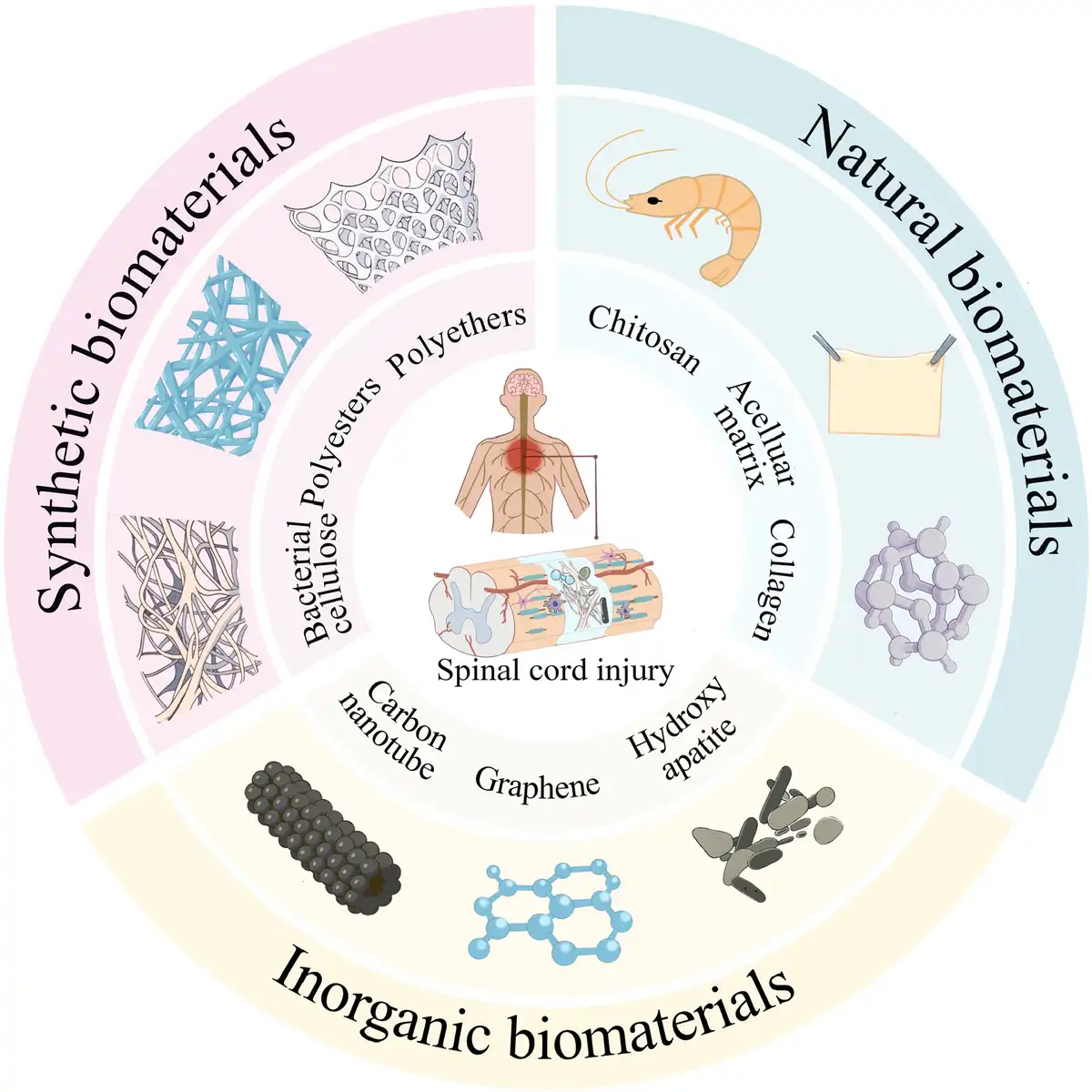
Over the past decade (2015–2025), bionic biomaterial strategies targeting the complex spinal cord microenvironment have achieved significant breakthroughs in SCI repair (Figure 1). The core strategy of these materials has evolved beyond mere physical filling; they now actively regulate the postinjury pathological microenvironment by mimicking the physical, chemical, and biological characteristics of healthy spinal cord tissue. This approach “induces” endogenous or exogenous cells to achieve nerve regeneration and functional reconstruction, thereby creating a regenerative microenvironment conducive to nerve regeneration.[2]
Figure 1.
Over the past decade (2015–2025), bionic biomaterial strategies targeting the complex spinal cord microenvironment have achieved significant breakthroughs in SCI repair.
2. Types and design of biomimetic materials for spinal cord microenvironment
Biomimetic materials facilitate nerve regeneration by emulating the composition and structure of the extracellular matrix (ECM), such as collagen, laminin, and fibronectin. An ideal biomimetic material should match the mechanical properties, degradation rate, and pore structure of native spinal cord tissue to promote cell migration, proliferation, and differentiation.[3]
2.1 Natural biomaterials
Collagen, a primary ECM component, bridges SCI gaps and promotes axon regeneration. Hyaluronic acid, a key constituent of the central nervous system, provides an appropriate microenvironment for neural stem cells via derivatives like hyaluronic acid-methyl methacrylate hydrogel. Chitosan exhibits antibacterial and wound-healing properties. Cross-linked chitosan hydrogels demonstrate efficacy in reducing glial scar formation and promoting neural functional recovery in SCI models.[4]
2.2 Synthetic polymers and composite materials
Poly(lactic-co-glycolic acid) offers controllable degradation rates and excellent mechanical properties. Polycaprolactone’s slow degradation profile makes it suitable for long-term nerve regeneration applications. Several polymer-based biomimetic products have advanced to clinical trials. Natural-synthetic hybrid materials, combining the bioactivity of natural components with the mechanical strength of synthetics, are also employed for spinal microenvironment reconstruction. Furthermore, functionalized nanomaterials like graphene, nano-hydroxyapatite, and carbon nanotubes enhance neural signal transmission and regulate neuroimmune balance following modification.[5]
3. Integration of active factors and cells
Controlled-release systems for neurotrophic factors—such as brain-derived neurotrophic factor (BDNF), neurotrophin-3 (NT-3), and nerve growth factor—provide sustained growth signals to the injury site, promoting nerve regeneration. Delivering cytokines, chemokines (eg, interleukin family), and regenerative factors via material carriers modulates postinjury inflammatory responses and tissue repair processes.[6] Microenvironmental biomimetic scaffolds also provide a three-dimensional (3D) culture environment for cells. Seed cells—including neural stem cells, umbilical cord stem cells, and Schwann cells—engage in nerve regeneration through synergistic interactions with these scaffolds.[7]
4. Core application strategies and progress
Biomimetic microenvironmental materials actively regulate the damaged microenvironment by replicating physical, chemical, and biological signals from healthy spinal cord tissue, thereby supporting nerve regeneration.
4.1 Bionic physical topological structure
Techniques like electrospinning, magnetic field induction, and 3D printing fabricate scaffolds with micro/nanoscale-oriented structures (eg, aligned nanofibers, microchannel hydrogels) that simulate the directionality of nerve fiber bundles in spinal cord white matter. These scaffolds provide “tracks” for regenerating axons, guiding them across the injury site to achieve functional neural circuit reconnection. They also deliver contact guidance cues, promoting the long-distance, directional extension of host and transplanted neurons/neural precursor cells across the injury scar.[8]
4.2 Regulation of the immune microenvironment
Materials with immunomodulatory functions, such as those loaded with M2-type macrophage-inducing factors or antifibrotic drugs or those that regulate the phenotype of microglia/macrophages by utilizing their own physicochemical properties (such as surface chemistry, stiffness)[9] inhibit the activation of proinflammatory (M1 type) macrophages, promote the polarization of anti-inflammatory/repair (M2 type) macrophages, inhibit the excessive activation of pericytes, fibroblasts, and astrocytes to form dense scars, and create a more permissive environment for regeneration.[10]
4.3 Reconstruction of the nutritional microenvironment
Carriers like intelligent hydrogels, microspheres, and nanoparticles enable sustained, gradient, or on-demand release of neurotrophic factors (eg, BDNF, NT-3, glial cell-derived neurotrophic factor [GDNF]) and axon guidance molecules (eg, Netrin-1, sonic hedgehog [Shh] signaling molecule) at the injury site. This provides essential bioactive signals for neuronal survival, axonal growth, and angiogenesis while regulating inflammation, mitigating secondary damage, and guiding precise axonal targeting.[11]
4.4 Conductive and electroactive materials: restoring neural signal conduction
Conductive scaffolds or coatings constructed from conductive polymers (eg, polypyrrole, polyaniline), graphene, or carbon nanotubes bridge disrupted neural pathways to restore signal conduction. Exogenous electrical stimulation further enhances neuronal differentiation, axonal growth, and synaptogenesis.[12]
5. Future research prospects
5.1 Multifunctional integration and intelligence
Given the limited efficacy of single strategies, future biomaterials will be highly integrated platforms capable of simultaneous structural guidance, immune regulation, temporally controlled multifactor release, and electrical signal conduction within a single system. Self-assembling smart materials that trigger drug release or structural changes in response to specific pathological signals are under development. Combining biomimetic materials with physical therapies like electrical stimulation, optogenetics, and magnetic field therapy will yield synergistic effects.[13]
5.2 “4D and 5D bioprinting” and spinal cord organoid technology
High-precision 3D bioprinting is expected to enable scaffolds capable of programmed transformations in both spatial structure (3D) and time (4D). Advancements in functional dimension or free-form surface manipulation (5D) will facilitate full life-cycle design, allowing precise simulation of the spinal cord’s dynamic injury-repair process. Scaffolds could initially feature dense structures to suppress inflammation, later transitioning to larger pores promoting cell infiltration and axonal growth. A 4D-designed shape-memory hydrogel scaffold transforms from a linear structure into a spiral configuration triggered by body temperature, conforming closely to the injury cavity and providing axially aligned guidance to promote axonal regeneration. Simultaneously, such scaffolds can be loaded with mesenchymal stem cells via microfluidic printing, enabling their targeted differentiation into neuron-like cells under reactive oxygen species (ROS) stimulation, thereby achieving a closed-loop “sensing-response-repair” system. Spinal cord organoids show great promise as a potential source for cell transplantation. With reliable and scalable cell sources, and containing multiple synergistic cell types, transplanted organoid fragments can potentially provide replacement neurons, supportive glial cells, and scaffold functions to reconstruct the microenvironment simultaneously, offering greater integrative potential than single-cell-type transplants. Furthermore, spinal cord organoids provide a novel platform for drug discovery and toxicity testing in an environment more closely resembling the human body. Using patient-specific induced pluripotent stem cells (iPSC)-derived spinal cord organoids, disease-in-a-dish models can be established to simulate pathological changes under specific genetic backgrounds or injury conditions, enabling high-throughput screening of candidate drugs that promote neuroprotection, axon regeneration, myelin repair, or inhibit glial scar formation.[14]
5.3 Integration of stem cell therapy and genetic engineering
Biomedical materials will serve as advanced cell delivery vehicles and “in vitro stem cell niches.” Material optimization will enable precise control over stem cell fate (survival, differentiation, integration). Coupled with gene editing (eg, clustered regularly interspaced short palindromic repeats [CRISPR]), silencing intrinsic inhibitory genes or enhancing pro-growth gene expression during cell delivery will significantly boost therapeutic potential.[15]
5.4 Collaborative reconstruction of the Neuro-Immune-Vascular network
Future research will emphasize synergistic interactions between neural regeneration, immune modulation, and vascular regeneration. Designing biomaterials that concurrently promote vascular regeneration (nutrient supply) and lymphatic vessel regeneration (clearance of metabolic waste and inflammatory factors) is crucial for establishing a truly healthy regenerative microenvironment.[16] Future material designs must increasingly address clinical requirements, including biodegradability, long-term biocompatibility, scalable manufacturing, personalized integration protocols, and surgical compatibility. Concurrently, developing more precise noninvasive imaging technologies to track material dynamics and repair efficacy in vivo is essential for clinical translation.
Funding
This work was financially supported by the National Natural Science Foundation of China (No. 82402801), Shandong Province Natural Science Foundation (Nos. ZR2023QH321 and ZR2024MH012), Shandong Provincial Education Department Project (No. 2024KJJ078), the Young Talent of Lifting engineering for Science and Technology in Shandong (No. SDAST2025QTA009), and the Open Foundation of Shandong Key Laboratory of Oral Tissue Regeneration (No. SDKQ202503).
Conflicts of interests
The authors declare that they have no conflicts of interest.
Author contributions
W.W. and M.S. conceived, organized, and wrote the manuscript. Z.Z. and S.G. organized and wrote the manuscript. H.S. and Z.W. conceptualization. B.N. conceptualization, funding acquisition, investigation, project administration, resources, and supervision. All authors discussed and approved the final manuscript.
References
- [1] Wang W, Liu C, He D, et al. CircRNA CDR1as affects functional repair after spinal cord injury and regulates fibrosis through the SMAD pathway. Pharmacol Res. 2024;204:107189.
- [2] Cao J, Zhang X, Guo J, et al. An engineering-reinforced extracellular vesicle-integrated hydrogel with an ROS-responsive release pattern mitigates spinal cord injury. Sci Adv. 2025;11(14):eads3398.
- [3] Qiu C, Sun Y, Li J, et al. A 3D-printed dual driving forces scaffold with self-promoted cell absorption for spinal cord injury repair. Adv Sci (Weinh). 2023;10(33):e2301639.
- [4] Sun Z, Chen Z, Yin M, et al. Harnessing developmental dynamics of spinal cord extracellular matrix improves regenerative potential of spinal cord organoids. Cell Stem Cell. 2024;31(5):772–787.e11.
- [5] Liu M, Zhang W, Han S, et al. Multifunctional conductive and electrogenic hydrogel repaired spinal cord injury via immunoregulation and enhancement of neuronal differentiation. Adv Mater. 2024;36(21):e2313672.
- [6] Tan Z, Xiao L, Ma J, et al. Integrating hydrogels manipulate ECM deposition after spinal cord injury for specific neural reconnections via neuronal relays. Sci Adv. 2024;10(27):eado9120.
- [7] Li J, Luo W, Xiao C, et al. Recent advances in endogenous neural stem/progenitor cell manipulation for spinal cord injury repair. Theranostics. 2023;13(12):3966–3987.
- [8] Koffler J, Zhu W, Qu X, et al. Biomimetic 3D-printed scaffolds for spinal cord injury repair. Nat Med. 2019;25(2):263–269.
- [9] Ge X, Zhou Z, Yang S, et al. Exosomal USP13 derived from microvascular endothelial cells regulates immune microenvironment and improves functional recovery after spinal cord injury by stabilizing IkappaBalpha. Cell Biosci. 2023;13(1):55.
- [10] Bhattacharjee M, Escobar Ivirico JL, Kan H-M, et al. Injectable amnion hydrogel-mediated delivery of adipose-derived stem cells for osteoarthritis treatment. Proc Natl Acad Sci U S A. 2022;119(4):e2120968119.
- [11] Zhang D, Wang W, Han S, et al. Nano-fiber/net artificial bionic dura mater promotes neural stem cell differentiating by time sequence external-oral administration to repair spinal cord injury. Theranostics. 2025;15(6):2579–2596.
- [12] Gao C, Li Y, Liu X, et al. 3D bioprinted conductive spinal cord biomimetic scaffolds for promoting neuronal differentiation of neural stem cells and repairing of spinal cord injury. Chem Eng J. 2023;451:138788.
- [13] Ma DZ, Fu C, Li F, et al. Functional biomaterials for modulating the dysfunctional pathological microenvironment of spinal cord injury. Bioact Mater. 2024;39:521–543.
- [14] Roth JG, Brunel LG, Huang MS, et al. Spatially controlled construction of assembloids using bioprinting. Nat Commun. 2023;14(1):4346.
- [15] Alves CRR, Ha LL, Yaworski R, et al. Optimization of base editors for the functional correction of SMN2 as a treatment for spinal muscular atrophy. Nat Biomed Eng. 2024;8(2):118–131.
- [16] Li YH, Zhang Q, Liu Z, et al. Microenvironments-modulated biomaterials enhance spinal cord injury therapy. Adv Funct Mater. 2024;34(46).
This article is available under Open Access.
© 2025 The Authors